Series 6: Phyllodes tumors still surprise us: 2026 updates.
Post 3 : Gross , Histopathology, & Sampling .
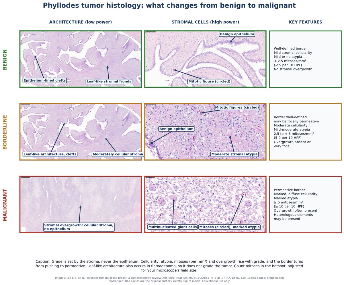
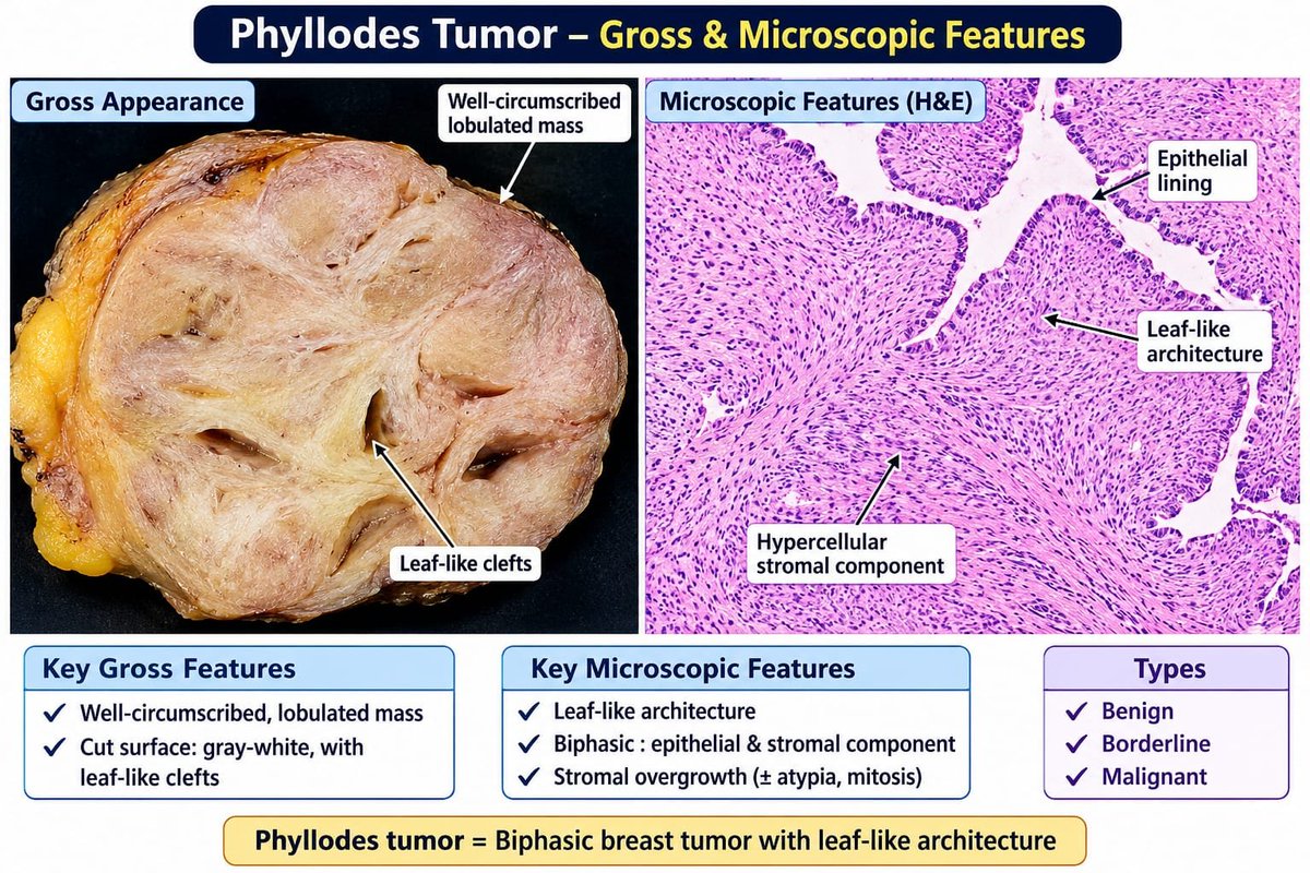
⏩Grossly : well- defined , bosselated, often multinodular masses with fleshy , cleft like cut surfaces . Tongues of tumor into the parenchyma suggest invasion , and hemorrhage and necrosis correlate with malignancy .
➖The key problem is heterogeneity . About 1/3rd of PTs container years that , taken alone , look like fibroadenoma . High grade foci and heterologous elements can be small.
⏩Sampling :
- at least 1 block per cm of the largest dimension
- 2 blocks per cm of the gross appearance is heterogeneous or the diagnosis is uncertain.
- sample margins extensively when breast conservation is planned
- use synoptic reporting
- sampling hasn't been standardized , so this is expert consensus .
⏩The core biopsy challenge:
A core biopsy says " fibroadenoma" How sure can you be ?
- accuracy across studies : 13-84%
- false negative rate 25-30% , underestimation 18.8-37.5%
- In one 455- lesion series, 19.7% of fibroepithelial lesions were upgraded to PT.
- Fewer than 1% of PTs are missed when radiologic pathologic correlation is good.
➖Clues towards PT :
- rapid growth , larger size , heterogeneous ultrasound
- prior fibroadenoma surgery
- stromal predominance or overgrowth
- bipolar nuclei in the background favor fibroadenoma, while stromal fragmentation points to borderline or malignant PT .
⏩Is WHO's " full house" too strict ?
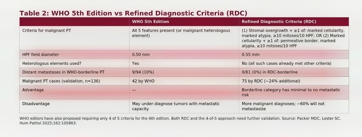
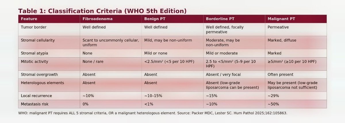
WHO 5th edition required all 5 features for malignant :
1. marked atypia
2. overgrowth
3. 5 or more mitoses/ mm2 , marked diffuse cellularity , and a permeative border.
⚠️The concern is undercalling . 44% ( 22 of 50 ) of PTs with distant metastases would have been called borderline .
⚫ Alternatives :
1. WHO 6th edition ( 2026): four of five features
2. Refined Diagnostic Criteria (RDC) : malignant if overgrowth plus one other features, or marked cellularity plus one other
In a 136 case validation :
-76% classified the same by both systems .
- RDC called 75 tumors malignant vs 42 by WHO
- WHO borderline tumors metastasized in 9 of 94 ( 10% ) , RDC borderline in 0 of 61.
The trade off : about 60% of RDC malignant tumors won't metastasize , so some patients may be over labeled . The authors say more validation is needed .
#BreastPathology #WHOClassification #WHO6 #RDC #DiagnosticPathology #PhyllodesTumor #Histopathology
Is this flat epithelial atypia (FEA)?
No, this is columnar cell hyperplasia
CK 5/6: It is negative like FEA
ER: Positive
Dr. Allison. 2026 Diagnostic Pathology Update #USCAP#pathology#PathX
Ovary – Changes to WHO Classification (6th edition, 2026)
Serous borderline ovarian tumors:
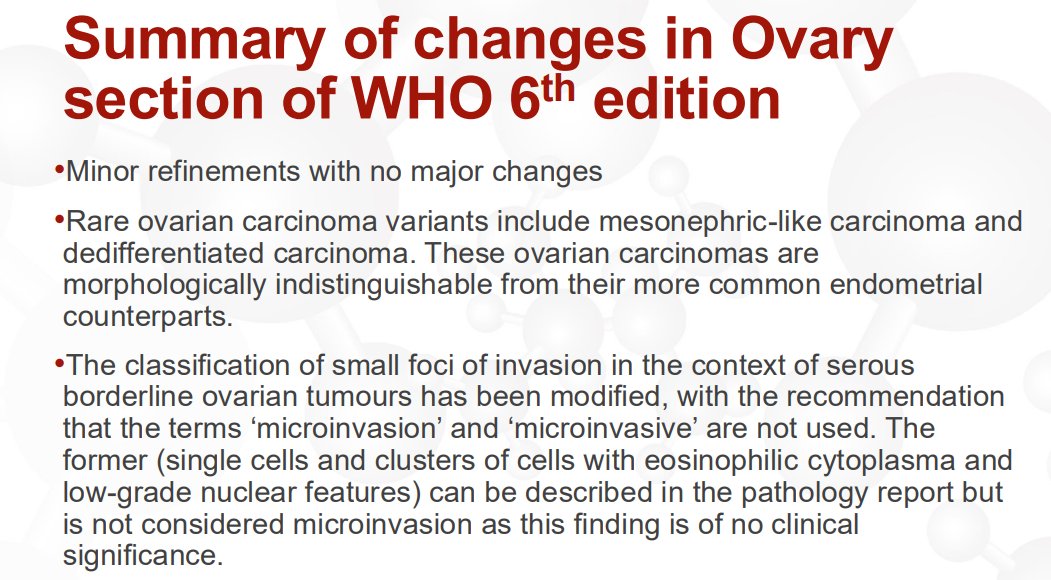
The terms “microinvasion” and “microinvasive” are no longer recommended
What was previously called microinvasion (single cells and clusters of cells with eosinophilic cytoplasm and low-grade nuclear features) has no clinical significance (see pic 2). It can be mentioned in a comment if desired; Dr. Gilks previously reported it but no longer does
What was previously called microinvasive carcinoma represents small foci of low-grade serous carcinoma (see pic 3). Report the size and extent of these foci and diagnose them as a small focus of low-grade serous carcinoma arising in a serous borderline tumor
Dr. Gilks. Gynecologic Pathology Masterclass 2026
#USCAP #pathology #pathX
How to differentiate between adenoid cystic carcinoma and basaloid squamous cell carcinoma?
p40 stains basal/myoepithelial cells only in ACC and is diffuse in SCC.
Pitfall ⚠️: SOX10 and MYB IHC can be positive in both.
Dr. Nishino - 2025 Diagnostic Pathologic Update #USCAP #pathology #PathX
Recognizing pT4a in CRC is important
➡️ Tumor continuous w/ serosal surface through loose inflammation/granulation tissue = pT4a
➡️ Tumor separated from serosa only by non-inflamed fibrosis = pT3
➡️ Tumor <0,1 mm from serosal surface, but not continuous through granulation tissue/loose inflammation = pT3 (as recommended by CAP), but these seem to behave like pT4a
Dr. Rish Pai, #HMAR_DIGESTIVE26
#gipath #pathtwitter
MMR proteins — the simplest way to remember
DNA replication often makes small mistakes (extra or missing bases).
The mismatch repair (MMR) system fixes these errors in two steps:
🔍 MSH2 + MSH6 → detect the error
🔧 MLH1 + PMS2 → repair the error
They work together as a single pathway — first detection, then repair.
If this system fails → errors accumulate → dMMR
And that’s where microsatellite instability (MSI) begins.
Bottom line:
Detection first → then repair
#MVOnco #Oncology #MedEd
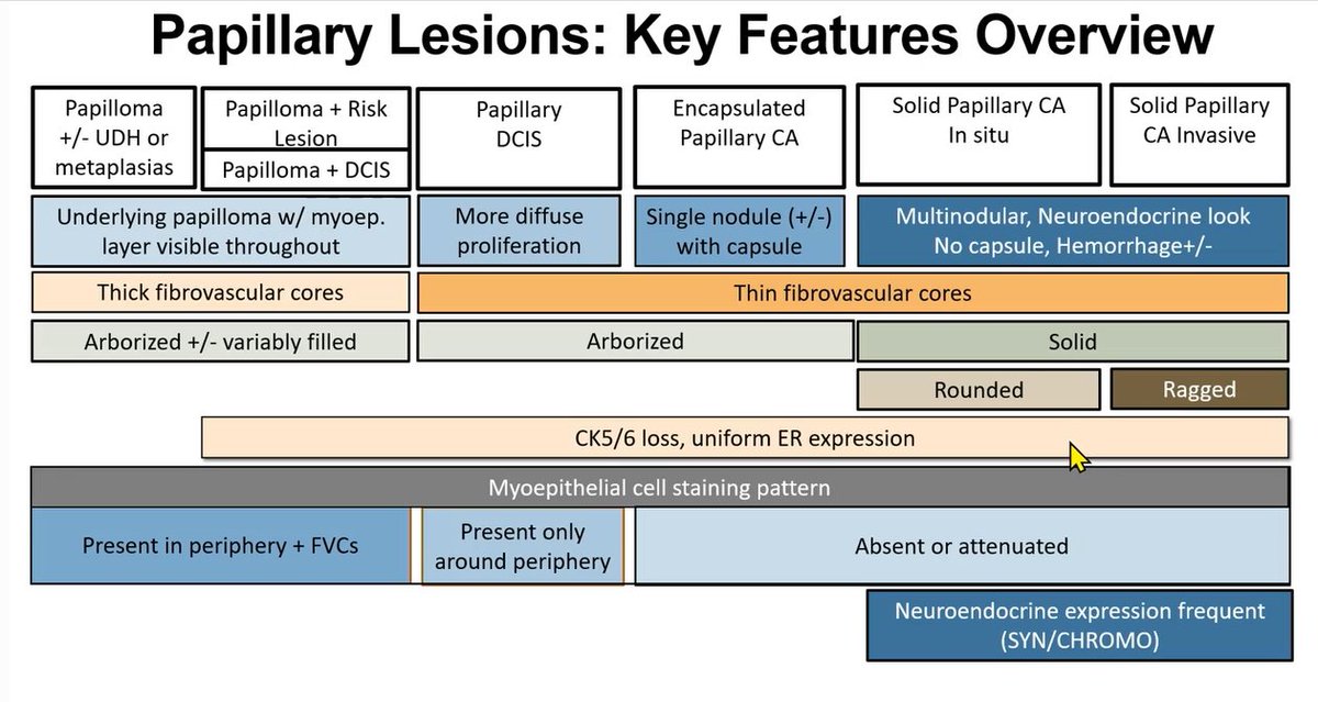
Question 2: The correct IHC profile is Strong, diffuse ER positivity; negative for CK5/6.
#PathTwitter#Pathology#BreastPath
WSI: https://t.co/dF0ROphc7O
Learn more: https://t.co/jEnYok7tYI
Stunning contrast between flat urothelial carcinoma in situ (right side of each image) and reactive urothelium (left side)
CIS➡️enlarged, irregular, & hyperchromatic nuclei
Reactive➡️small, uniform nuclei w/vesicular chromatin + intraepithelial acute & chronic inflammation
Breast
Histologic Variants of Invasive Lobular Carcinoma (ILC)
-Many of these variants coexists and could be difficult to separate one from the other
New variants (WHO 6th edition)
-ILC with extracellular mucin
-ILC with solid papillary growth
Dr. Sahin #USCAP25#pathology #PathX
Urothelial carcinoma with micropapillary features displaying multiple small to medium sized nests lacking fibrovascular cores, situated within lacunar spaces
Neuroendocrine neoplasms can be separated in 3 groups-IARC/WHO Taxonomy
Epithelial NEN
Non-epithelial NEN
Paraganglioma-like NEN
Dr. Mete #USCAP25#pathology#PathX