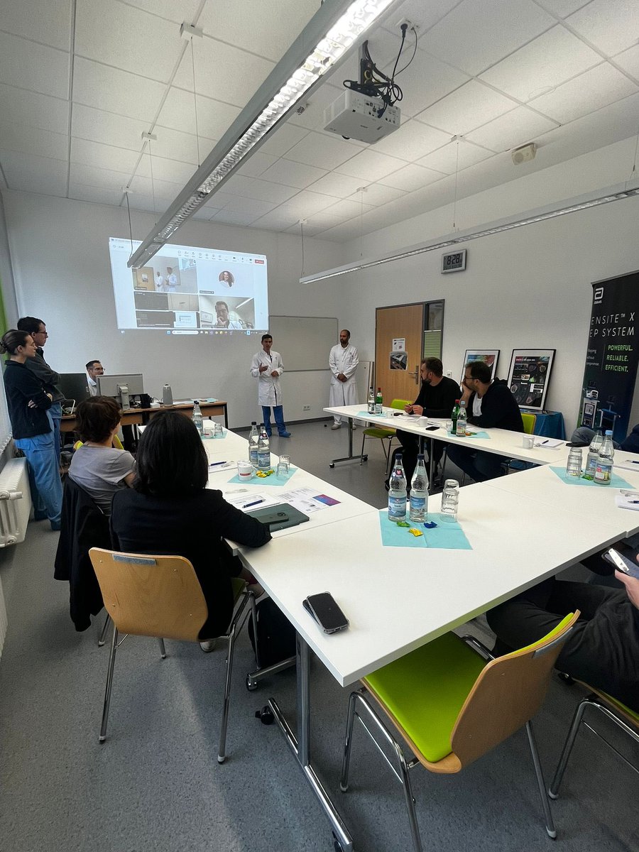
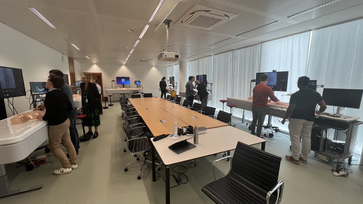
How cool is this?!
First-ever Over-the-Air (OTA) software update — Installed within 1 hour of regulatory approval! No downtime, no delays — just seamless upgrades. Kudos @AbbottCardio for always improvising .
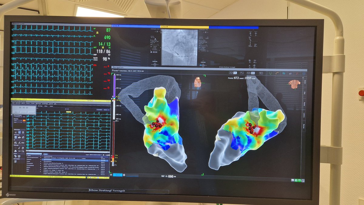
First Ensite 3.1.1 with Contact Index assessment— Precision with the Farawave catheter + ICE = efficacy and safety alongside efficiency #EPeeps
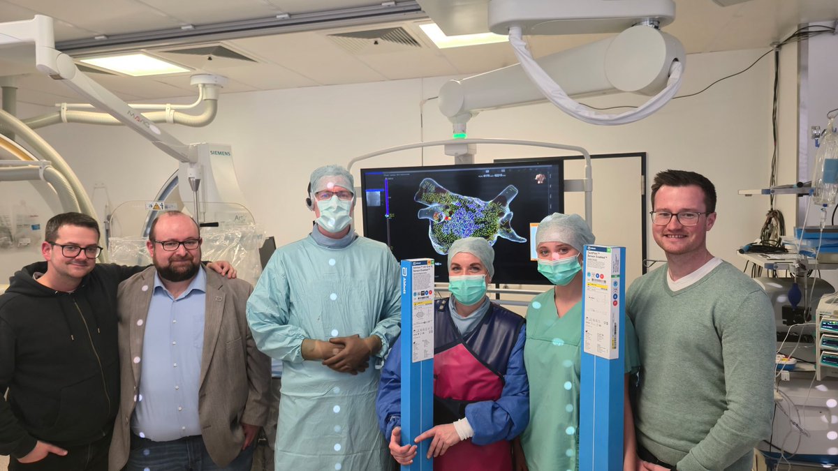
First experience with #HDGridX and #TactiFlex at the #HKZ Rotenburg. Fast Mapping with very good signals. Thanks for Support and teaching @NDalladas@AbbottCardio and Nico Küchenthal and Tim Lehmann
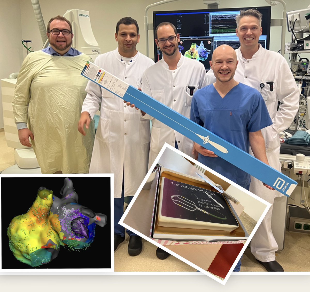
Today we introduced the new and now available #Abbott Advisor HD-Grid X as a first case in DACHPL. It demonstrated convincing fast and accurate electroanatomical mapping!
Thanks to the whole Team, especially to @ChrSteeb and @melhamriti@PhiSo_de, @chris_sohns, @VanessaSciacca_
First European cases of #HDGridX. Amazing anatomy, no need for shaving and immediate collection, no shifts, 100% time high confidence acquisition, "just" real anatomy. Look LA anatomy without NO editing and its merging with CT. @laiallorca1@OriolMartinT@hospitalclinic
Monday started with First-in-Human Use of “ViewFlex X SE” ICE Catheter with @AbbottCardio
I've always loved using the ViewFlex ICE for its exceptional image quality, but now with seamless ICE integration into Ensite X, this is a total game-changer that enhances precision like never before.
Some features that stood out to me
🔹 ICE Integration with Grid and FAM Mapping - Real-time visualization that aligns perfectly with 3D GridX and FAM maps. This precision ensures more efficient targeting of critical anatomy, improving procedural accuracy.
🔹 Smooth Transition to the Left Atrium
Despite its softer shaft, the ViewFlex X SE provided excellent maneuverability and control, allowing smooth navigation to the left atrium while maintaining consistent imaging quality.
🔹 Maintaining Outstanding Image Quality
For those of us who’ve always trusted the ViewFlex ICE platform for its superior imaging, this new catheter doesn’t disappoint—it adds seamless integration without compromising clarity.
#3DMapping #PrecisionMedicine #ViewFlexXSE #Innovation #EPWorkflow @StBernards #epeeps #ArrhythmiaResearchGroup
With multi-wavefront mapping in VT ablation we are always looking for new ways to visualize core concepts. Emphasis mapping on the activation window with ILAM highlights the narrowest isochrones to visualize WADLs which co-localize with the 3D boundaries of the VT. (Thread 1/6).
Check up our latest paper illustrating the value of Peak Frequency Map for Brugada Syndrome
https://t.co/ndxcgplpAF
Panel A illustrates pre-ablation maps, while Panel B illustrates post-ablation maps.
Bei den Herztagen unseres Partners Abbott im CCH traten gestern mehrere Teams im Torwandschießen an. ⚽ Am Ende siegreich: Das Trio um Pauline Machtens und Amelie Woelki sowie Ex-#HSV-Keeper René Adler. 🙌
#nurderHSV [1/2]
What is the mechanism of this arrhythmia? Is this micro Vs. macro reentry? What are the anatomic structures involved?
Interesting questions that continue to rise.
Credits to super mapper @JuanBurnTerm
Happy Friday! #AblateAF@AbbottCardio
👇Idiopathic epicardial and focal VT; have you seen this before❓👇
A young patient with normal LVEF, MRI, and other diagnostic findings presented with incessant refractory and highly symptomatic tachycardia for several months. The patient had previously undergone an unsuccessful ablation attempt externally (endocardial + CS without effect).
With the first RF pulse in the epicardium, the tachycardia was terminated permanently🤩🤩
#EPeeps It is wonderful that the patient can now return to a calm and normal daily life😊😊
@Phiso_de@MBraunEP@VanessaSciacca_@chris_sohns@MoneebKhalaph@Cardioschool@steeb_chr @AleSpoken @AbbottNews #CardioTwitter @AGEP_DGK@_JanzenD
👇Rare WPW Syndrom: Accessory pathway between the left atrial appendage and the left ventricle👇
#EPeeps Very young patient with incessant orthodromic AVRT and status post 3 unsuccessful external ablation attempts. Very challenging anatomical conditions due to severe kyphosis.
The earliest atrial insertion of the AP is located in the LAA. After the first RF pulse in the LAA, the tachycardia terminates and simultaneously blocks the antegrade limb of the AP 🤩🤩
Mapping performed with #HD_Grid and #Ensite_X
@Phiso_de@VanessaSciacca_@chris_sohns@MoneebKhalaph@MBraunEP@Cardioschool @AleSpoken @gut_and6253 @AbbottNews@AGEP_DGK@YoungDgk #Cardiotwitter



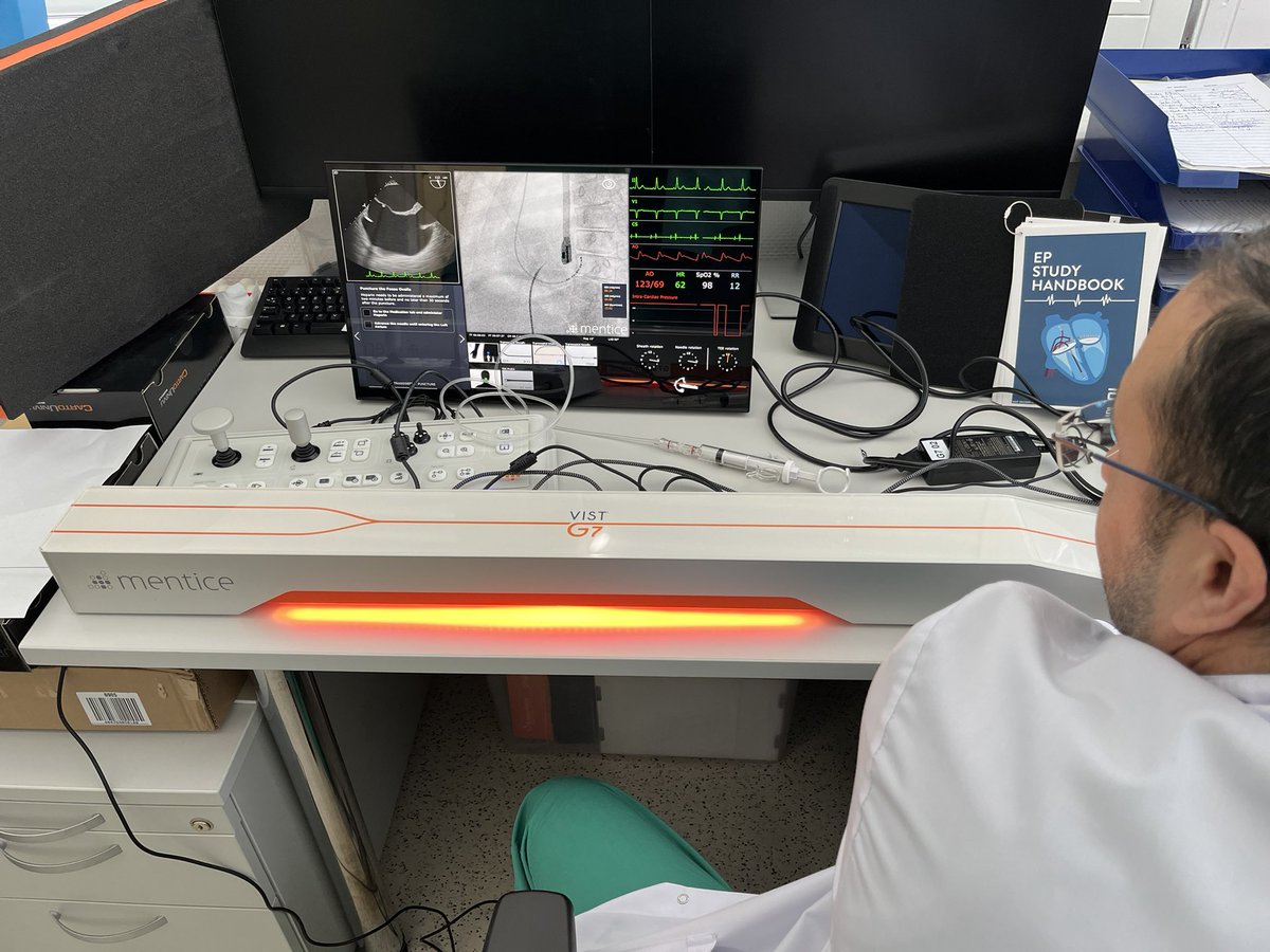









![hsvfrauen_'s tweet photo. Bei den Herztagen unseres Partners Abbott im CCH traten gestern mehrere Teams im Torwandschießen an. ⚽ Am Ende siegreich: Das Trio um Pauline Machtens und Amelie Woelki sowie Ex-#HSV-Keeper René Adler. 🙌
#nurderHSV [1/2] https://t.co/KWZQFRegZV](https://pbs.twimg.com/media/GYdyPqwWQAA1JW5.jpg)