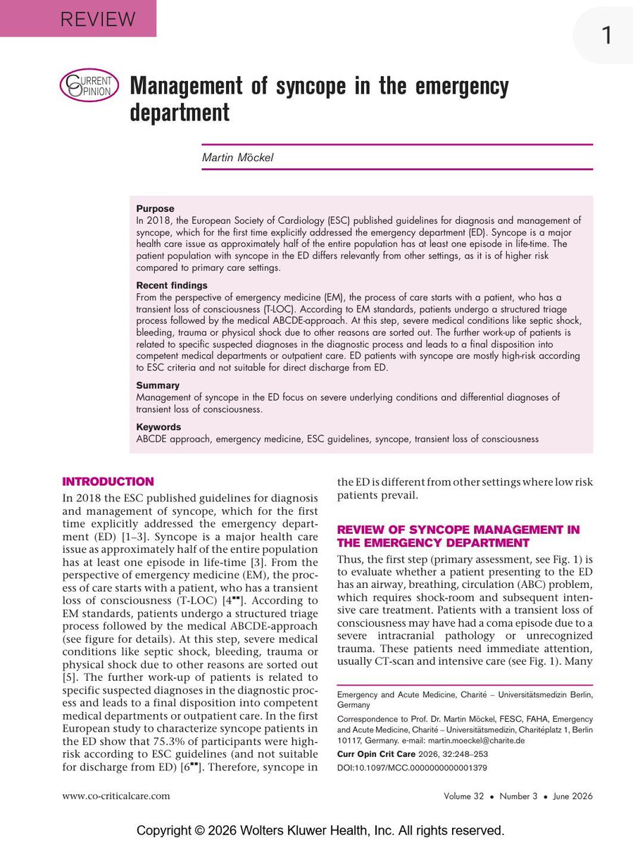
🧠 How should we manage syncope in the Emergency Department?
Syncope is one of the most frequent reasons for emergency department (ED) visits, yet its management often focuses on identifying the cause rather than identifying who is at immediate risk. This updated review reinforces an important concept: the ED should evaluate transient loss of consciousness (T-LOC), not just syncope.
The proposed approach is a simple four-step pathway:
1. Primary assessment using the ABCDE approach. Immediately identify airway compromise, shock, trauma, intracranial emergencies, or sepsis before considering syncope as the diagnosis.
2. Structured diagnostic evaluation. Every patient should undergo vital signs, ECG, focused laboratory testing, and targeted imaging when clinically indicated. A normal ECG is one of the strongest indicators of low risk, whereas abnormal ECG findings should immediately raise suspicion for cardiac syncope.
3. Risk stratification. According to European data, approximately 75% of ED patients with syncope fulfill ESC high-risk criteria, meaning that only a minority are appropriate for direct discharge. From an emergency medicine perspective, the author argues that intermediate-risk patients should generally be managed similarly to high-risk patients.
4. Disposition. Low-risk patients can usually be discharged with outpatient follow-up, whereas high-risk patients require monitoring, further investigations, and treatment directed at the underlying cause. The review's management algorithm clearly summarizes this decision-making process.
Several practical messages deserve particular attention.
Contrary to common teaching, bradyarrhythmias appear to be a more frequent cause of unexplained cardiac syncope in the ED than ventricular tachycardia. Because most clinically significant arrhythmias are detected within the first hours, 6 to 12 hours of ECG monitoring is generally sufficient for most patients with suspected cardiac syncope.
The review also highlights important differential diagnoses that should never be overlooked. Pulmonary embolism should be considered when clinically suspected, although routine screening is not recommended because its prevalence is much lower than initially reported. Severe aortic stenosis should prompt focused echocardiography, and clinicians should actively search for infection or sepsis, particularly in older or frail patients where syncope may be the presenting symptom.
Perhaps the most important clinical takeaway is that syncope is not a diagnosis but a presentation. The emergency physician's task is not simply to explain why the patient fainted, but to rapidly distinguish patients who can safely go home from those harboring potentially life-threatening disease.
Reference 📚
Möckel, M. (2026). Management of syncope in the emergency department. Current Opinion in Critical Care, 32(3), 248–253. https://t.co/qSo0JWkiVW
💉Acute pancreatitis is no longer just about fluids and pain control.
Our understanding of acute pancreatitis has changed dramatically over the past decade. What was once considered a disease driven primarily by pancreatic autodigestion is now recognized as a complex interaction between calcium dysregulation, mitochondrial dysfunction, impaired autophagy, innate immunity, inflammatory signaling, and multiple forms of programmed cell death. These mechanisms ultimately determine whether a patient experiences a mild, self-limited illness or progresses to persistent organ failure.
This comprehensive 2026 review highlights how advances in molecular biology are reshaping clinical practice. Early management still relies on aggressive supportive care, but risk stratification is becoming increasingly sophisticated. Traditional scores such as BISAP, APACHE II, SOFA, CTSI, and the revised Atlanta Classification remain essential, yet their greatest limitation is that they often describe disease severity after the inflammatory cascade is already established rather than predicting it early enough to change outcomes.
Perhaps the most exciting development is the emergence of artificial intelligence. Machine learning models consistently outperform conventional scoring systems for predicting severe acute pancreatitis, organ failure, ICU admission, and mortality. Rather than relying on a few static variables, these models integrate laboratory trends, imaging, physiological parameters, and clinical evolution to generate dynamic risk estimates that may eventually support real-time bedside decision making.
The review also reinforces several important practical messages. Gallstones remain the leading global cause of acute pancreatitis, followed by hypertriglyceridemia and alcohol, although regional differences are substantial. Serum lipase should be preferred over amylase for diagnosis because of its superior sensitivity and specificity, while contrast-enhanced CT remains the cornerstone for assessing complications, ideally after the first 72 hours when necrosis becomes more accurately defined.
For intensivists, the future of pancreatitis management will likely move beyond supportive therapy toward precision medicine. Biomarkers, explainable artificial intelligence, transcriptomics, metabolomics, and immunophenotyping may soon identify patients who require individualized interventions before irreversible organ dysfunction develops.
Acute pancreatitis remains one of the most challenging inflammatory diseases encountered in critical care. The next major breakthrough will probably not be a new drug, but earlier recognition of the patient who is about to deteriorate.
Reference
Hu, Q., Hu, Y., Tan, C., Yang, Y., Su, H., Huang, Z., Tang, W., Wang, R., Liu, J., & Wan, M. (2026). Acute pancreatitis: Mechanisms and therapeutic approaches. Signal Transduction and Targeted Therapy, 11, 15. https://t.co/GR6eeFh2Jv
¿Gastritis, úlceras o infección por Helicobacter pylori? 🦠
La ACG ha actualizado sus recomendaciones de tratamiento, resumidas en este paper de JAMA.
1️⃣ Para pacientes sin tratamiento previo, la terapia cuádruple con bismuto durante 14 días es la opción recomendada como primera línea.
2️⃣ No se recomienda usar empíricamente regímenes con claritromicina, por el problema de la resistencia antibiótica.
3️⃣ Si BQT no es viable, se sugieren alternativas como terapia triple con rifabutina o terapias con PCAB, como vonoprazan, según el caso.
4️⃣ Es clave confirmar la erradicación con prueba de aliento, antígeno en heces o biopsia al menos 4 semanas tras completar el tratamiento.
La guía se centra en adultos de Norteamérica; en otros contextos, los patrones locales de resistencia pueden cambiar la elección del tratamiento.
#Hpylori #Helicobacterpylori #Gastroenterología #Microbiología #ResistenciaAntimicrobiana #Antibióticos #Medicina #DivulgaciónCientífica @SEMicrobiologia
🎯 Helicobacter pylori (H. pylori) infection treatment GUIDELINES
🕯️ Triple therapy for Helicobacter pylori (H. pylori) infection typically consists of a proton pump inhibitor (PPI) and two antibiotics.
🎯 Amoxicillin, clarithromycin, and omeprazole is the most commonly used regimen worldwide for the eradication of H. pylori. The combination has a cure rate of about 80%.
#MedEd #MedX #MedTwitter #GastroTwitter #infections #pharmacy #IDTwitter #ClinicalPearls
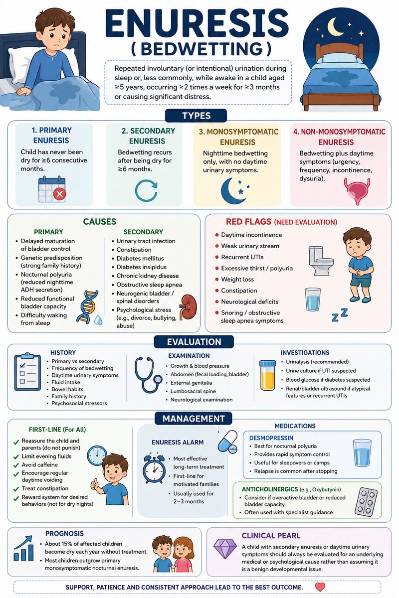
Bedwetting isn’t just “a phase.”
Enuresis can be a symptom of bladder immaturity, constipation, diabetes, urinary tract disease, sleep disorders, or psychological stress.
Knowing the difference between primary and secondary enuresis can change management.
Here’s a quick evidence-based guide 👇
#MedTwitter #Pediatrics #FOAMed #MedicalEducation #Enuresis
🌀 7 CAUSES OF VERTIGO
Vertigo is a symptom, not a diagnosis.
When a patient complains of “dizziness,” the key question is:
❓ Is this true vertigo (a false sensation of movement) or something else?
Correctly distinguishing peripheral from central causes is essential, as some causes are benign, while others may represent a neurological emergency.
🔑 Common Causes of Vertigo
1️⃣ BPPV (Benign Paroxysmal Positional Vertigo)
• Most common cause
• Brief episodes triggered by head movement
• No hearing loss
• Positive Dix-Hallpike test
• Treated with the Epley manoeuvre
2️⃣ Vestibular Neuritis
• Acute severe vertigo lasting days
• Often follows a viral illness
• Nausea and vomiting common
• No hearing loss
3️⃣ Ménière Disease
• Recurrent episodes of vertigo
• Fluctuating hearing loss
• Tinnitus and aural fullness
• Usually unilateral
4️⃣ Vestibular Migraine
• Vertigo associated with migraine features
• Photophobia, phonophobia, aura
• May occur without headache
5️⃣ Labyrinthitis
• Vertigo + hearing loss
• Often post-viral
• Auditory and vestibular symptoms occur together
6️⃣ Acoustic Neuroma (Vestibular Schwannoma)
• Progressive unilateral hearing loss
• Tinnitus
• Gradual onset imbalance or vertigo
7️⃣ Central Causes 🚨
• Stroke
• Multiple sclerosis
• Cerebellar tumours
• Brainstem lesions
⚠️ Red Flags Suggesting a Central Cause
🚨 Diplopia
🚨 Dysarthria
🚨 Limb weakness
🚨 Severe ataxia
🚨 New headache
🚨 Persistent neurological deficits
🩺 Clinical Approach
✔️ Take a careful history
✔️ Examine for nystagmus
✔️ Perform the HINTS examination when appropriate
✔️ Assess hearing
✔️ Look for focal neurological signs
💡 High-Yield Pearl
Peripheral vertigo is common, but never miss a central cause.
Remember:
🧠 Acute vertigo + neurological signs = Stroke until proven otherwise.
📚 Save this post and visit our website for innovative high-yield medical notes:
https://t.co/Ivljom2bsM
#Vertigo #Neurology #BPPV #MeniereDisease #vestibularneuritis
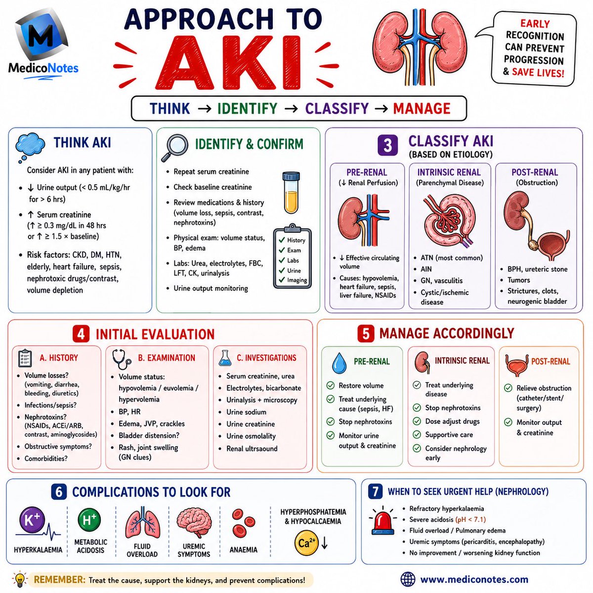
🩺 Approach to Acute Kidney Injury (AKI)
AKI is a medical emergency—recognize it early, identify the cause, and intervene before irreversible kidney damage occurs.
AKI is defined by a rapid decline in kidney function, resulting in rising serum creatinine and/or reduced urine output. The key to management is determining whether the cause is pre-renal, intrinsic renal, or post-renal.
📚 Stepwise Approach to AKI:
1️⃣ Think AKI
• Rising creatinine or oliguria (<0.5 mL/kg/hr).
• Review previous renal function and identify high-risk patients.
2️⃣ Confirm the Diagnosis
• Repeat renal profile.
• Perform urinalysis and monitor urine output.
• Review medications, recent contrast exposure, and comorbidities.
3️⃣ Classify the Cause
🟢 Pre-renal: Hypovolemia, sepsis, heart failure, cirrhosis.
🔵 Intrinsic: ATN, glomerulonephritis, AIN, vasculitis.
🟠 Post-renal: Urinary tract obstruction (BPH, stones, malignancy).
4️⃣ Investigate Systematically
• Assess volume status and blood pressure.
• U&Es, bicarbonate, urinalysis, urine sodium/osmolality.
• Renal ultrasound if obstruction is suspected.
5️⃣ Treat the Underlying Cause
✔️ Restore perfusion in pre-renal AKI.
✔️ Stop nephrotoxic drugs (NSAIDs, ACEi/ARB where appropriate).
✔️ Relieve urinary obstruction promptly.
✔️ Correct electrolyte abnormalities and monitor fluid balance closely.
💡 High-Yield Exam Pearls
✔️ ATN is the most common intrinsic cause of AKI.
✔️ Always exclude post-renal obstruction with renal ultrasound.
✔️ Hyperkalaemia, severe metabolic acidosis, pulmonary oedema, and uraemic complications require urgent nephrology review and consideration of dialysis.
✔️ Treat the cause, not just the creatinine.
📖 Want more concise, exam-focused nephrology notes?
Visit https://t.co/2UQyCbb6in and explore our Nephrology & Urology book—designed for medical students, MRCP, USMLE, PLAB, AMC, and everyday clinical practice.
❤️ Save this post for revision.
📤 Share it with your colleagues.
💬 What investigation do you always order first when assessing AKI?
#AKI #AcuteKidneyInjury #Nephrology #InternalMedicine #MedicalStudent
After H. pylori treatment:
• Continue PPI daily for 4–6 weeks if ulcer is large (>1 cm) or complicated.
• Confirm eradication with stool antigen/PCR or biopsy ≥4 weeks after antibiotics and ≥2 weeks after stopping PPI.