@Dr_Brian_Cox @GIJamesMD Super interesting! Yes, as @Dr_Brian_Cox says, p16 can be a sign of Rb loss. My colleagues had a paper showing that neuroendocrine carcinomas in the GI tract typically have Rb loss or HR-HPV but not both. p16 is positive in both situations. Do you have Rb stain or HR-HPV?
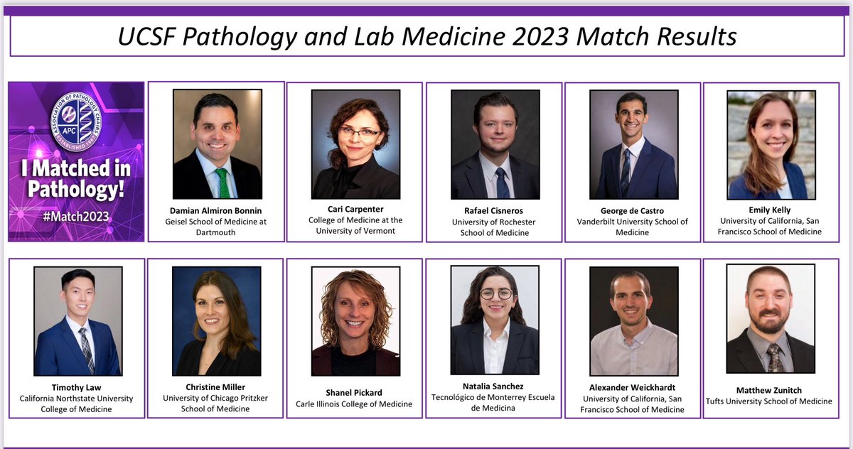
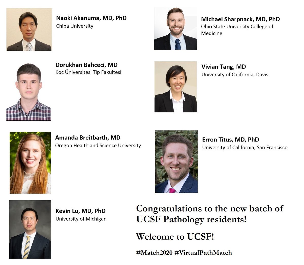
Can’t think of a better first tweet than to proudly announcing the newest outstanding additions to the UCSF Pathology family!!!! #Match2020#VirtualPathMatch#Path2Path#UCSF
Anastomosing hemangiomas are just plain fun to spot. Sarah Umetsu @neuthunter and her colleagues have even found GNAQ and GNA14 mutations in these cuties.
UCSF has an unexpected opening in our Surgical Pathology Fellowship for 2019-2020. The experience includes a wide range of in-house cases, consultation cases, and intraoperative frozen section diagnosis. Any trainees that are interested should email me ([email protected]).
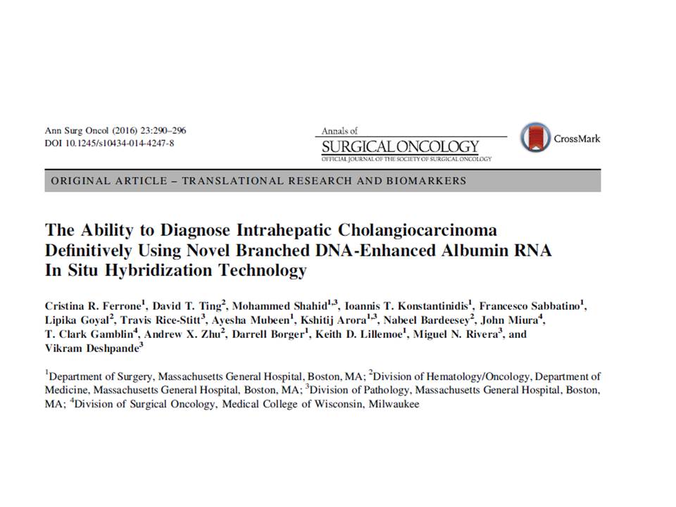
@slusagar Albumin ISH is great, but will be positive in both HCC and intrahepatic cholangiocarcinoma, so not helpful in this setting. I find it most useful in distinguishing intrahepatic cholangiocarcinoma from a metastatic adenocarcinoma to the liver.
Super lovely example of anastomosing hemangioma of kidney. It even has extramedullary hematopoiesis and hyaline globules. These tumors are so cute and can be foolers for angiosarcoma.
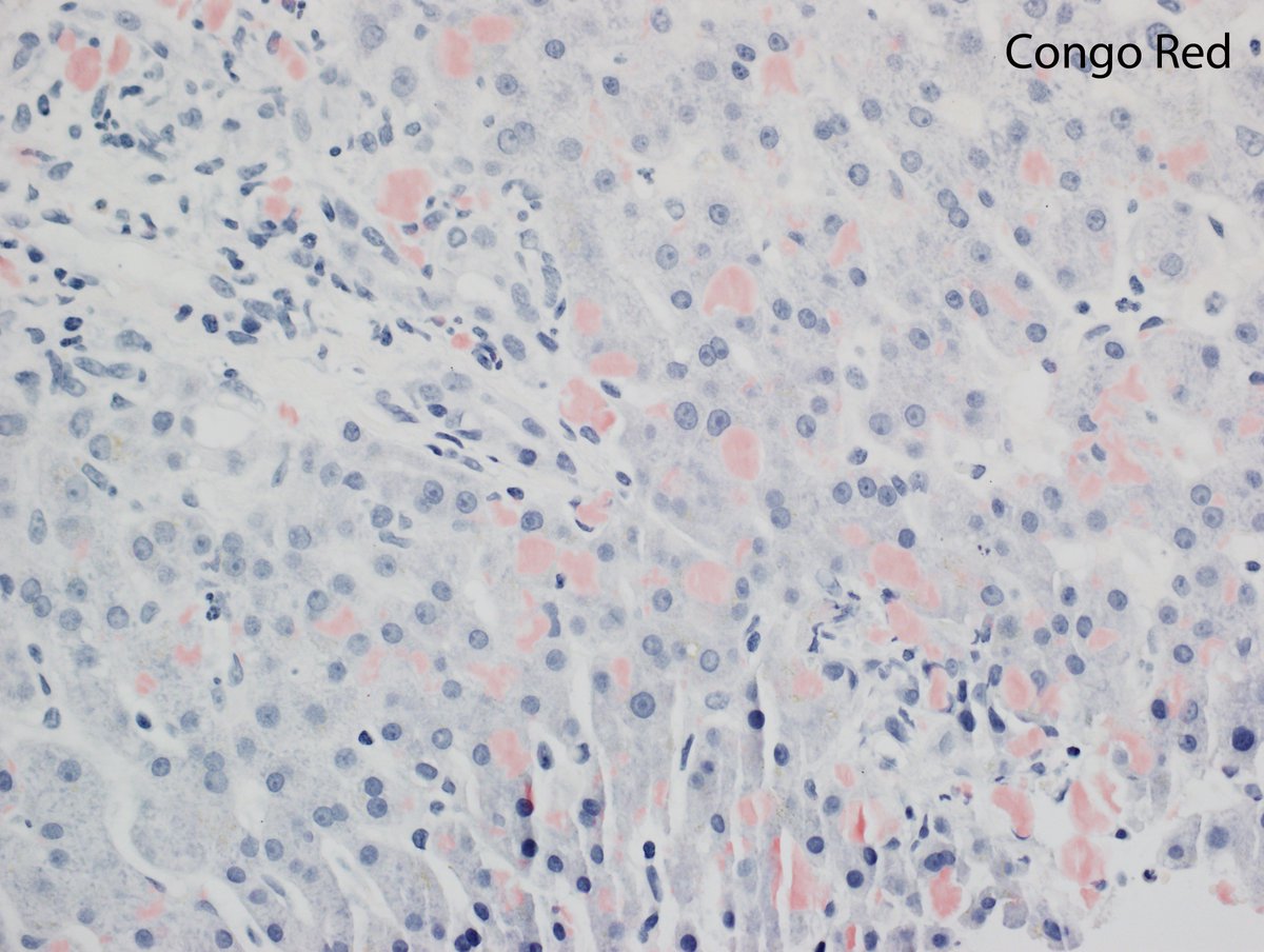
A more subtle case of hepatic amyloid. In the liver, this globular pattern of amyloid is highly specific for ALECT2, an unusual type of amyloid associated with patients of Hispanic ethnicity. The globules are seen in the sinusoids, portal tracts, and around central zones.
Impressive example of hepatic amyloidosis. 84 year old man presented with liver failure and hepatomegaly. Kappa restriction shown by immunofluorescence. Patient later found to have a plasma cell neoplasm.
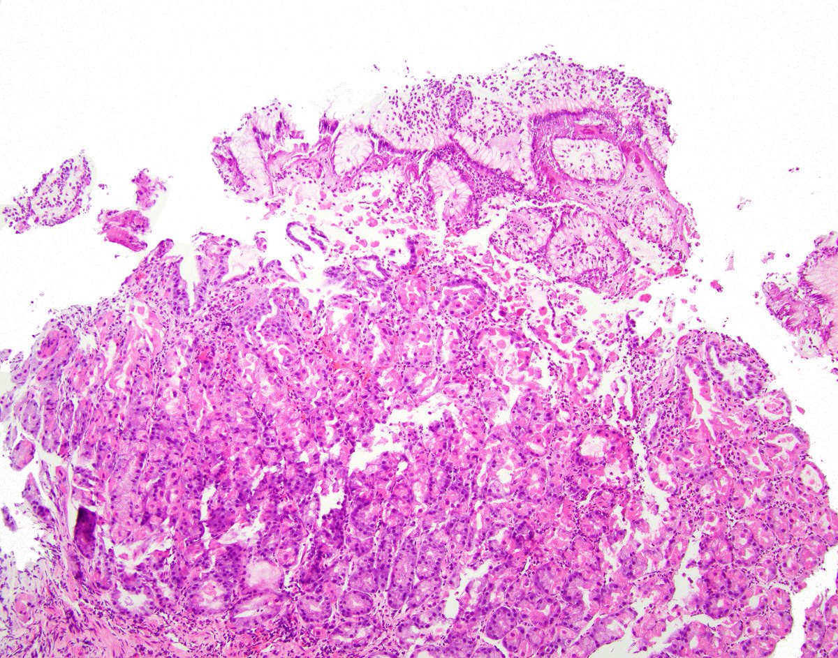
Yummy case of doxycycline gastropathy. Check out the fibrinoid change in the tiny vessels that are pointed out with arrows. I was just proud to be able to draw arrows in photoshop.