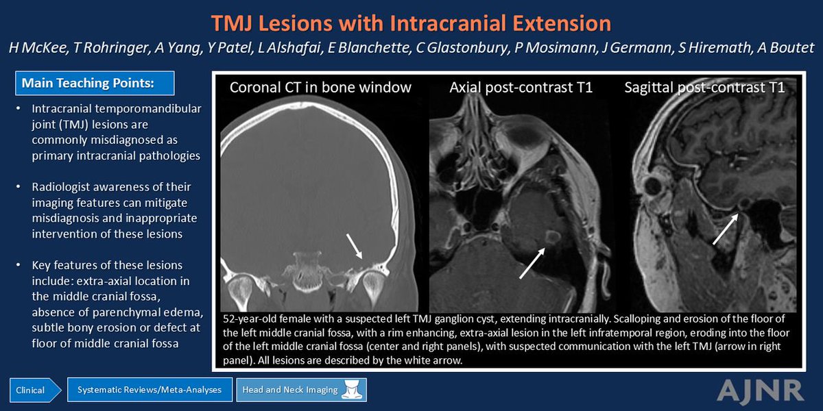
"Temporomandibular Joint Lesions with Intracranial Extension: Illustrative Cases from a Systematic Review of the Literature and Our Institution"
https://t.co/YupbYklulL
@HayleyAMcKee
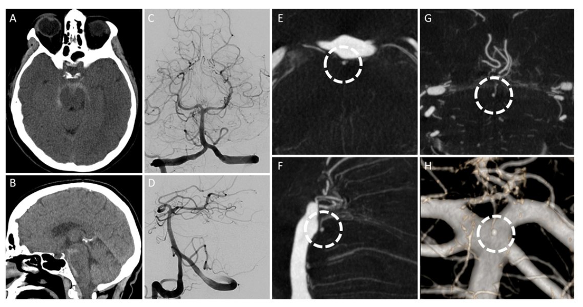
PICA dissecting aneurysms require careful planning and often require trapping. Bypass is frequently needed but good collateral circulation may obviate the need for it. This video was prepared by Dr Pachon from our PNT/NSI lab. @MariaJPachonL@MayoClinicNeuro@MayoClinic@cvsection@AmirSigari@AbhiBathiniMD@DeviPatraMD@jacquesmorcosmd
*Used with permission of Mayo Foundation for Medical Education and Research, all rights reserved.*
The gateway to the “Sistine Chapel of the brain” is the tentorial fissure separating cerebrum & cerebellum. Pineal & midbrain pathologies are made even more spectacular w/gravity retraction in sitting position. Long and awkward reach is addressed w/ a chair w/armrests raised high…
Check out our latest publication in Surgical Neurology International @SNInt:
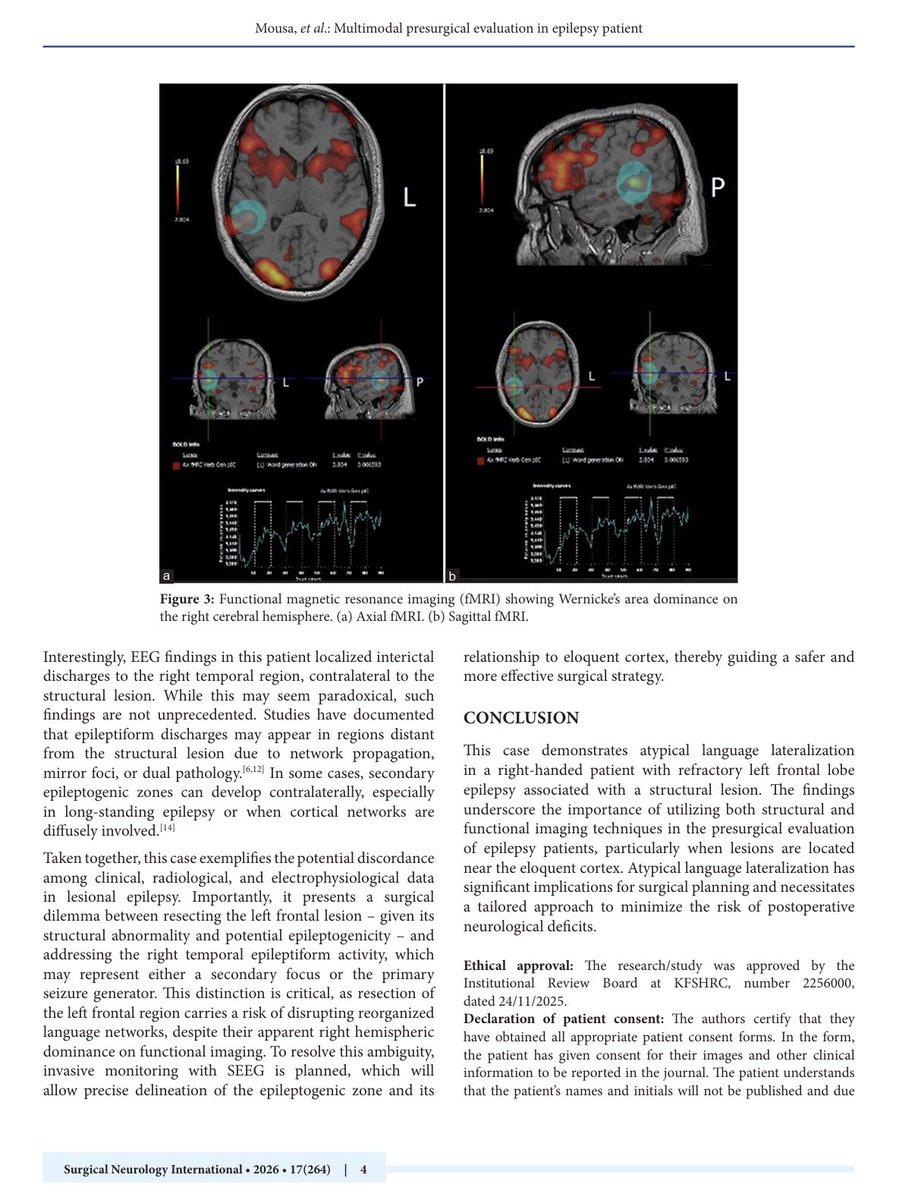
“Atypical Language Lateralization and Contralateral Epileptiform Activity in a Right-Handed Patient with a Left Frontal Cortical Lesion: A Multimodal Presurgical Evaluation”
🔗 Link: https://t.co/iU0pGeo6ug
🔹In this case report, we present a unique presurgical epilepsy evaluation highlighting atypical right-hemisphere language dominance in a right-handed patient with a left frontal cortical lesion, emphasizing the importance of multimodal assessment in epilepsy surgery planning.
🔹Key insights:
• Functional MRI demonstrated atypical right-sided dominance for both Broca’s and Wernicke’s areas
• EEG revealed contralateral right temporal epileptiform activity despite a left frontal lesion
• Multimodal integration of imaging and electrophysiology was critical for individualized surgical planning
Special thanks to Dr. @Afnan_Alkhotani for her continuous support and mentorship throughout this work.
#Neurosurgery #Epilepsy #fMRI #EEG #EpilepsySurgery #BrainTumor #Neuroimaging #LanguageLateralization #SurgicalNeurology #Research
#Neurology
Extradural clinoidectomy is a key skull base technique that involves removal of the anterior clinoid process before opening the dura.
This approach is important in the treatment of select anterior skull base pathologies, like meningiomas. By working extradural first, the tumor can be devascularized early, making resection safer and more controlled. It also allows for early decompression of the optic nerve and provides improved visualization of critical neurovascular structures once the dura is opened.
#SkullBaseSurgery #Neurosurgery #Meningiomas #OpticNerve #NeurosurgicalApproach #SurgicalAnatomy #NeuroOncology #SkullBase
In this operative video, @AaronCohenGadol demonstrates the orbitozygomatic approach for resection of a craniopharyngioma.
The area of the lamina terminalis where the tumor is protruding is identified, and the thin roof is dissected as far posterior as possible from the chiasm. The operative route is limited and removal can be challenging, especially when filling the posterior third ventricle. The tumor is removed piecemeal through the sub-frontal operative trajectory. Debulking allows mobilization of the mass into the resection cavity despite the limited exposure within the lamina terminalis.
Learn more here: https://t.co/d16p1BV8Ez
#Neurosurgery #Craniopharyngioma #DrCohen #SurgicalExcellence
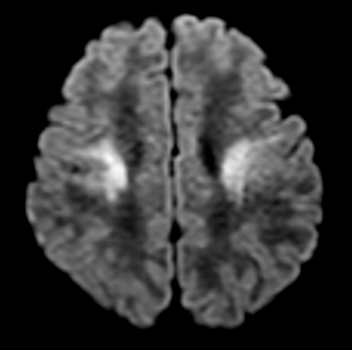
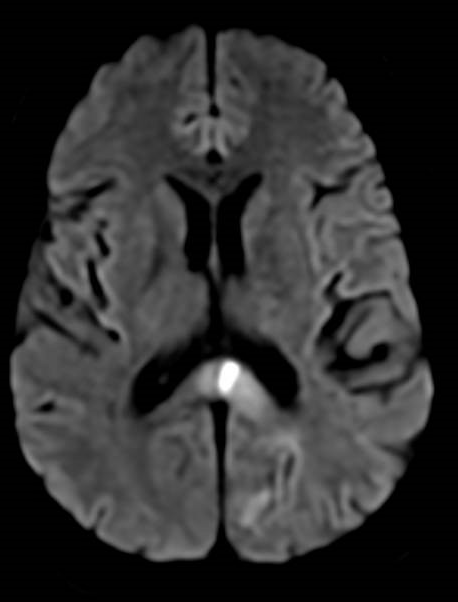
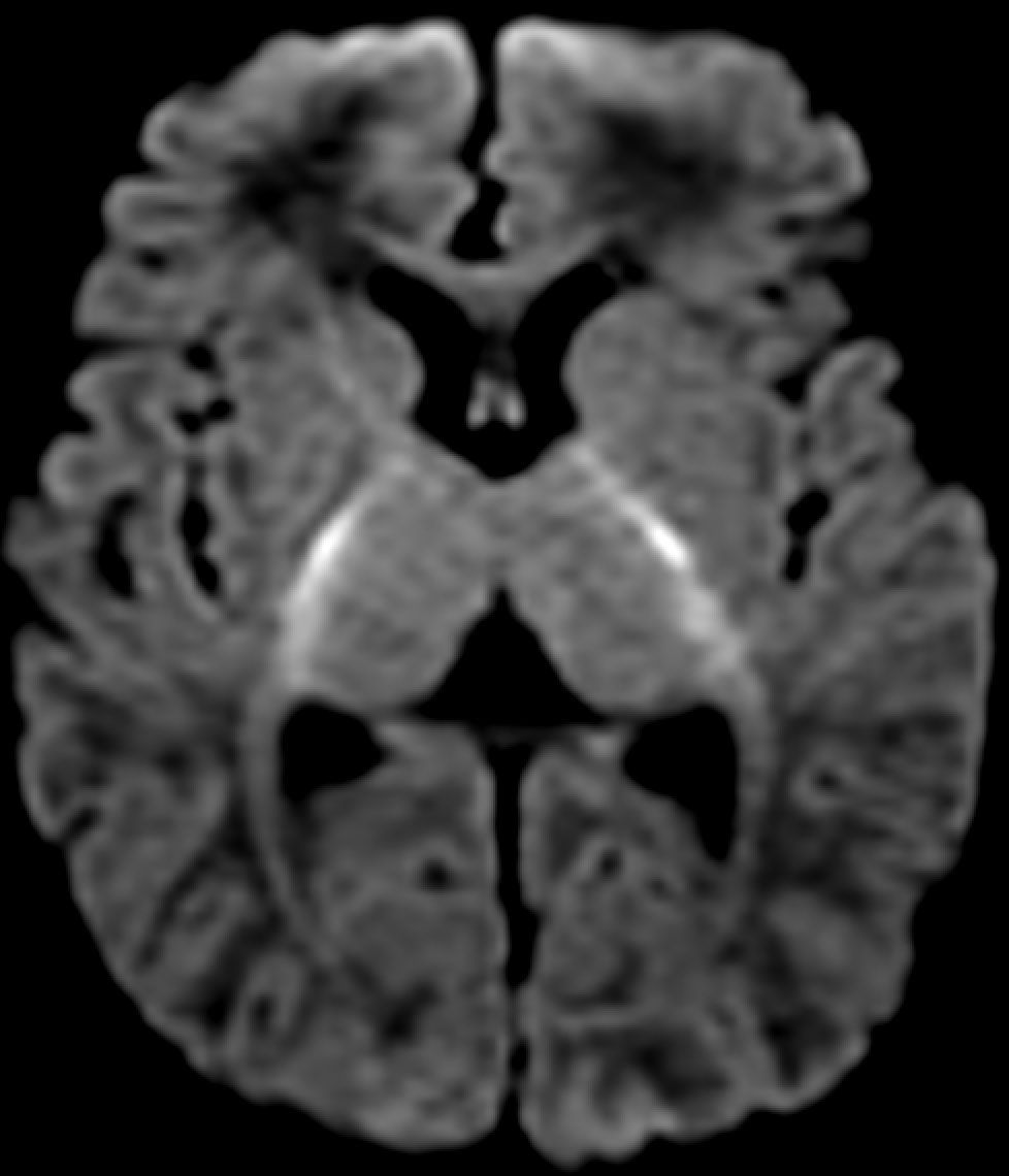
A 66-year-old man with a longstanding history of severe metabolic syndrome presents to the ED with acute neurological deficits.
MRI DWI (axial) demonstrates restricted diffusion in multiple brain regions, as shown below.
Based on these imaging findings, what are his likely symptoms and signs?
Drop your thoughts below 👇👇
#Stroke #Neuroradiology #FOAMed
🏥Adult patient with a slowly progressive, painless skull swelling over several years. No focal neurological deficits. Cosmetic concern becomes the main reason for consultation.
👨⚕️What would you consider in the differential?
#NEUPrac Fluorescence of Cladophialophora Bantiana After Administration of 5-Aminolevulinic Acid Hydrochloride: Case Report https://t.co/IUmMXUiSJR by Mitchell et al @michiganstateu@CNS_Update@dgolubMD
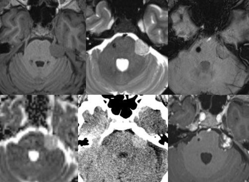
🩺This neonate presented with hypotonia, lethargy, feeding difficulties, and respiratory distress, followed by seizures.
🩻There is bilateral symmetric diffusion restriction in the peri-Rolandic white matter, posterior limbs of the internal capsules, dorsal midbrain, central tegmental tracts, and middle cerebellar peduncles.
🔬The patient was found to have biallelic pathogenic IBA57 variants, causing multiple mitochondrial dysfunction syndrome (MMDS).
💡MMDS is caused by mutations in genes encoding for proteins involved in mitochondrial iron-sulphur cluster assembly, leading to multi-enzyme mitochondrial dysfunction. MMDS is severe, progressive, and ultra-rare, unfortunately with no curative treatment.
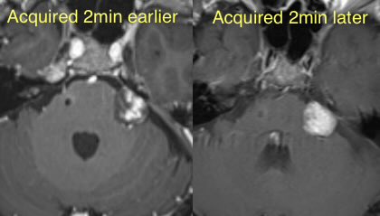
I operate on a dozen carotid body (CB) tumors each yr & rarely see the glomus vagale tumor. Least common of the head/neck paragangliomas, occurring 1/10th the frequency of CB tumors, it arises from inferior nodose ganglion of vagus nerve, midway b/w carotid bifurcation & skull base. It’s an upward reach above hypoglossal nerve & thru digastric muscle…
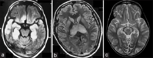
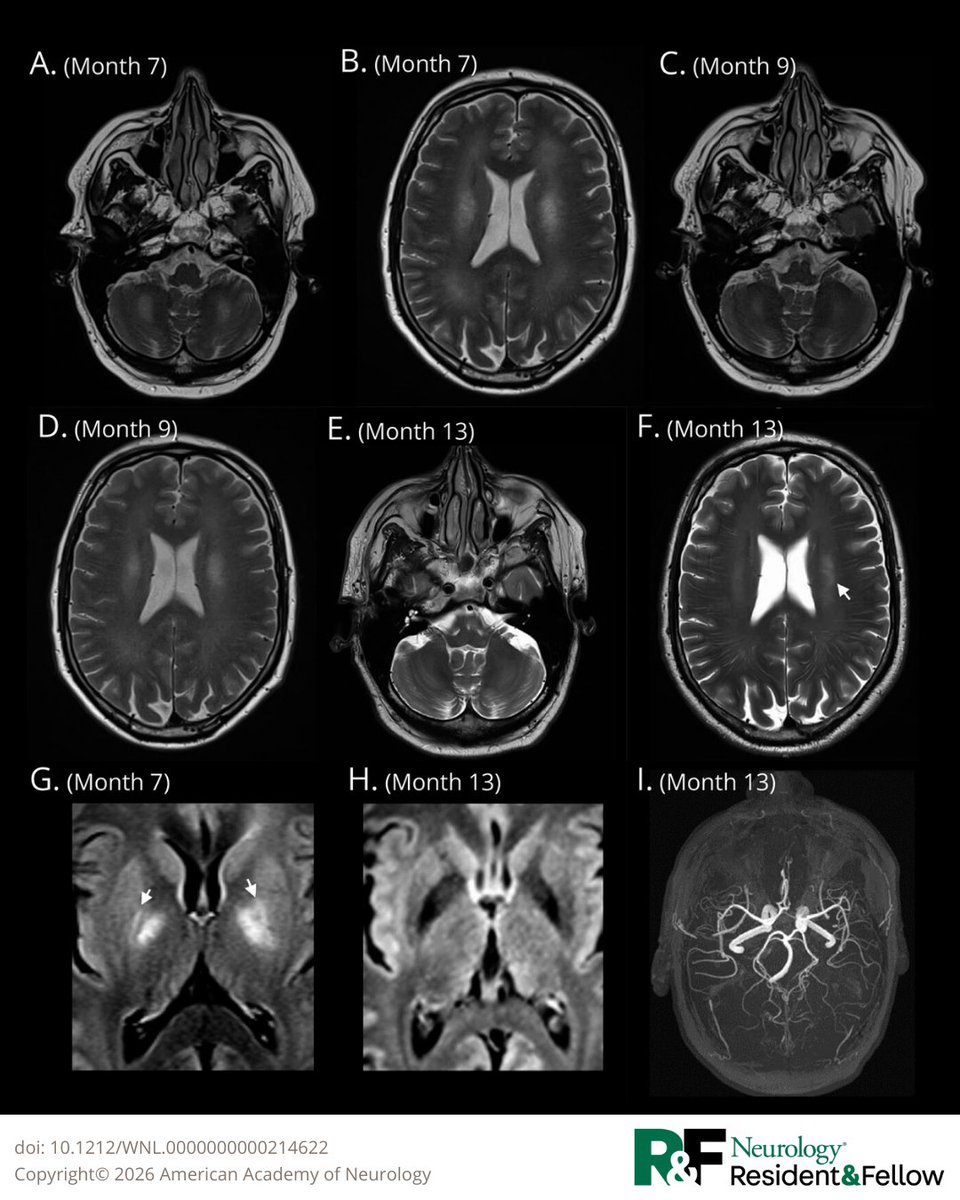
🧙♂️69-year-old man with subacute cognitive decline affecting work performance. Initial improvement after steroids, followed months later by seizures, then relapse with nonfluent aphasia and rapidly progressive left hemiparesis.
👨⚕️What differential diagnoses would you consider?
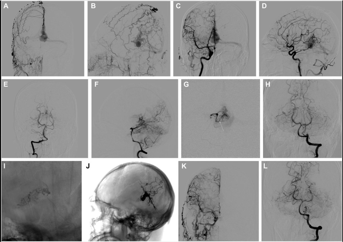
#NEUNew Intracranial Dural Arteriovenous Fistulas With and Without Pial Artery Supply: Analysis of Treatment Outcomes https://t.co/CqPWkfRuV7 by Su et al at Xuanwu Hospital, Capital Medical University, Beijing @CNS_Update@visishs@dgolubMD