A Cork nurse working with epileptic children came up with a plan for @CUH_Cork that has saved the HSE €260,000 and freed up hundreds of bed days in just two years.
Neurology nurse specialists are transforming care for those with neurological conditions and their familes, as well as saving money and freeing up hospital beds. @HSELive needs more of them.
Full story via @thejournal_ie at
https://t.co/HbXbJDZMYK
The #myneurologynurseandme awareness campaign shines a spotlight on the vital role of neurology nurse specialists in supporting people living with neurological conditions across Ireland.
Specialist neurology nurses are at heart of neurological services, proving expert clinical support, educating and empowering patients and improving outcomes for people with neurological conditions 💜
Lovely few days with this @BPNA_org team of ED, Paed neuro, general paed clinicians!
Focussing on those critical first few hours when children present with neuro probs!! Updated course in the pipeline for Sep 25 👀
“Time is brain!”
Age isn’t just a number!
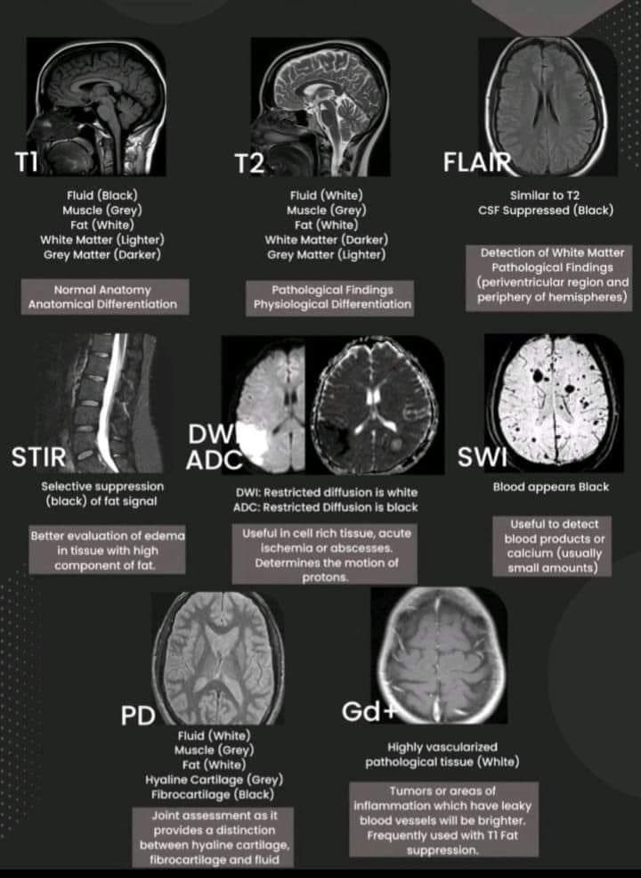
Do you know how to tell the age of blood on MRI?
This post shows you how to date blood on MRI so that the next time you see a hemorrhage, your guess on when it happened will always be in the right vein!
No one likes the crazy mnemonic about babies & daddies
But if you look at the mnemonic, you will notice that T1 signal is all you need to tell if blood is acute, subacute or chronic.
Remember 2 basic MRI rules:
(1) Remember T1 loves protein. More protein = brighter on T1. I remember this bc T1 looks like the T & I in protein
(2) T2 loves water. Fluid is bright on T2. This is easy bc there’s a 2 in H2O.
ACUTE BLOOD
--First hours, basically blood just poured out of the artery.
--How does T1 feel about acute blood? Acute blood has water w/little protein
--Get some love from T1 for the little protein, isointense on T1.
SUBACUTE BLOOD
--Subacute = blood gets oxidized.
--Why an apple or a steak turns dark when it’s left out
--Hemoglobin becomes methemoglobin.
--Cells will begin to lyse & water content is lost = more dense protein.
--More protein means HIGH T1 signal.
CHRONIC
-- Proteins lyse, including heme, releasing iron
--Iron clumps to make ferritin/hemosiderin.
--Neither T1 or T2 sequences like metals like ferritin/hemosiderin.
--Remember: metal doesn’t mix well w/protein (cuts right through it) or water (together water & iron make rust).
Hopefully this post will age well!
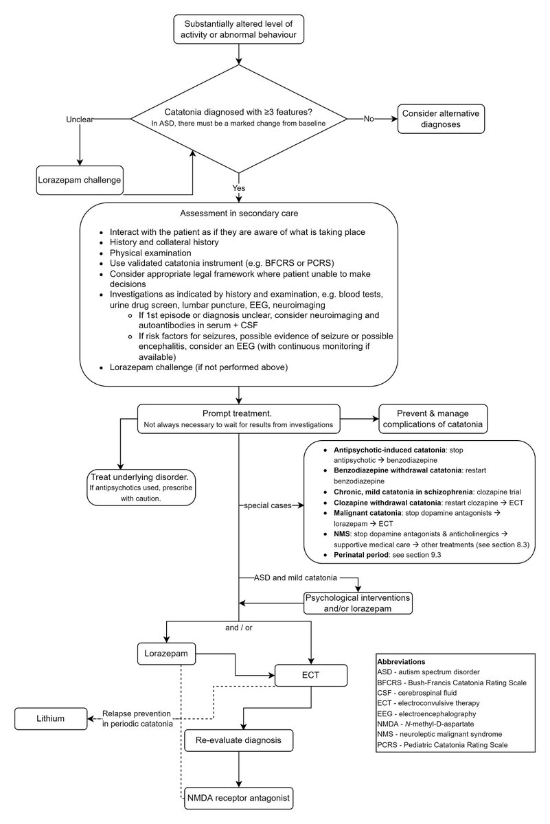
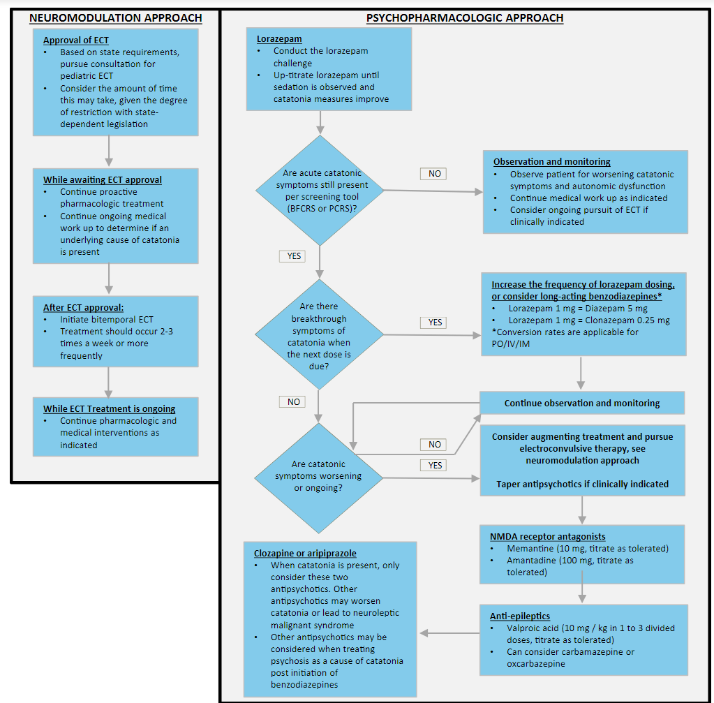
📢Our new British Association for Psychopharmacology guidelines on CATATONIA are now out!📢
💠22 experts
💠3 continents
💠>500 references
Read the full #OpenAccess guideline here: https://t.co/7w5b211JkQ.
@BAPsych#catatonia
1/Radiologist not answering the phone?
Just want a quick read on that stat head CT?
Here's a little help on how to do it yourself w/a thread on how to read a head CT!
Veins are like defense, arteries like offense.
Arteries get all the glory & attention, but you're nothing w/o veins
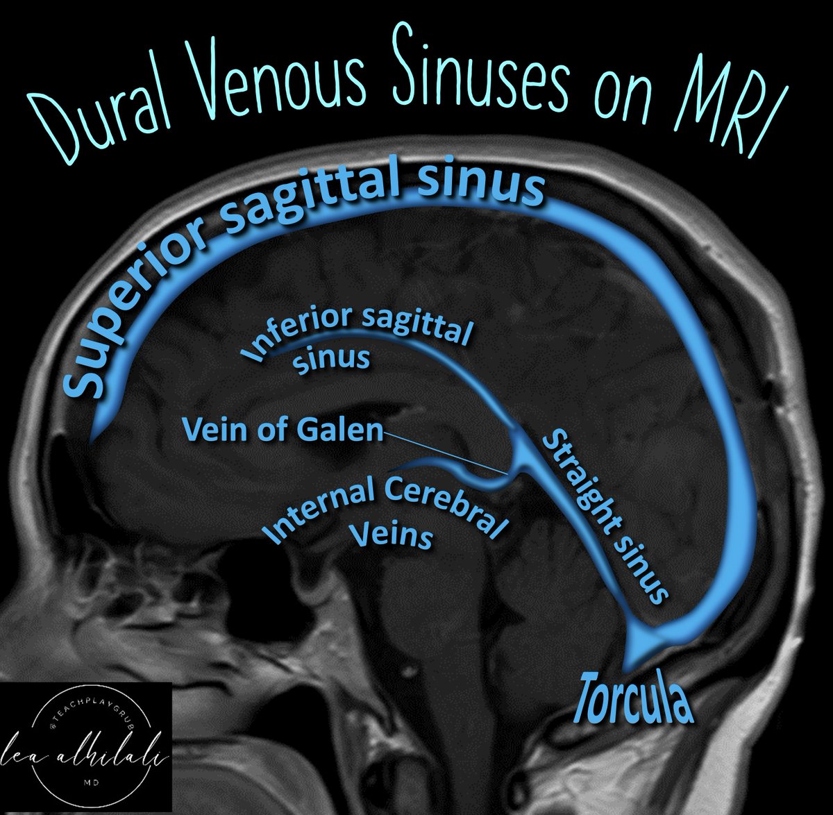
It's important to check dural venous sinuses on every MRI, not just venograms
If you're not looking at veins on brain MRIs, you're missing things!
Here's the anatomy you need to know in the sagittal plane:
▶️Superior sagittal sinus:
Appropriate name—it goes along the sagittal midline superiorly!
▶️Inferior sagittal sinus:
Also appropriate name--it goes along the sagittal midline inferiorly!
▶️Straight sinus:
Appropriate name—it runs a straight along the tentorium
🌟Remember, tents need straight poles to hold them up so there is a straight sinus beneath the TENTorium
▶️Internal cerebral veins:
Appropriate name, comes from the more internal or central part of the cerebrum
▶️Vein of Galen:
Connects the internal cerebral veins to the straight sinus
🌟I call it the Vein of Get-In—it’s how the deep venous system GETs IN to the straight sinus
▶️Torcula:
Where all the deep and superficial system meet
🌟I think torcula sounds like Dracula & Dracula drinks all the blood—so this is where all the blood goes!
Now when it comes to dural venous sinus anatomy, you will always be in the right vein!
Say bye to questions about bilateral thalamic lesions!
Feeling bipolar about bilateral thalamic lesions?
Unfortunately, the differential for bilateral thalamic lesions isn’t binary!
But here’s an easy mnemonic that can help you to remember: THALAMIC!
Here's how it works:
T = Tumor (glioma)
H = Hypoxic/ischemic encephalopathy
A = Artery of Percheron Infarct
L = Loss of thiamine (Wernicke’s)
A = ADEM
M = Metabolic/Toxic
I = Internal cerebral vein thrombosis. Infection (west nile)
C = Creutzfeld-Jacob
Now you’ll never bypass a bilateral thalamic diagnosis with this easy mnemonic!
White matter is the poor, stepchild to gray matter. We know cortex is diverse, but so is WM.
3 main WM fiber types:
Association: Cortex to cortex
Commissural: Hemisphere to hemisphere
Projection: Cortex to caudal structures
#medtwitter#neurotwitter#neurology#neurorad#radres
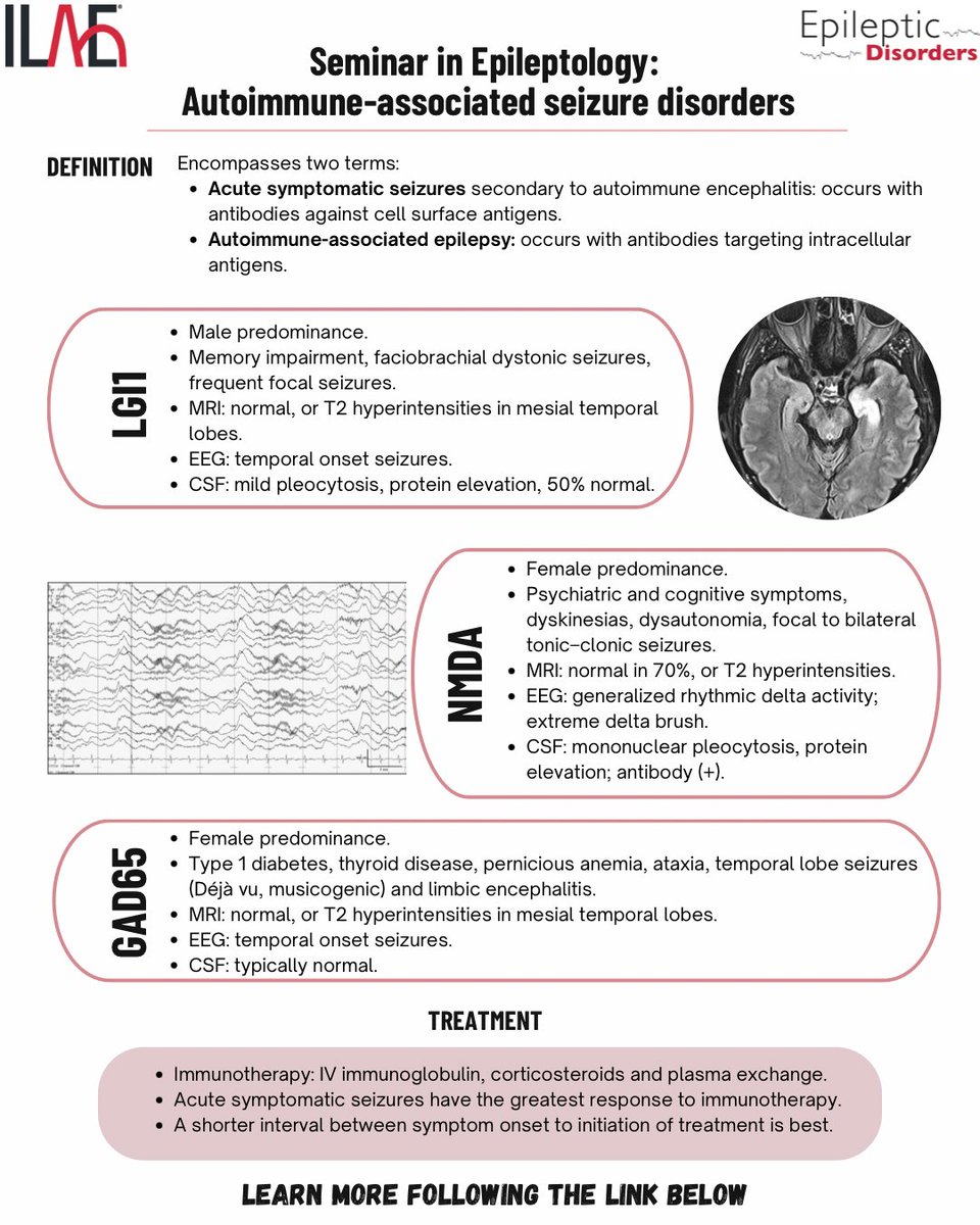
In this Epileptology Seminar, Smith et al. review the pathophysiology, clinical presentation & outcomes of common autoimmune-associated seizure disorders.
@mariajpol@RoohiKatyal@fabnascimen@SBeniczky @WileyNeuro @WileyHealth@IlaeWeb https://t.co/F45LEi32DK
Pity the fool who doesn’t know pituitary anatomy!
On a brain MRI, do you just look at the size & stalk & move on?
Do you know the pitfalls of pituitary anatomy?
Check out this review here & the latest on pituitary imaging in @theAJNR:
https://t.co/q2pUewNYOw
Pituitary gland is literally an odd couple living together:
▶️Anterior pituitary
🔸Epithelial is origin & acts like a normal gland.
🔸Adenomas are from here. Remember Adenoma & Anterior both start w/A
🔸Comes from the oral cavity! (Rathke’s pouch)
⏩Two parts:
🔸Pars tuberalis around the bottom of the stalk. Remember it’s around the tube of the stalk!
🔸Pars distalis is the bulbous lobe part (pars distalis is the distal part of the gland
🟡Somatrophs (growth hormone) are 50% of the gland & are posterior
🟡Lactotrophs (prolactin) are 20% & are anterior
🚨Remember first you breast feed before you grow���lactrotrophs are in front of somatotrophs
▶️Posterior pituitary
🔸Neural tissue from the hypothalmus
🔸Hypothalamic neuroendocrine cell axons terminate in the posterior pituitary
🔸Direct connection to the hypothalamus via the pituitary stalk
⏩Two parts:
🔸Pars nervosa. "Lobe" part (Remember, you would NERVOUS if you lose the pars nervosa bc you wouldn’t get any of the hormones)
🔸Infundibulum. Funnel-shaped connection to the hypothalamus. Remember inFUNNELdibulum
So hopefully this little pitstop has helped you to remember pituitary anatomy!!