"Signet-ring overload" - This truly impressive peritoneal effusion is involved by a signet-ring cell carcinoma, suspected to be of urothelial/bladder in origin. The mucicarmine stain on the cell block appears so photogenic. (Cytospin and CB)
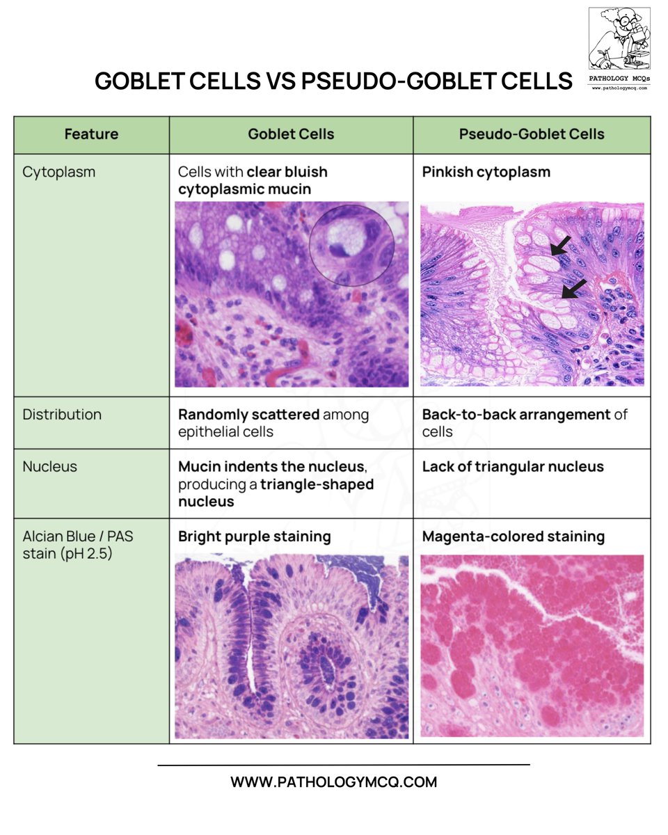
True goblet cells contain acid mucin,show a triangular nucleus, and stain Alcian blue positive🔵
Pseudo-goblet cells are actually gastric foveolar cells with neutral mucin, showing PAS magenta staining🟣
#Pathology#Histopathology#PathologyMCQ#GIPathology#BarrettEsophagus
@DrCycloPath@pepeheffernan@IARCWHO Thank you for such a masterful explanation. That's an excellent way to learn and differentiate between each diagnosis.
"Coffee beans" in a liver FNA from a patient with history of "adult granulosa cell tumor", diagnosed by my able fellow @AmerSwid, (smears and cell block).
IHCs with FOXL2, SF1, Inhibin and Calretinin confirms the dx.
Now it’s cryptococcus. Characteristic birefrigent capsule, round to oval, thin walls. Sometimes you can even picture a “sunshine” appearance. Isn’t cytology beautiful ?? Happy Valentine’s you all !
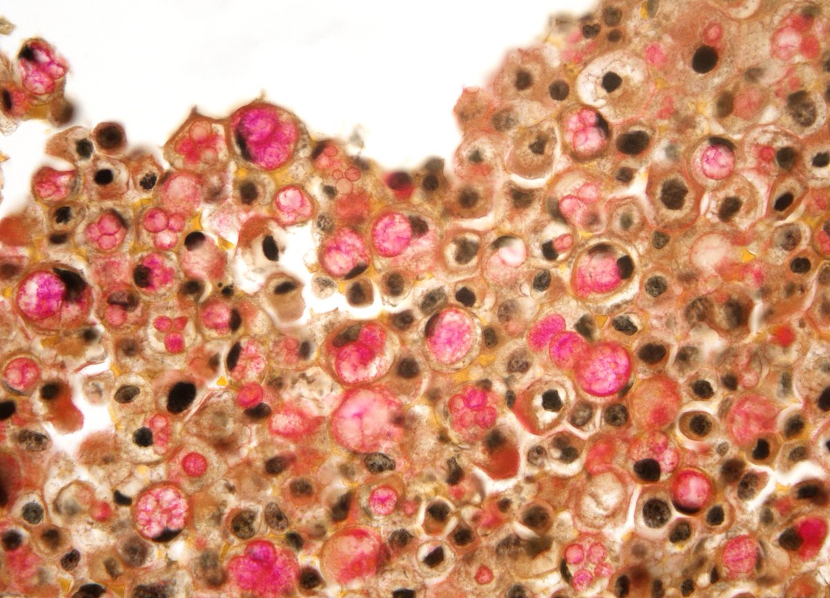
A combination of intracytoplasmic mucin and hydropic degeneration, resulting in cells that looks like "Big Balloons" - "Metastatic Pancreatic Adenocarcinoma" in ascitic fluid. How often do I personally ask for a mucicarmine stain? - almost never.
Postmenopausal bleeding in a 60-year-old woman.
Her pap smear is shown. What is your interpretation?
A) Negative for malignancy
B) High grade
C) Squamous cell carcinoma
#CytoPath#GynPath#PathTwitter#Pathology
🔬 Succinate Dehydrogenase (SDH)-Deficient Renal Cell Carcinoma ~ Can you see the cytoplasmic inclusions with eosinophilic or pale flocculent material? #GUpath#KidneyTumors#Pathology#Urology
Diffuse right frontal tumor with no enhacing in a 28yo woman.
Gemistocytic astrocytoma
Something to remember:
* >20% gemistocytes are required to be considered a major tissue pattern
* it's associated with a focal gain of chromosome 12p
#Neuropath
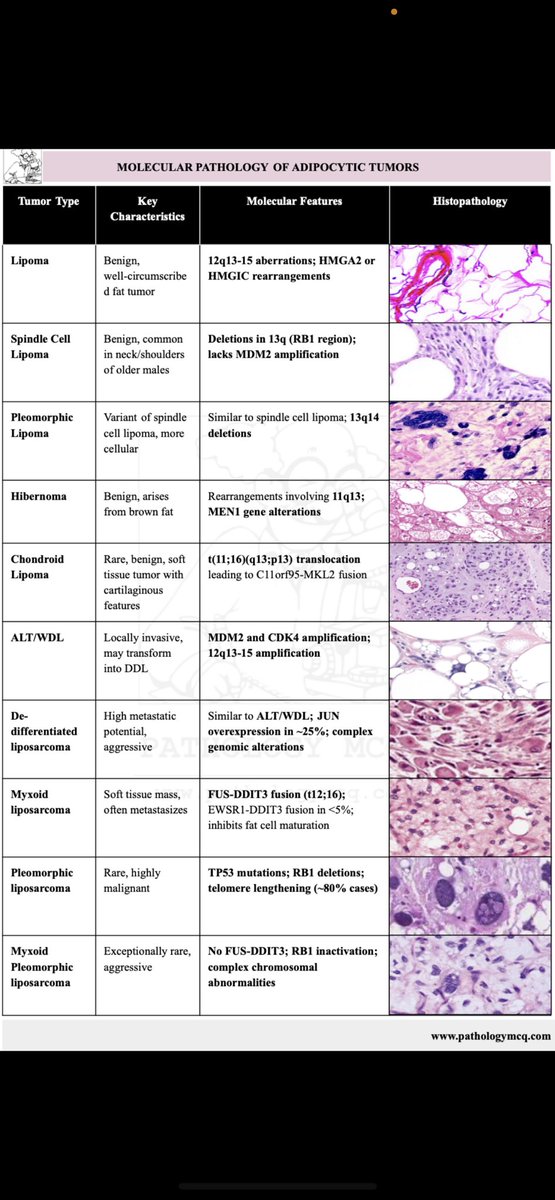
🔬✨ Dive into the molecular pathology of adipocytic tumors! Understanding the key characteristics, molecular features, and histopathology of these fascinating entities is vital for diagnosis and research. 🧠💡
Perfect for those preparing for #NEETSS and #FRCPath! 🩺 #pathboards
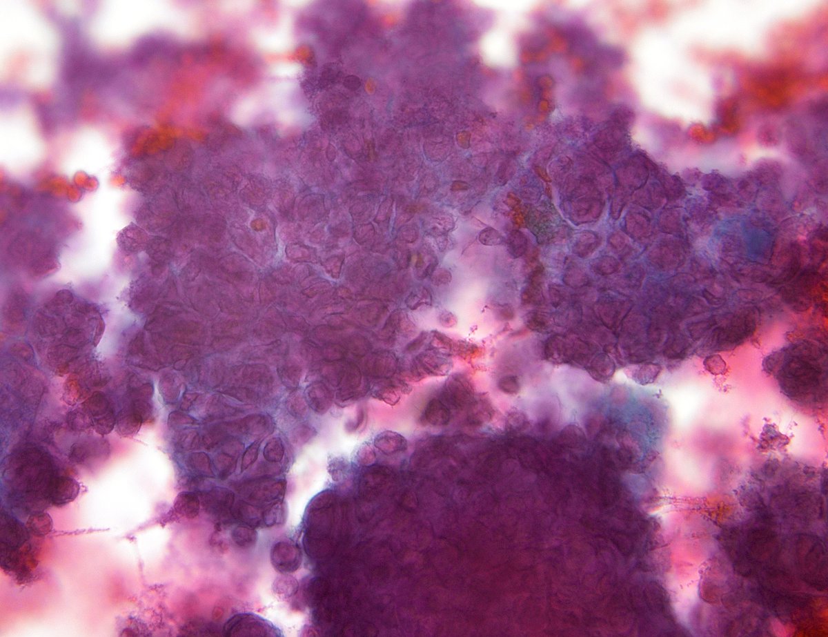
Ascitic fluid positive for malignancy. Easy to recognize.
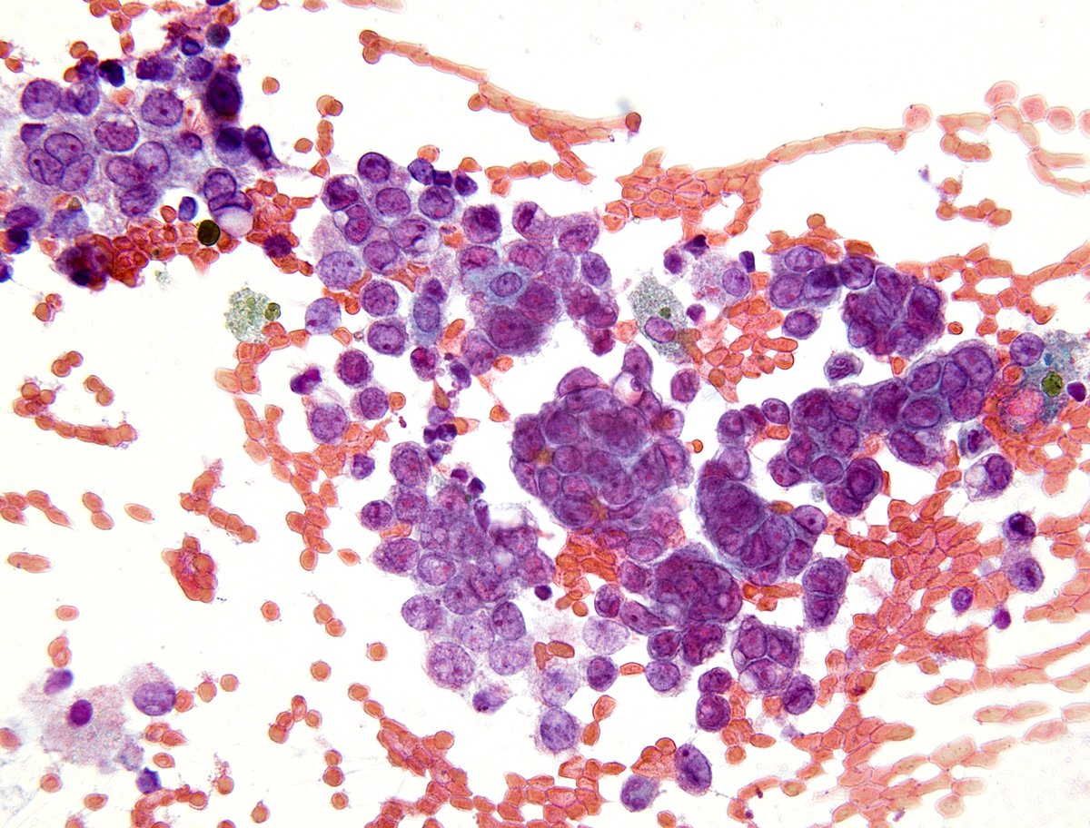
In PDAC, It's not uncommon to see isolated cells that can be quite anaplastic. The same can occur when there is peritoneal involvement, as in this ascitic fluid of a patient with PDAC 🔬❤️🩹