Is trying to remember toxic encephalopathy patterns making you toxic?
Does memorizing metabolic encephalopathy patterns put you into a metabolic coma?
Don't become encephalopathic!
Here is the cheat sheet of the encephalopathy patterns you NEED to know & how to remember them!
➡️Corpus Callosum:
🔸Lesions here are called cytotoxic lesions of the corpus callosum or CLOCC.
����Usually transient from cytotoxicity, especially viral infections.
🔸Remember they are commonly viral & transient bc CLOCC lesions are on the CLOCK
➡️Central Pons:
🔸Usually osmotic demyelination
🔸Remember this bc the T2 hyperintensity is in the shape of O for Osmotic
➡️Dentate:
🔸Usually flagyl toxicity
🔸Remember this bc the wavy appearance of the dentate looks like a FLAG for FLAGyl
➡️Corticospinal tract:
🔸Usually from cobalamin deficienty
🔸Remember cortiCOspinal for CObalamin
➡️Symmetric Gray Matter:
🔸Differential is EXTENSIVE
🔸Remember the differential list for deep gray is deep!
➡️Cortical Gray Matter:
🔸Typically hypoglycemia & hyperammonemia
🔸Remember it looks like the brain has been coated in shimmering sugar = related to glucose
🔸Hyperammonemia has about 1000 Ms in it & the undulated cortex looks like an M
➡️Symmetric White Matter:
🔸Commonly from chasing the dragon (heroin) & methotrexate
🔸Remember that the puffy white matter signal looks like the smoke from a dragon
🔸Remember methotrexate as methoTRACTSate = affects many white matter TRACTS
➡️Posterior subcortical edema:
🔸This is PRES or posterior reversible encephalopathy
🔸So if it’s all POSTERIOR, think POSTERIOR reversible encephalopathy
➡️Asymmetric White Matter:
🔸This is usually demyelinating from chemotherapy
🔸Demyelinating lesions have a C shaped pattern of enhancement
🔸So if you see a C enhancing think of C-hemotherapy
Now you know all the toxic/metabolic encephalopathy patterns.
So hopefully now remembering the patterns of toxic/metabolic won’t seem so diabolic!
🩺Clinical & neuroimaging 🧠 clues to differentiate 🧩autoimmune encephalitis & its mimics🔎
Current opinions in Neurology 📖
10.1097/WCO.0000000000001489.
https://t.co/Xeh9opjZuC
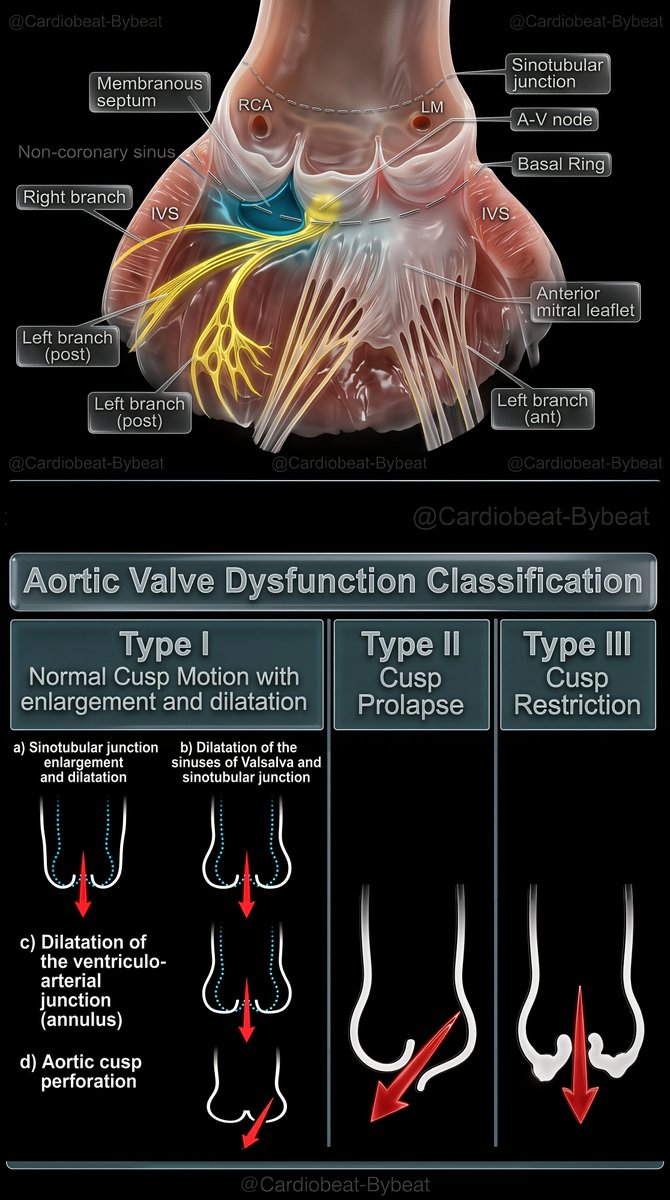
🫀 Aortic Valve Dysfunction Classification
Type I – normal cusp motion with root/annulus dilation ± perforation; Type II – cusp prolapse; Type III – cusp restriction.
Precise anatomy guides valve-sparing repair strategy. 🔬📚 #Cardiology#AorticValve
there’s an Odyssey movie. Love it or hate it, the poem is still better.
I built a free reader for it — Greek with facing English, tap any word for the grammar, every place mapped, the route animated, Flaxman’s engravings. Share and enjoy.
https://t.co/aPWvdTPCVB
Having trouble remembering what to look for on scans for Alzheimer’s dementia?
Want the key findings to stay in your memory?
Medial temporal atrophy (MTA) score quantifies hippocampal volume loss
It rates the amount of CSF around the hippocampus as a marker of volume loss
CSF surrounds the hippocampus like a snake surrounding its prey—how far the snake has gone in getting its prey determines the MTA score:
MTA-1: Choroidal fissure opens, looks like the snake has started to slither in
Remember: the number one looks flat like a slightly opened choroidal fissure
MTA-2: Temporal horn widens, looks like the snake has surround the hippocampus
Remember: the number two look like the CSF curving around as the temporal horn opens
MTA-3: Moderate hippocampal atrophy, looks like the snake has begun to squeeze the hippocampus
Remember: the number three has a tight curve, so it looks like it is crushing the hippocampus & it looks small (atrophied)
MTA-4: Severe hippocampal atrophy, looks like the snake has crushed the hippocampus
Remember: Number four has only a tiny hole in it, so the hippocampus has to be super tiny & atrophied to fit inside!
Hopefully, this will help you commit this rating system to memory so you’ll never forget what you need to say for Alzheimer’s!
🚨HILO🚨
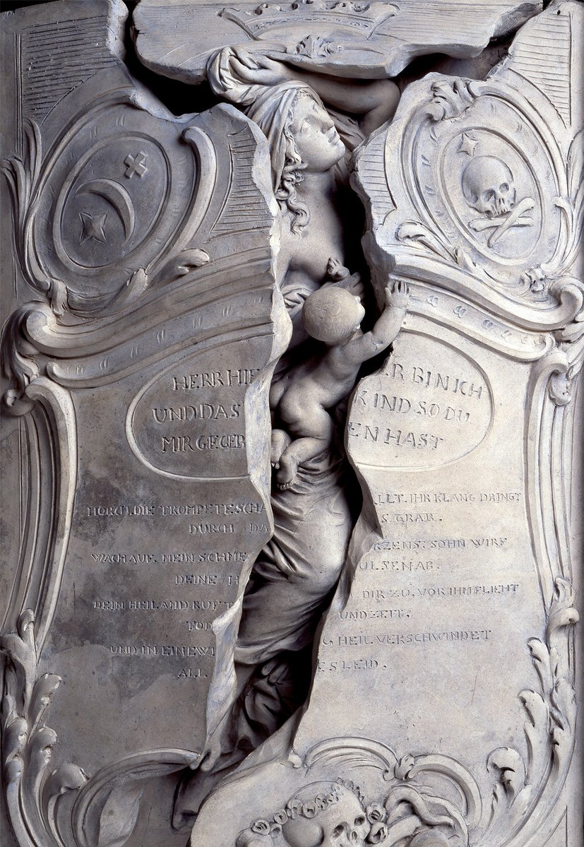
La más bella metáfora de la resurrección jamás labrada en piedra se esculpió en condiciones dramáticas para una madre y su hijo recién nacido que no sobrevivieron al parto.
A día de hoy sigue siendo uno de los monumentos funerarios más admirados del mundo...👇
Median nerve infiltration using a long-axis approach.
This shower effect is achieved by placing the needle's bevel on the nerve's upper pole. A safe and efficient approach to treat mild and moderate carpal tunnel syndrome. #MasterecoMSK
Calcifying pulmonary metastases. Remember causes with mnemonic BOBCAT: Bone sarcomas (most common); Ovary, Breast & Colon; Any primary post-chemotherapy; Thyroid & Testicular. Rare case of metastatic epithelioid hemangioendothelioma, h/o chemotherapy. #FOAMrad#chestrad#radres
GUYON CANAL = ULNAR TUNNEL
begins at proximal border of 💥pisiform bone & ends distally at the hook of 💥hamate.Protect ulnar nerve&artery.
●Compression of ulnar nerve at this canal 👉Guyon canal syndrome.
●Compression of ulnar artery at this canal👉
Hypothenar hand syndrome.
Left-ventricular trabeculations are a vexing feature in LV imaging for #function or #prognosis. An Expert panel paper just published in @JACCJournals provides clarity. a 🧵
Open access full paper here: https://t.co/Gu7RTvSNPx
#heart#cardiotwitter#whyCMR
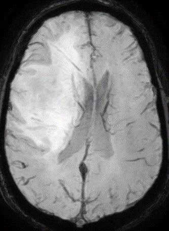
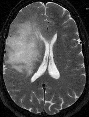
Tumor? Or Tumefactive Demyelination? Can you figure it out? Patient is a 55 y/o M with no PMH.
Additional images in thread.
Answer in a few hours. 🧠
#Neurology#Neurosurgery#radres#MedTwitter@TheASNR
Meniscal Ossicle
• Focus of bone within meniscus substance.
• Posterior horn,medial > lateral meniscus.
• Range from tiny to > 1 cm.
• Ovoid or triangular.
• MR follows normal marrow signal on all sequences.
♦️Always correlate MR suspected meniscal ossicle with radiograph
It's incredible how standard #WhyCMR sequences can miss huge things; the key is to customize protocols and be at the console (presential or remotely) in challenging cases | Standard SAX CINE in a patient with HCM suspicious based on EKG | May be mil-mod LVH
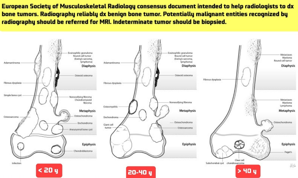
🛑I love these 3 illustrations that based on the age & location of bone tumors offered the differential diagnosis.👍👏
https://t.co/fVuhWLsXT3
🌟This wonderful document that I put its link above, prepared by the European Society of Musculoskeletal Radiology to help radiologists
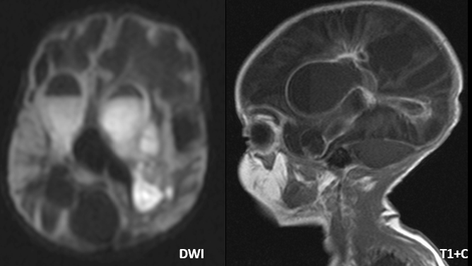
Tip of the day 💡: When the clinician asks you, 'is there active bacterial infection here?' rely on the diffusion weighted imaging (pus restricts) compared to the post contrast T1.