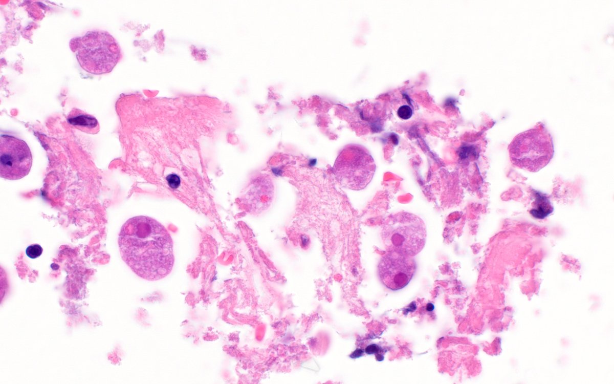
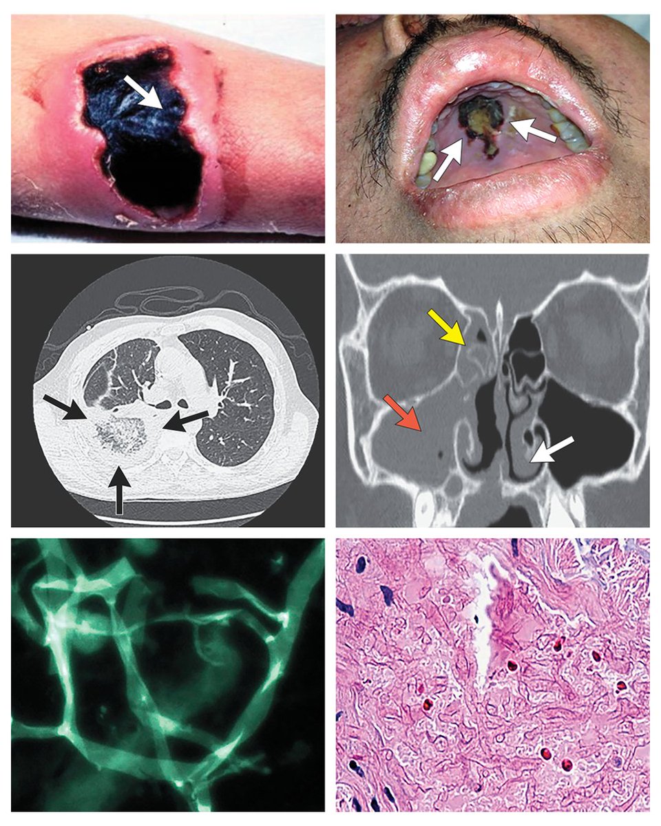
Mucormycosis is a rapidly progressive, invasive fungal infection that causes severe disease. The latest epidemiologic data, risk factors, diagnostic strategies, and treatment approaches are discussed in a new review. 👉 https://t.co/FjWD1FcZxe
Early diagnosis and initiation of antifungal therapy significantly reduce mortality among patients with mucormycosis and hematologic cancers, from approximately 80% to 40%. Accurate diagnosis of mucormycosis begins at the bedside with evaluation of symptoms and signs in patients at high risk (seen in figure). Although early symptoms and physical findings in patients with mucormycosis are often nonspecific, they carry important diagnostic significance in patient populations with high risk. Periorbital cellulitis, diplopia, focal palatal necrosis, and necrotic wounds in patients at high risk should cause serious concern for mucormycosis and lead to prompt assessment. Maintaining a high index of suspicion for mucormycosis in such patients can be the impetus for obtaining fluids and tissues for diagnostic analysis, ordering imaging procedures, initiating antifungal therapy, requesting surgical assessment, reversing immunodeficiencies, and correcting metabolic abnormalities.
Learn more in the Review Article “Mucormycosis” by Dimitrios P. Kontoyiannis, MD, ScD, and Thomas J. Walsh, MD, from the University of Texas M.D. Anderson Cancer Center (@MDAndersonNews), Center for Innovative Therapeutics and Diagnostics, and University of Maryland School of Medicine (@UMmedschool): https://t.co/FjWD1FcZxe
Weekend digest: Name the pathogen
- Painful cellulitis on wrist of adult preparing raw Tilapia
- Rapid onset fever and lymphangitis within 24 hours of puncture injury
- Positive blood cultures within 24 hours of symptom onset
Current answer is Purpureocillium lilacinum.
Can be confused with blasto
Differentiated by presence of elevated (1,3)- beta-D-glucan and presence of hyphal elements in tissue biopsy.
#Parasite case 785 is up, donated by Idzi Potters; This object fell out of a patient’s nose! 😳
https://t.co/qseRwvxLZg What is your identification?
Answer later this week! #mayopath#pathology#PathBugs
The answer to the #Parasite Case 784 is up: Cuterebra sp. Larva (L3). Read more here: https://t.co/wmJcD4iX4U the classic morphology.
#mayopath#pathology#PathBugs
Weekend Digest
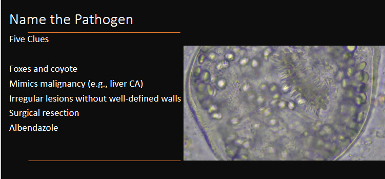
Name the Pathogen
Five Clues
1. Foxes and coyote
2. Mimics malignancy
3. Irregular lesions without well-defined walls
4. Surgical resection
5. Albendazole
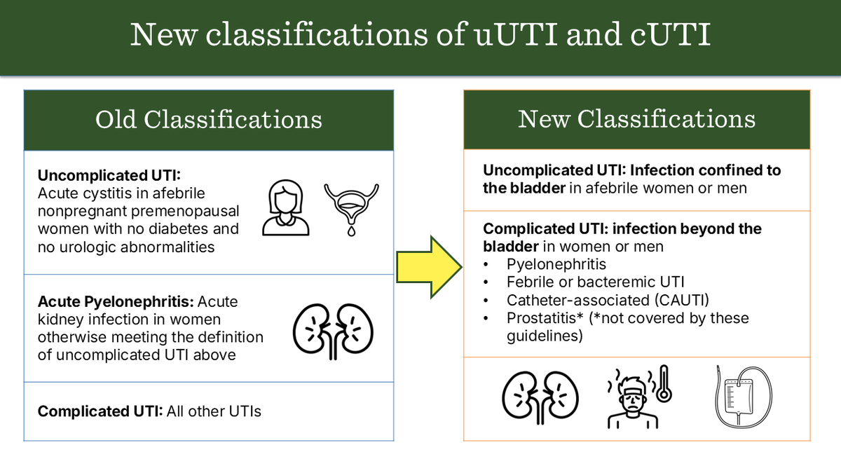
🚨IDSA has released the first guidelines for the management and treatment of complicated urinary tract infections (cUTIs)!
The new guidelines simplify the prior definitions of uncomplicated UTIs and cUTIs, & address treatment for cUTIs.
View guidelines: https://t.co/jauYIl0Zyv
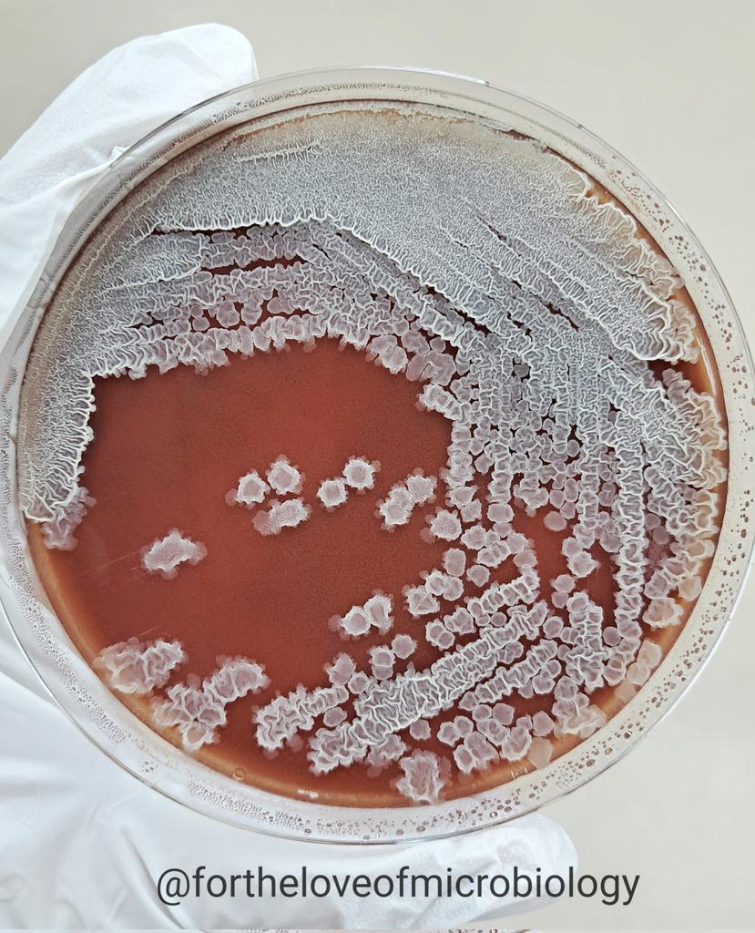
Bacillus subtilis colony on chocolate agar showing characteristic large, spreading, and wrinkled growth with a dry, ground-glass appearance.
The colony exhibits lobate edges and a highly filamentous, frost-like surface—typical of motile Bacillus species.
B. subtilis is a Gram-positive, spore-forming rod widely found in soil and the environment. Though typically considered non-pathogenic, it may occasionally be isolated in clinical specimens and should be interpreted with caution, particularly in immunocompromised patients or device-related infections.
#Fortheloveofmicrobiology #clinicalmicrobiology #mmidsp #microrounds #IDpath #ASMClinMicro #STEM #WomeninMicrobiology #ClinMicro #microbiologypakistan #PathBugs #crittersontwitter
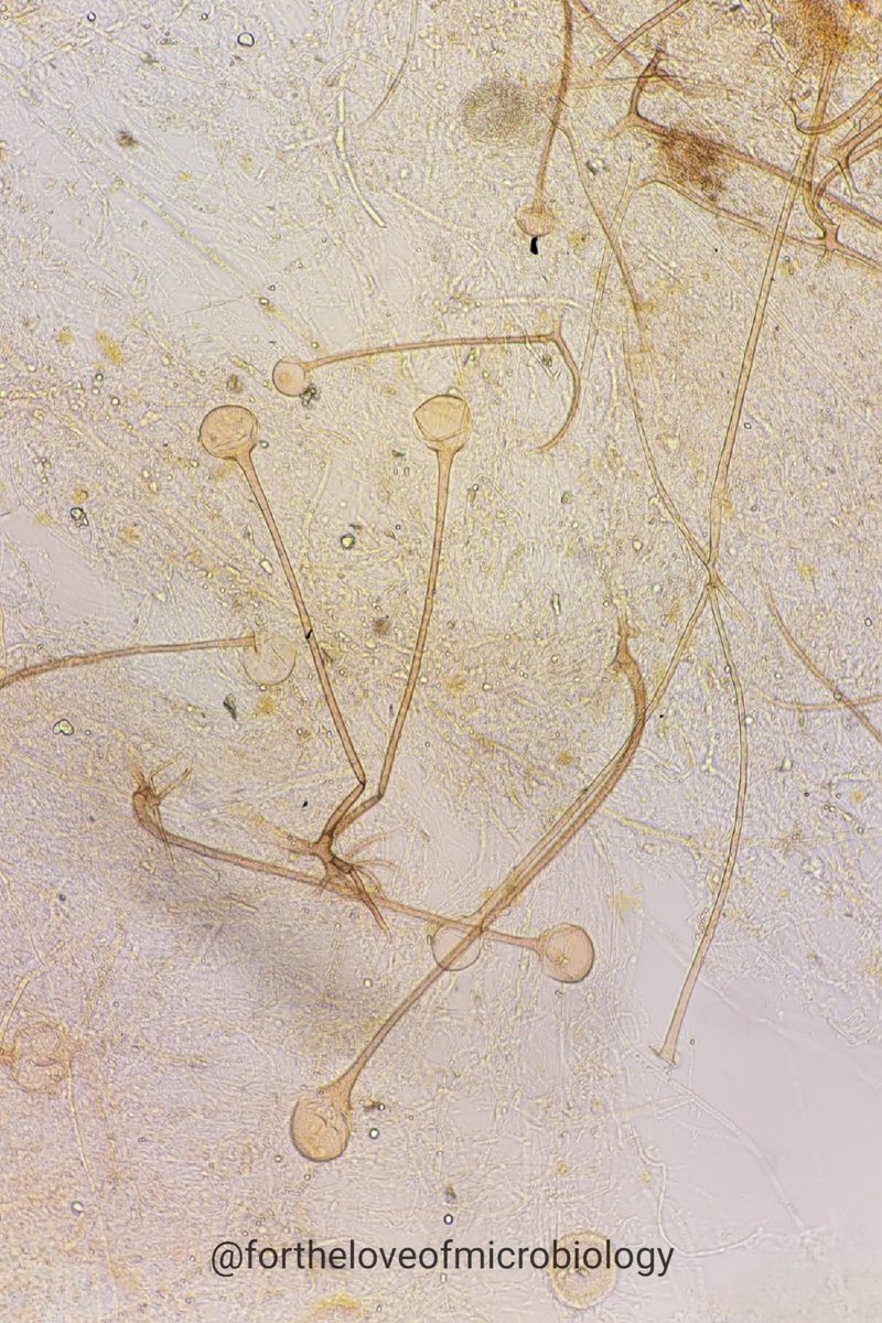
🔬 KOH Mount – Nasal Scraping
A rare and striking view of Rhizopus in full sporulation — directly from a clinical sample!
This image captures classic microscopic features of Rhizopus, a member of the Mucorales order and a known agent of mucormycosis. Under the microscope, you can clearly see:
✔️ Sporangiophores – upright stalks bearing sporangia
✔️ Sporangia – round, spore-filled sacs at the tips of sporangiophores
✔️ Rhizoids – root-like structures at the base, typically found opposite the sporangiophores (a key feature distinguishing Rhizopus from other genera like Mucor)
These morphological features are diagnostic and provide rapid presumptive identification on direct microscopy — crucial for initiating timely antifungal therapy.
🧫 Stain: 10% KOH
📍 Sample: Nasal scraping
📸 @fortheloveofmicrobiology
#Microbiology #MedicalMycology #Mucormycosis #Rhizopus #KOHMount #InfectiousDiseases #LabLife #ClinicalMicrobiology #Microscopy #IDMed #Fungus
#Fortheloveofmicrobiology #clinicalmicrobiology #mmidsp #microrounds #IDpath #ASMClinMicro #STEM #WomeninMicrobiology #ClinMicro #microbiologypakistan #PathBugs #crittersontwitter
Weekend Digest
Name the pathogen
H&E stain of heart tissue of a 27M who developed severe intractable heart failure 2 months after allogeneic bone marrow transplant for acute leukemia.
Clue: consumption of food / water contaminated with feces of snakes
Photo credit: CDC
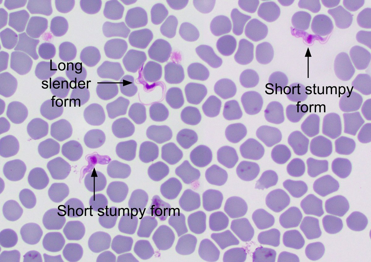
The answer to the #Parasite Case 780 is up: Trypanosoma brucei causing Human African Trypanosomiasis (HAT). Check out the full discussion on my blog, with pointers on what to look for in clinical cases. Thanks to Dr. Mutnal for this classic case! https://t.co/h3goiazSLn
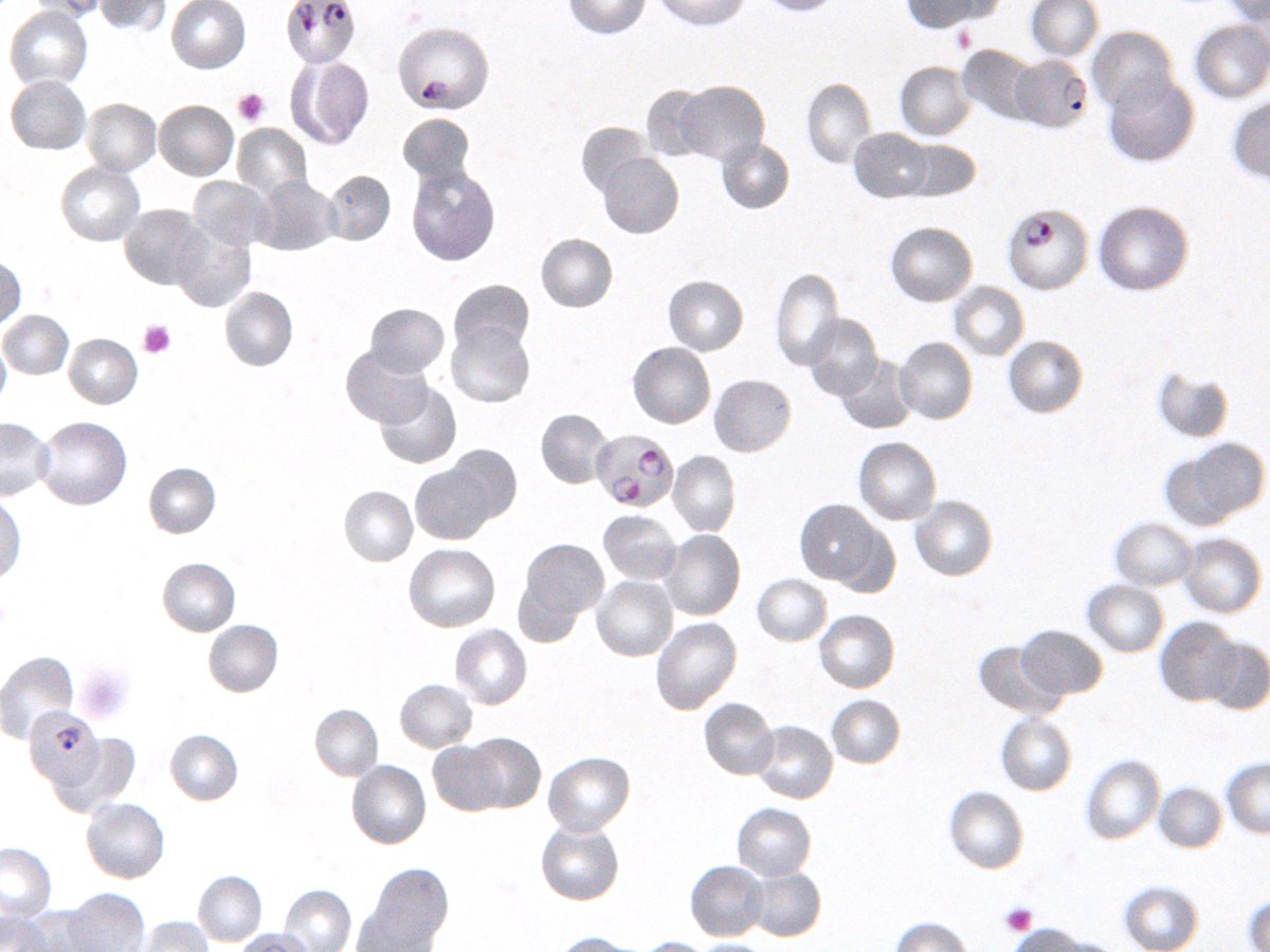
#Parasite case 779 is now up donated by Dr. Richard Bradbury: Giemsa-stained blood films and urine from a febrile patient in The Gambia recently treated with quinine. What is this condition and its cause? https://t.co/q0XHANymQe Ans later this week! #mayopath#pathology#PathBugs
The #Parasite case 778 is now up: https://t.co/eSkqAsYPev - colon biopsy from a patient with abdominal pain and diarrhea. Objects seen are 20 microns. What is your diagnosis?
Answer later this week!
#mayopath#pathology#PathBugs
Weekend Digest
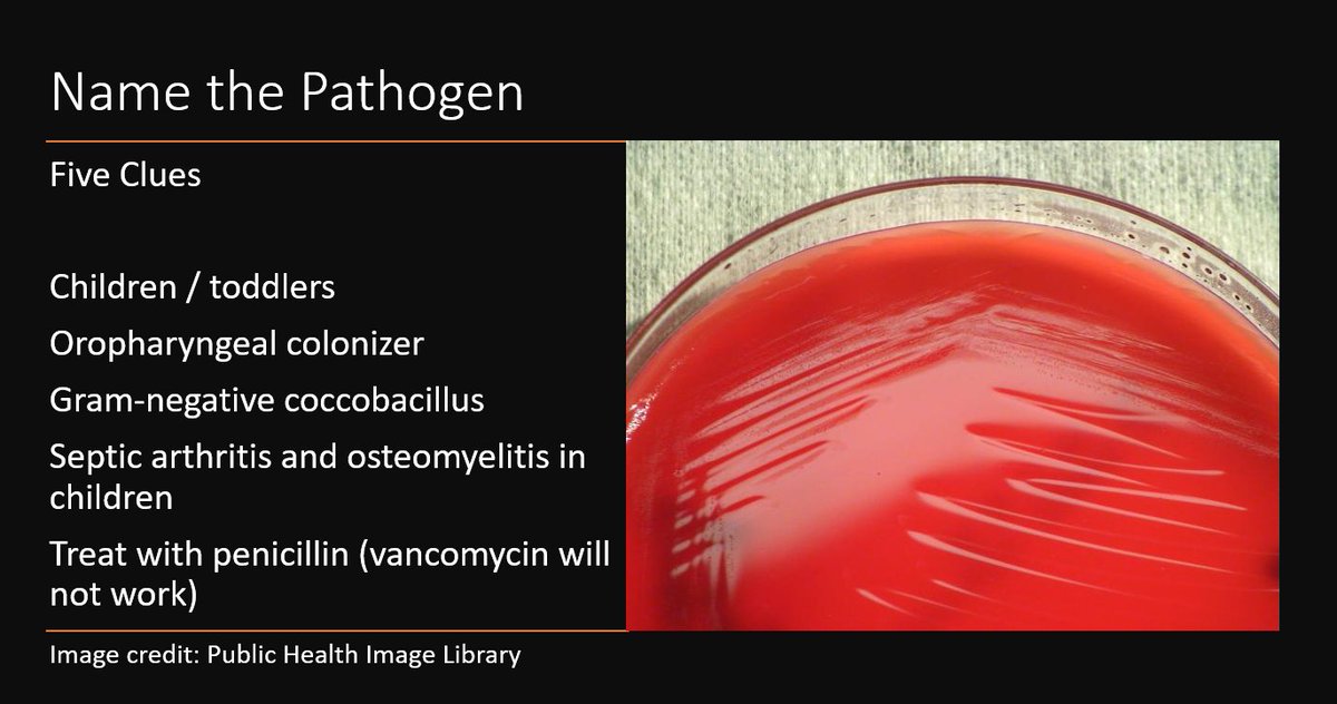
Name the Pathogen
Five Clues
1. Toddlers / children
2. Oropharyngeal colonizer
3. Fastidious Gram-negative coccobacillus
4. Septic arthritis and osteomyelitis in children
5. Treat with penicillin (vancomycin will NOT work)
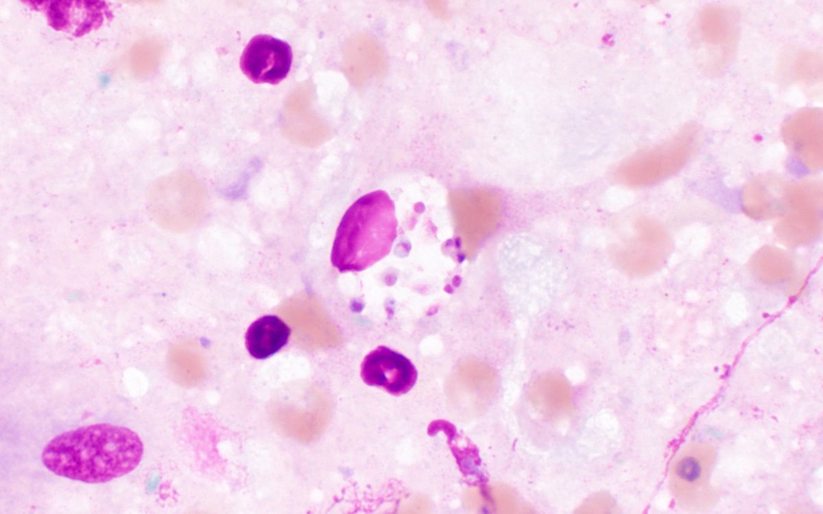
The #Parasite case 776 is now up: brain biopsy from a man with untreated HIV. Specimen appeared necrotic and bloody. Show are Giemsa-stained touch preps. ID? Answer later this week!