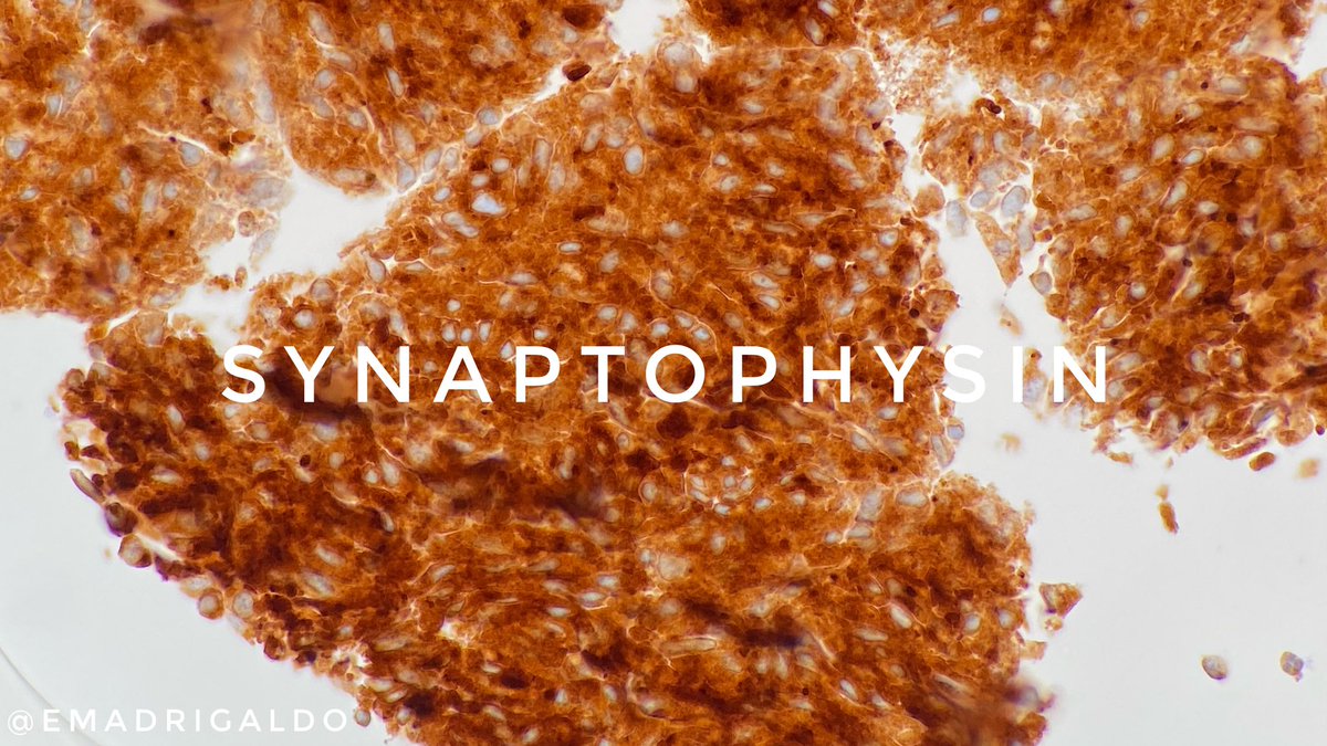
Case of the week. The enlarged splenectomy specimen of a 44-year old man shows multiple variably sized blood-filled nodules throughout the cut surface (A-D). Immunophenotype: CD163+ (subset of cells), CD34+ (subset of cells), CD8 negative in red pulp sinuses.
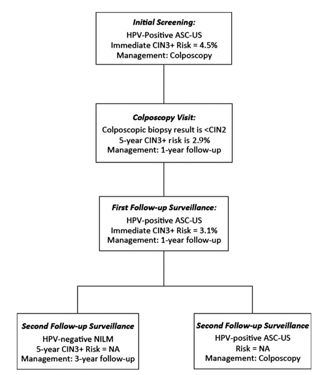
Recently found out about the new 2019 ASCCP guidelines for cervical cancer screening, with a shift toward risk-based clinical action thresholds: https://t.co/rt9gutRAzb
✳️Important to understand how our path diagnoses impact patient management!
#CytoPath#GynPath Thanks @cjvand!
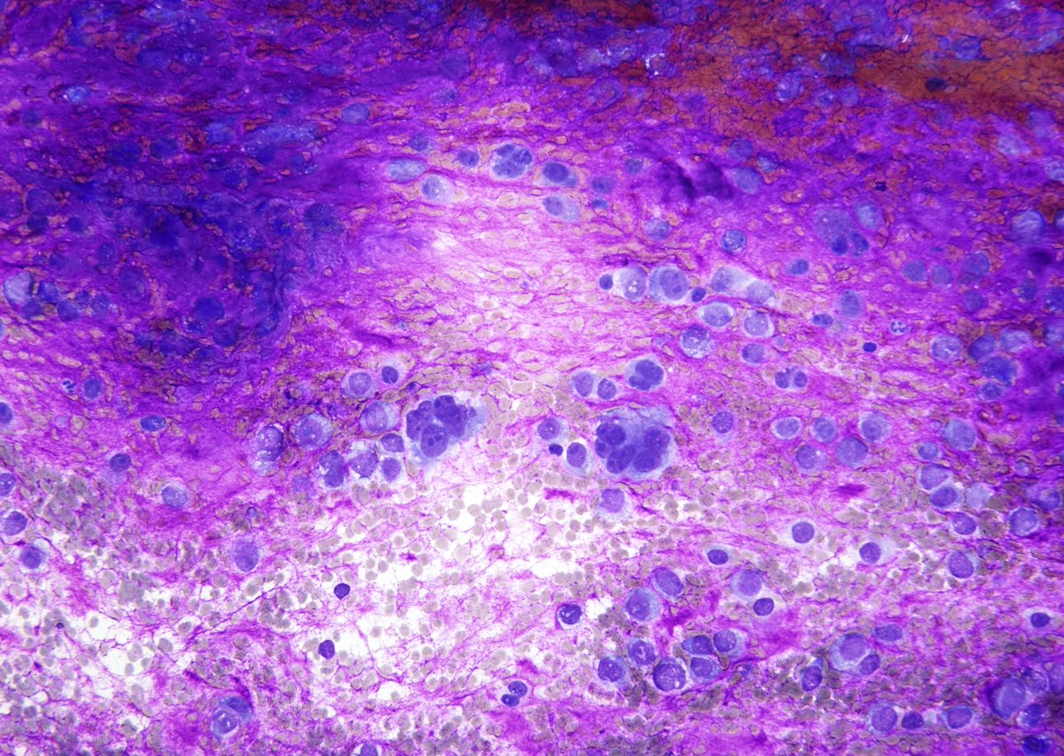
Textbook case of intestinal #amebiasis:
Biopsies taken from a colon with many ulcers displaying "poached egg" appearances. Histologic examination shows the presence of E. histolytica in both trophozoites and cysts form. #PathTwitter#GIpath#microbiology#pathharryspotter
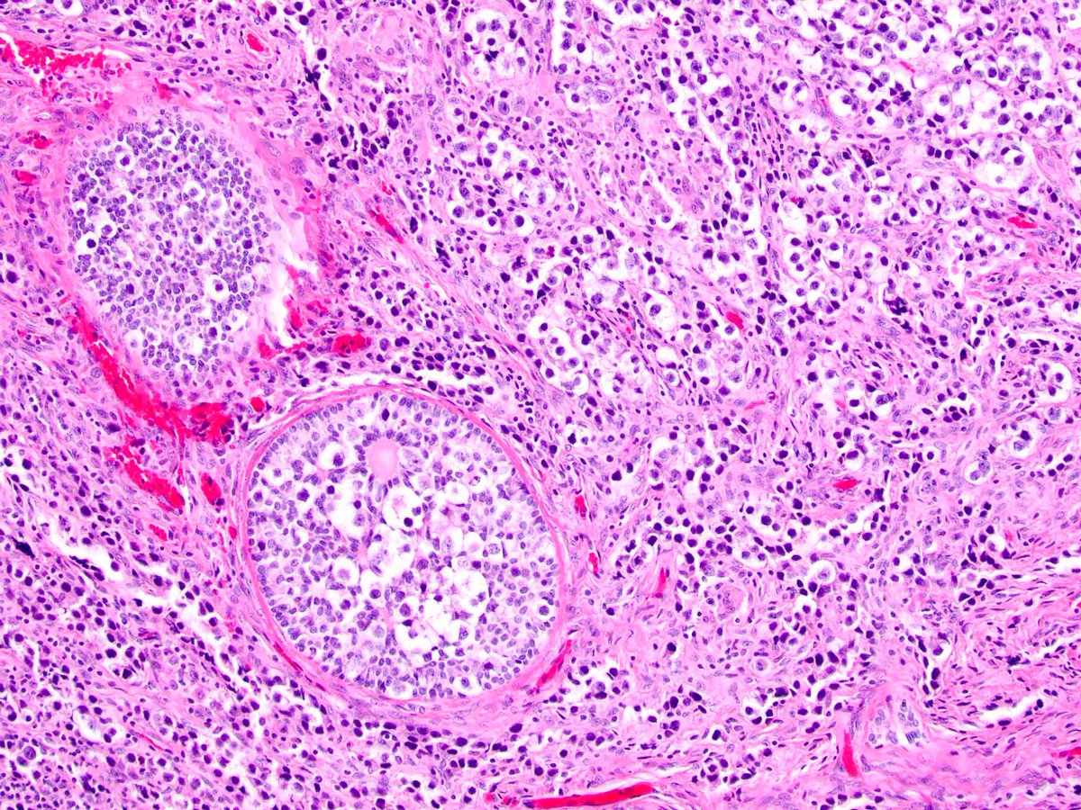
Typical FNA picture of an adenoid cystic carcinoma with uniform basaloid cells, microcystic architecture, and abundant hyaline globules. Check out the new @Pathoutlines article on malignant salivary gland FNA! https://t.co/v5kTNgHo1Z #cytopath#ENTPath#FNApath#FNAfriday
On low power, majority of you would think "PA from the Parotid". However, most would change the opinion on high power and once I disclose that the FNA came from a femoral lesion in a young male. Immature osteoid from "Osteosarcoma" appears extremely "myxoid".
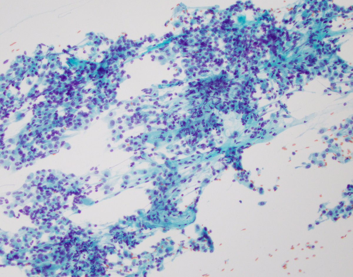
FNA clear cell renal cell carcinoma: Cells w abundant cytoplasm and w small cytoplasmic vacuoles. Can be difficult on liquid based preps where the cells r bland & may mimic histiocytes (low N:C, eccentric nuclei). The smears r helpful, w large vascular cohesive groups #cytopath
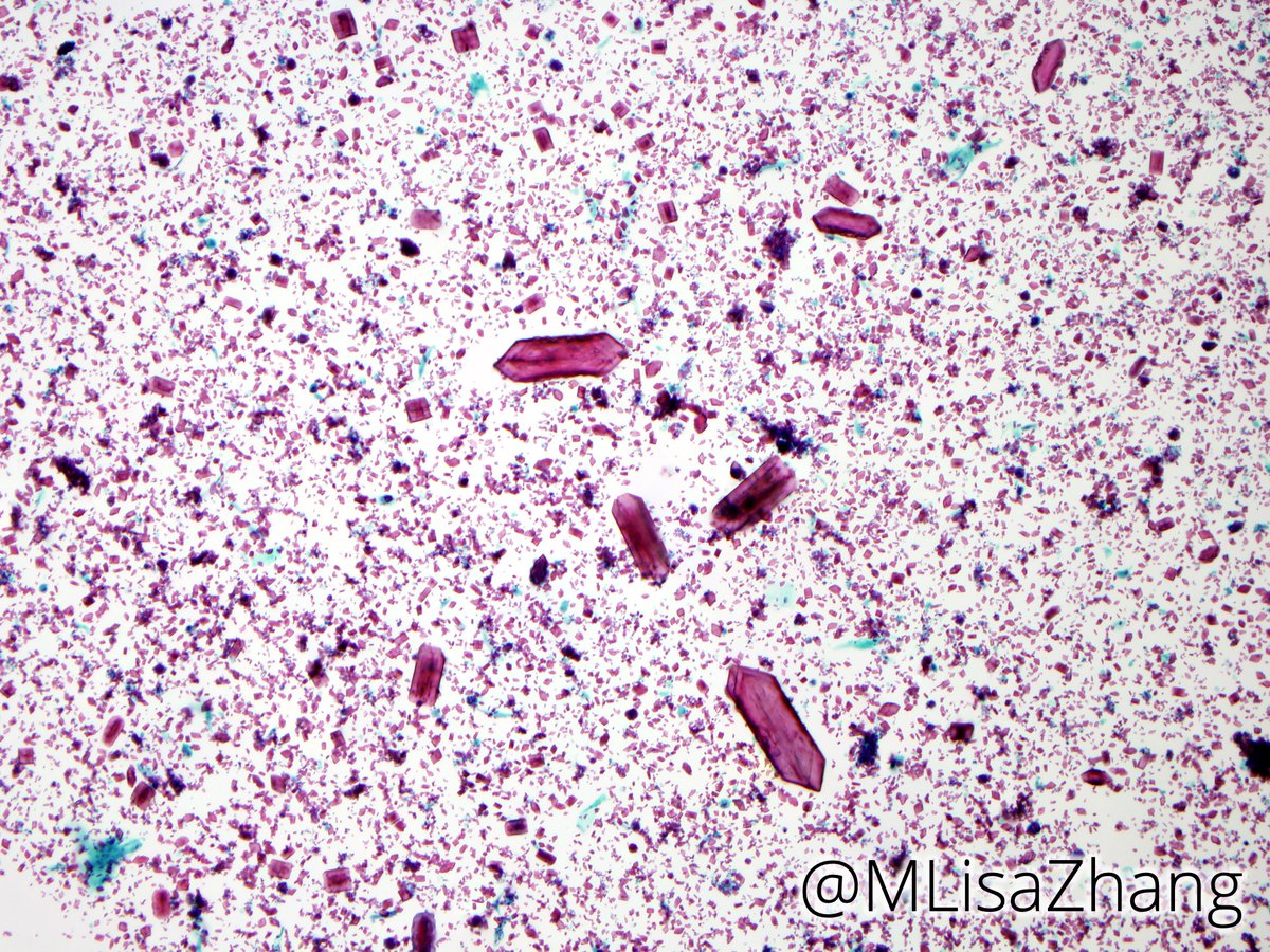
Parotid FNA with "plate-like" amylase crystals 💎 , associated with sialadenitis.
VS "floret-like" tyrosine crystals, which can be seen in pleomorphic adenomas and other neoplasms.
Feels like trivia fodder for @Sara_Jiang! 🤫
#FNAFriday#CytoPath#ENTPath#Pathology#pathboards
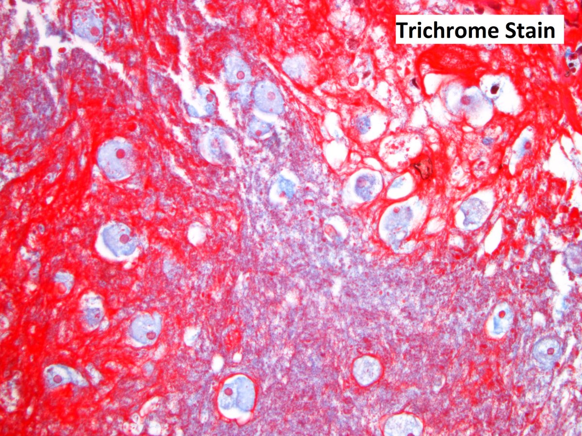
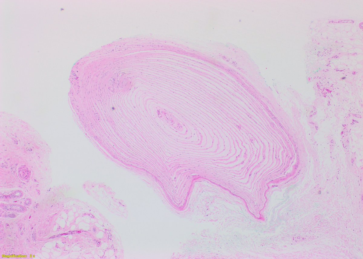
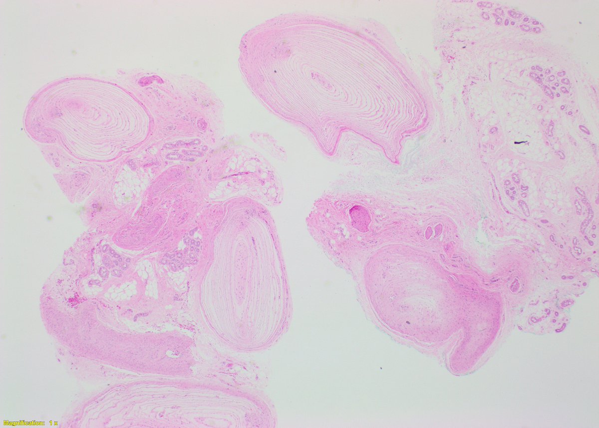
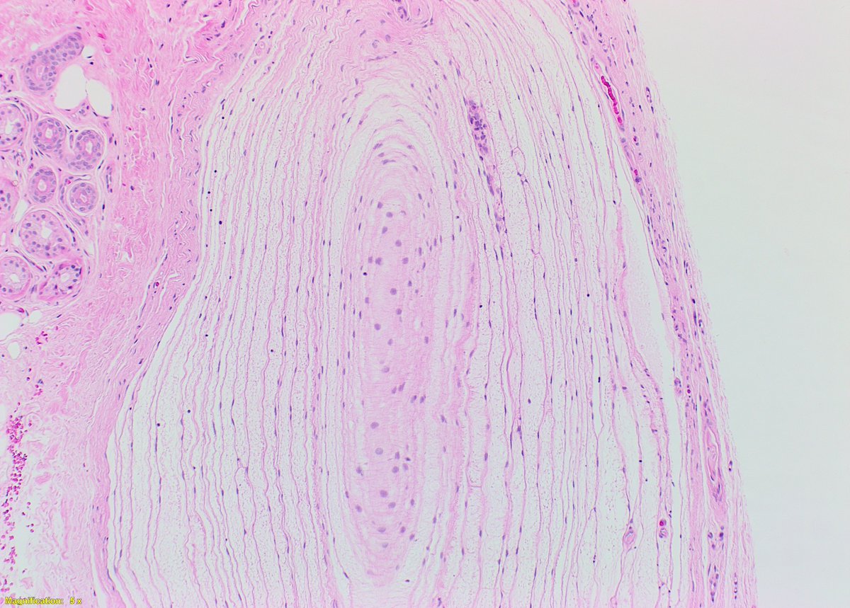
Today we had a nice example of a Pacinian Corpuscle Neuroma. It is a benign lesion composed of prominent pacinian corpuscles with variable patterns in the dermis. Typical corpuscles measure 1.0 - 1.5mm, these were slightly larger at 2mm. #dermpath#dermatology@PathBoards
Despite looking like a low-grade Gleason adenocarcinoma of the prostate, this is a case of adenosis. It can present with small glands, smooth luminal borders, and nucleoli. The key here is the PIN-4 stain demonstrating basal cells. Scary! #pathology@PathBoards@UNMC_PathMicro