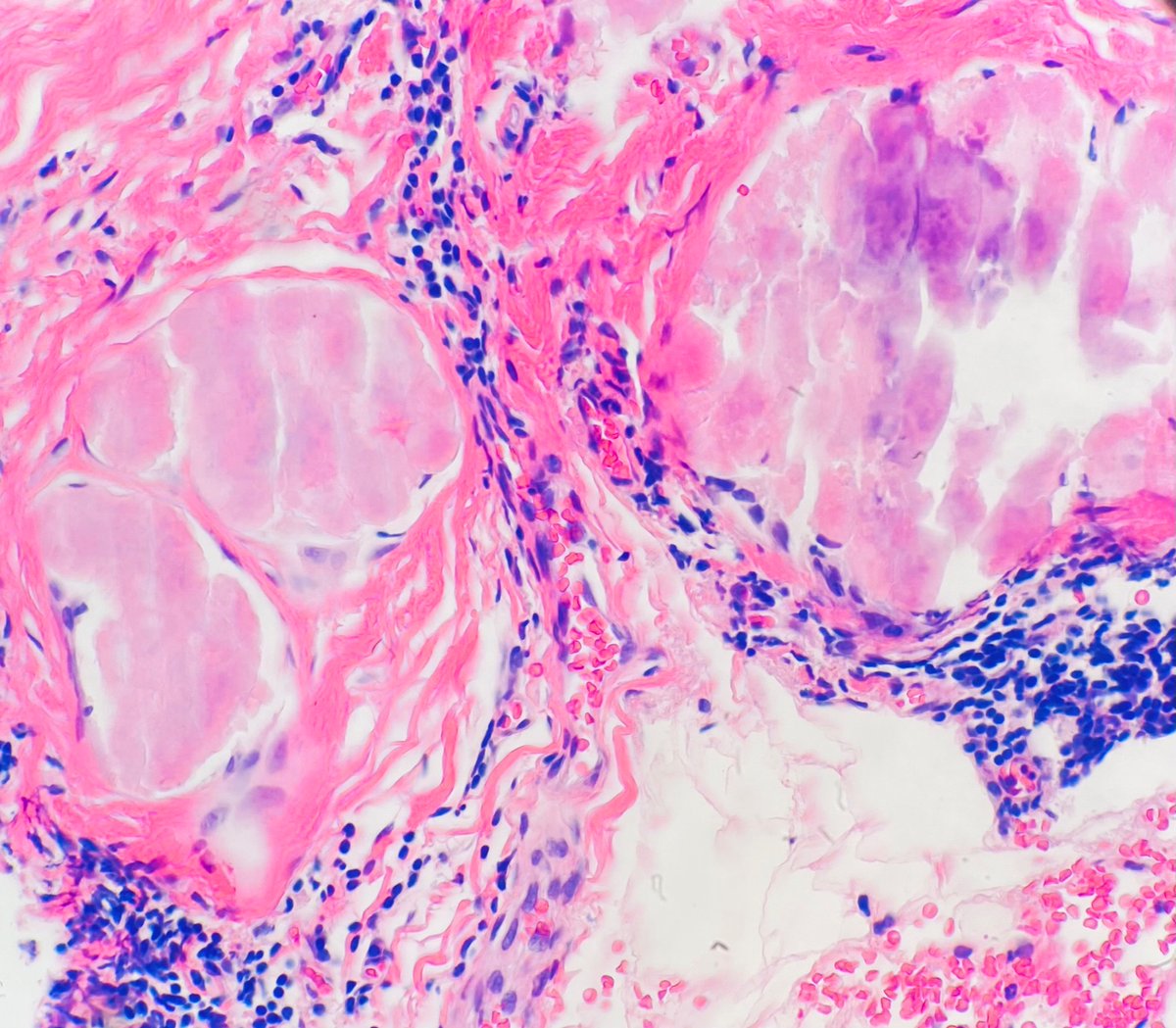
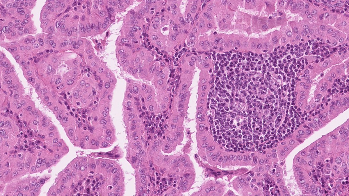
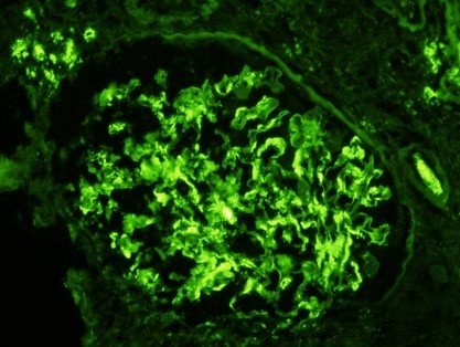
Primary localized amyloid of the breast #breastpath#PathTwitter#MedTwitter#pathology
🍏Detected on screening mammography as clusters of calcifications
🍏 25% do not develop a hematologic disorder or systemic amyloidosis
🍏Apple-green on polarized light
Balloon Cell Melanoma:
🔬Rare variant of MM in which cells have abundant clear cytoplasm
🔬IHC: HMB45+,S100+,MelA+
🔬DD: balloon cell nevi(lack atypia/mitosis),other clear cell tumors
🔬Prognosis: similar to other MM types @PathSpotters#PathTwitter#path#derm@ColumbiaPath
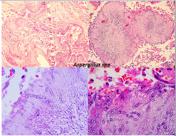
Chronic necrotising aspergillosis is locally invasive is seen in patients with immunodeficiency or chronic lung disease. Aspergilloma is a fungus ball that develops in a pre-existing cavity in the lung parenchyma. Acute angled dichotomous branching hyphae found in necrotic tissue
✅Bronchiectasis. In addition to respiratory complications, children also develop pancreatic insufficiency & lose the exocrine pancreas leading to malabsorption and steatorrhea, as well as GI complications. https://t.co/DkmTH9B1c4
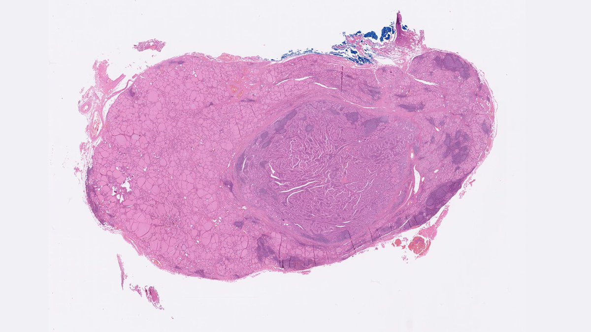
Warthin-like papillary thyroid carcinoma.
🔬Papillae lined with oncocytic cells, with prominent lymphocytic infiltration of stalks. Tumor nuclei are enlarged, irregularly sized with grooves and occasional pseudo-inclusions. Background of lymphocytic thyroiditis.
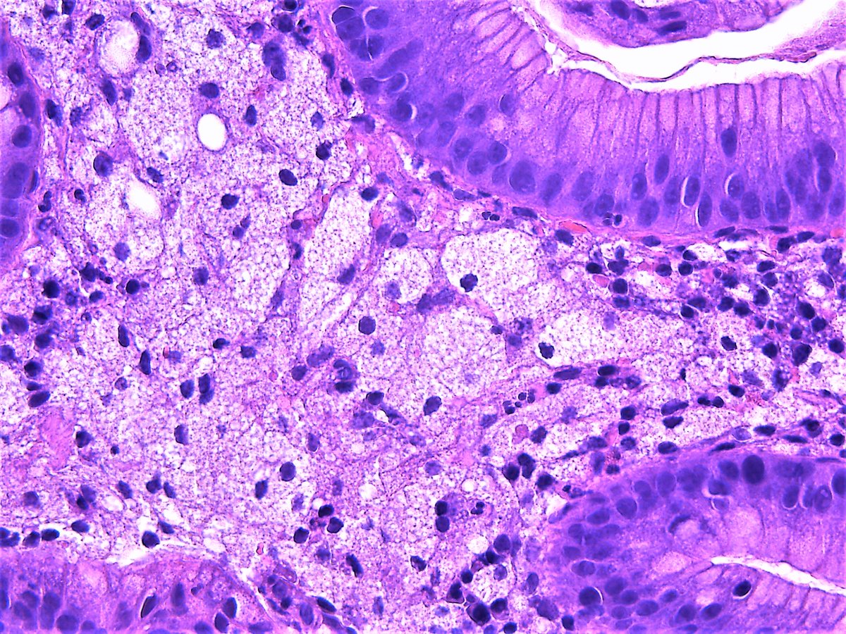
Mucinous metaplasia of the fallopian tube
- Can be associated with Peutz-Jeghers syndrome and mucinous neoplasms at other sites
- Should exclude mucinous neoplasia elsewhere in female genital tract/GI
#pathresidents#gynpath
Nodular melanoma: large profusely bleeding “skin tag" present for 3 months in the intergluteal fold. Positive for S100, Melan A, and HMB 45.Classic features: dermal-invasive melanoma with lack of significant radial growth phase (i.e. junctional component)
Metastatic breast IDC with lobular growth pattern in the pleural cavity: Cytology showed abundant atypical cells with cannibalism. Cell blocks were positive for MOC31, GATA-3,��GCFDP-15, and negative for mucicarmine, ER, PR, HER2.
Multifocal oncocytoma/oncocytosis of parotid.
🔹multiple nodules, some circumscribed and some not.
🔹some can be clear cell rich.
🔹p63 should show an incomplete outer layer around the nests.
But wait…aren’t these supposed to be golden brown grossly? See final pic.