Our work on "Lateral cell polarization drives organization of epithelia in sea anemone embryos and embryonic cell aggregates" is published today.
We thank the @NIH and @AmherstCollege for supporting our work!
We thank you for reading it!
https://t.co/QKUjehwgQy
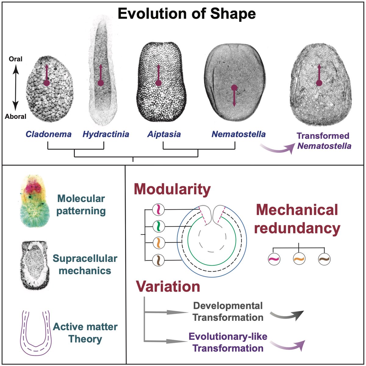
The evolution of shape diversity through the lens of physics—explored in our new paper! A fruitful collaboration between our lab @EMBL, Salbreux’s lab @unige & others, led by @BailleulRichar1—now independent at @ENS_ULM—& Nicolas Cuny. https://t.co/Dzwbeg3sGM
1/4 🚨 a brand new @biorxiv preprint from the lab! We charcterized both transcriptionally, morphologically and functionally (phagocytosis) immune cells from #Nematostella! We are convinced that our data supports the existence of such specialized cells! https://t.co/Fi2kmZ1N6R
🧵
We recently uploaded a new preprint exploring the mechanism of organ remodeling during dormancy.
https://t.co/CKtauianUx
Yuya, a student in my group, used the fly model to identify a novel role of BubR1 and Mad2 in pausing stem cells in a “ready-to-go” state, enabling the remodeling of the Drosophila midgut. #Drosophila
🌊🪸 A mesmerizing moment in marine #science! Institute researchers, led by Investigator @Gibson_Lab & Postdoc @EmmaRangelH, recently captured the stunning moment of successful #coral spawning— follow along as they share insights into this event & their coral #research. 🧵 (1/3)
📣 Looking for a reliable research assistant for the new year! We are at beautiful western Massachusetts and work together with undergraduates on sea anemone embryos. No extensive research experience required. Please share!
Thank you
@AmherstCollege
EMBL scientists have used sea anemones to study how animals restore their shape following major injuries.
Their results show how, in regenerating animals, systemic molecular and cellular changes help in regaining the original shape.
https://t.co/VMoYBCQLqU
“When it rains, it pours” the motto for this semester’s DevBio class on the last day of lab and first snowy day of fall. @AmherstCollege
For the love of #development
Excited to share my PhD work!🎉
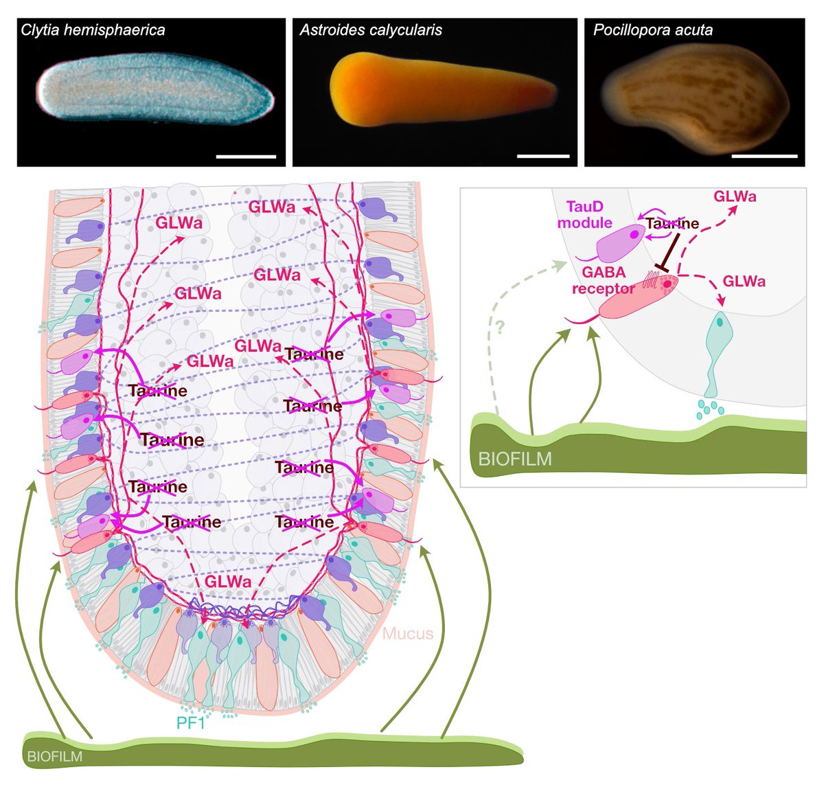
By characterizing and comparing the cells at the aboral end of the planula larva in 3 cnidarian species, we identified shared molecular features and propose a model for settlement regulation in cnidarians 🪼🪸. @Clytia_Vlfr
https://t.co/rob5gRtHP6
🔬✨ New insights into tissue regeneration! Our study uncovers a novel mechanochemical mechanism in Hydra, revealing how mechanical forces and Wnt signaling work together to create intricate tissue patterns. #Cnidarians#Hydra#Biology#Regeneration https://t.co/3Dk6IFFGsj
Our work on "Lateral cell polarization drives organization of epithelia in sea anemone embryos and embryonic cell aggregates" is published today.
We thank the @NIH and @AmherstCollege for supporting our work!
We thank you for reading it!
https://t.co/QKUjehwgQy
Cnidarians are supposed to be diploblastic. In this new ms we show that in the sea anemone Nematostella the interplay of Wnt, MAPK and Notch signaling controls the segregation of endodermal, mesodermal and ectodermal identities. https://t.co/DvH4jLRVwF
EXCITED to unroll this 🧵 on our new story on the metabolic licensing of animal development... How did we even get here..?
https://t.co/Z5mhTtLGde
Early embryo is thought of a pre-set machine that's set to go once it's fertilized - check out this gorgeous 📽️from Phil Keller!
Very happy to share the first article from my postdoc work in the Tomancak lab! https://t.co/0gmwblw1Wb. We studied the self-organization of actin in aggregates made from Hydra cells. Thread below (1/9)