ED POCUS Case 🍐
A 38 year old male presented to ED overnight with sudden onset epigastric pain radiating to his back.
He had no other associated symptoms. His observations, bloods and ECG were normal.
Enter POCUS...
@davemaca1yahoo1 This was one of the few articles I found with regards to small subpleural consolidations
https://t.co/ooJRWBLHrN “LUS was able to identify all the cases of bronchiolitis with concomitant bacterial pneumonia, of which 5/25 were subcentimetric pneumonia”
@davemaca1yahoo1 Hi Dave, I agree with what you’re saying, more studies need to be done, however I forgot to add that this infant had sats of 88% on room air when asleep and paeds informed me that they would have started abx given focal signs and day 5 of illness
🔍 Ultrasound-Guided Lumbar Puncture – an essential POCUS technique for improving procedural success in CSF sampling, especially in ↑BMI or anatomically challenging patients with spinal deformities or prior surgeries, minimizing traumatic taps, needle redirects, and complications to enhance diagnostic accuracy and patient safety in emergency, neurology, and critical care environments.
Best annotated and explained video I’ve seen 🤩
⚡ Employ pre-procedural static marking to locate optimal L3-L4/L4-L5 interspace, boosting first-pass success rates to over 90% in difficult cases and reducing overall attempts. 🩺
📏 paramedian sagittal views for hyperechoic spinous processes (crescent-shaped with shadowing) and transverse views for midline identification, ensuring precise needle trajectory and depth estimation. 📐
🚨 Switch to curvilinear probes in obese patients for better penetration; this halves failure rates compared to landmark-based methods, particularly when BMI exceeds 30. 📈
🔍 Incorporate real-time guidance to avoid bone contacts and minimize traumatic LPs (e.g., RBC counts <400/mm³), with meta-analyses showing fewer complications like post-dural puncture headaches. ⚠️
🤝 Integrate ultrasound with clinical assessment (e.g., palpation, patient positioning in sitting/lateral recumbent) for holistic management, supported by randomized trials in adult and pediatric populations across ED and ICU settings. 👥
PoCUS can help in diagnosis of chronic gastric outlet obstruction by
⭐️Finding evidence of gastromegaly
⭐️ Constant bubbling of gas in gastric fluid "effervescence sign"
Illustrative example of a hockey stick mitral valve (rheumatic mitral valve stenosis).
#POCUS#echofirst#FOAMed
🔗 Mutarelli et al. JACC Case Rep. 2024 PMID: 39534628
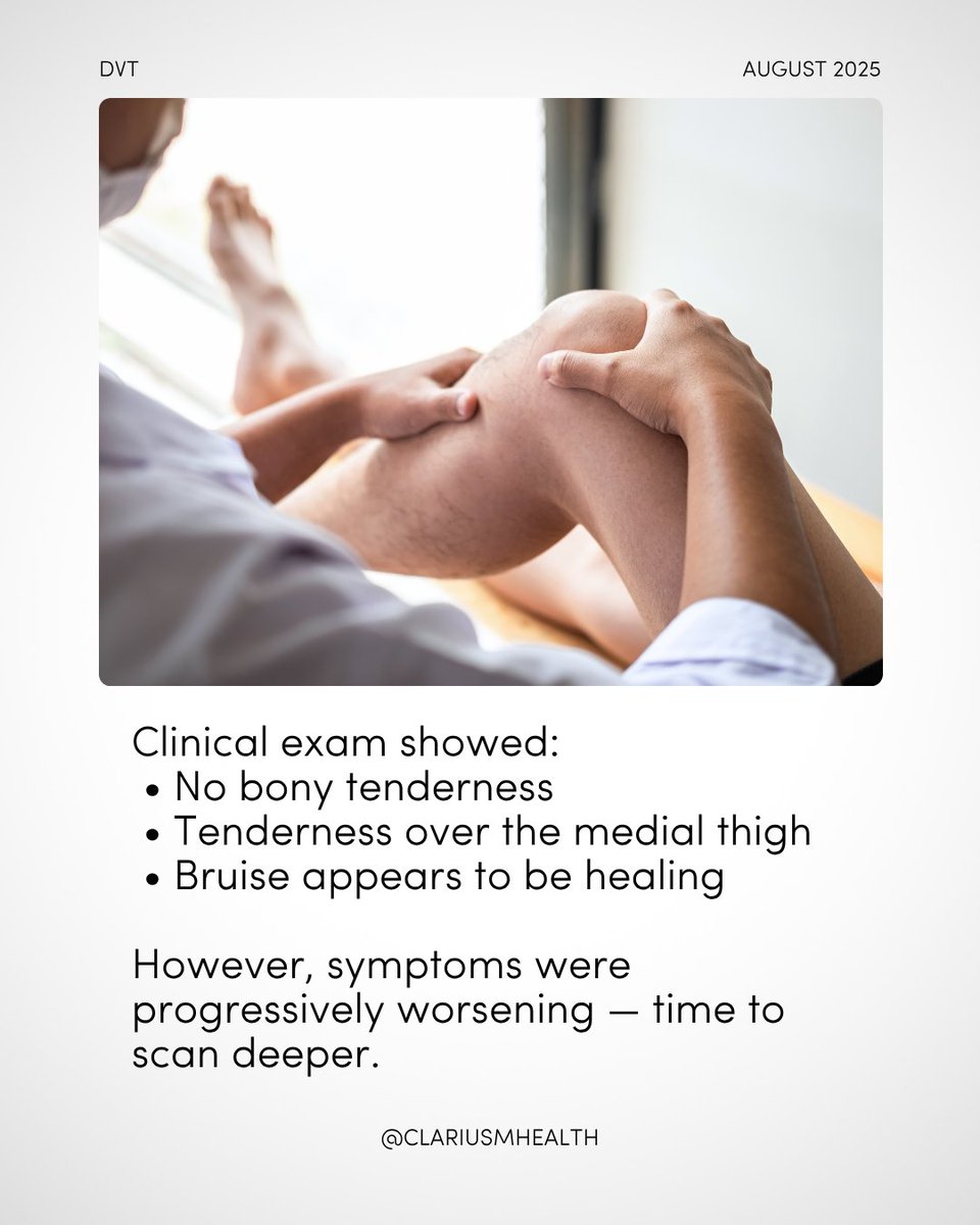
🦵 Thought it was a sprain. It was a DVT.
25F with thigh pain + bruising after a fall. No fracture.
POCUS = big SFV clot → thrombus to popliteal fossa. Later dx: genetic coagulopathy → lifelong anticoagulation.
🧠 Tip: Don’t rule out DVT in young pts. Trust your probe.
🚲 A 66-year-old trauma patient developed sudden painless vision loss after a bicycle accident. CT and external eye exam were unrevealing.
🔍 Using ocular POCUS, emergency physicians identified an irregular, echogenic lens, which is consistent with a traumatic cataract. The diagnosis was later confirmed by Ophthalmology.
💡 POCUS is proving to be a powerful tool in the early detection of ocular injuries, especially when standard imaging or clinical exams fall short.
🔗 Read the full case: https://t.co/dtez5CZG1S
#POCUS #OcularPOCUS #Trauma #EmergencyMedicine #Ultrasound
3/ Take home points 📝
1. PoCUS allowed me to quickly identify a complication of cholecystitis
2. Ultimately a CT was done, but PoCUS allows the radiologist to prioritize the scan + focus on looking for complications.
#POCUS#MedTwitter Images via PalHD3 @clariusmhealth
PoCUS case of the week 🍐
- 61F presenting with 3/7 hx of fevers and RUQ pain with vomiting.
- Bloods - CRP 311, obstructive LFTs.
- Findings: - Asymmetrical thickening of the GB wall (13mm at its thickest)
- Multiple gallstones
- Sonographically Murphy's+ve
2/⚠️ My main concern here was why there was so much asymmetrical thickening. Decision made to CT given findings and high CRP.
CTAP was performed which was reported as "appearances may also reflect an intramural abscess formation from an underlying microperforation."
2/ Treatment of Morel-Lavallée lesions vary depending on the size and symptoms :
-Small asymptomatic lesions can be managed conservatively with decompression bandaging.
-Medium lesions may need drainage.
- Large lesions may need open debridement.
PoCUS image of the week☄️:
- Morel-Lavallée lesion in a thigh of a 15 year old
- Closed degloving injury separating fascial layers often occurring after high energy trauma
- Hypoechoic initially and can contain internal debris
#pocus#clariushd@clariusmhealth#msk#foamed
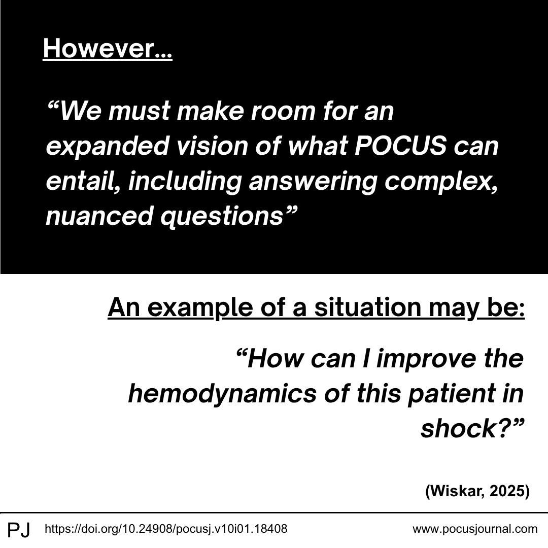
👩⚕️🔍 Point-of-care ultrasound (POCUS) is evolving.
While traditionally used to rule in or out specific diagnoses with yes-or-no answers, POCUS now enables complex, multi-organ assessments—integrating advanced scans and clinical data to guide nuanced decision-making.
This expansion is reshaping how we approach patient care at the bedside.
🩺 Read more about the future of ultrasound in clinical practice: https://t.co/fc8dxCeFmD
#POCUS #ClinicalUltrasound #PointOfCare #MedicalInnovation #UltrasoundEducation #MedEd #HealthcareTech
@cliffreid Unfortunately not. I’ve had to recently remove an ECG sticker with a patients hair still stuck to it from one of our SonoSite machines…
Might be worth including equipment hygiene and maintenance in those who are beginning their POCUS journey?
@cianmcdermott MAPSE was around 6mm I believe. Formal ECHO interestingly had LV VTI at 12.8cm and visually estimated EF at 20-25% with severe eccentric hypertrophy!
ED PoCUS Echo🫀
Thought I'd share some nice views from my night shift!
57yo male NFW presenting to ED with 2wk history of SOBOE and lying flat.
PLAX view:
- Sinus rhythm
- Visually estimated EF severely reduced (eyeballing method)
#ECHO#POCUS
3/ EPSS 🫀
- Measurement of the distance between the anterior tip of the mitral valve and IVS during early diastole.
- Rapid bedside measurement.
- EPSS >7mm is considered a sign of reduced EF.
- ⚠️Pitfalls - can be falsely elevated in valvulopathies
Happy scanning 🫀
#FOAMed