So excited to share that our new review article is now online in Diagnostic Histopathology!🎉
📝 “Recognizing parasitic helminths in tissue: histopathological insights into fascioliasis and echinococcosis” 🔬
https://t.co/XivNEVqWH0
#pathology#histopathology#echinococcosis
A proposal for a new testicular sex cord-stromal tumour classification system from a group of experts who represent two international pathology organisations. #gupath#openaccess https://t.co/xkMCD3IHL0
🔬 IAP Review of the Week
This week's educational pearl covers the practical approach to eosinophilic renal neoplasms:
• Pattern recognition
• Focused IHC
• Molecular testing
Content courtesy of Dr Mahmut Akgul.
Look at all the medications in this tiny portion of a gastric biopsy. The drugs present tell us that the patient has renal disease. Of course, microcrystalline cellulose is just pill filler. Kayexalate reduces hyperkalemia and Renvela reduces hyperphosphatemia. #UMiamiPath
🚨 ISUP Presents.....
🔬 Dr. Seema Kaushal on “A Rosetta Stone with Missing Glyphs: Immunohistochemistry as a Molecular Surrogate”
📅 21 July 2026 at 10am EST via Zoom
Register here: https://t.co/pGVl5d4WPd
Nice work! ✨
✅ Lymphocytic (Diabetic) Mastopathy
Keloid-like stromal fibrosis with prominent perilobular lymphocytic infiltrates, typically seen in patients with insulin-dependent diabetes and other autoimmune diseases.
#PathX#PathTwitter#breastpath
Breast - Infiltrating epitheliosis
Key histologic features (Eusebi & Millis)
• Predominantly florid usual ductal hyperplasia (UDH) with focal squamoid areas
• Scleroelastotic stroma distributed throughout the lesion (not limited to a central nidus like radial scar (RS)/CSL)
• Frequent desmoplastic reaction + keloid-like fibrous bands
Unlike classic UDH, ducts show jagged/irregular edges with epithelium “flowing out” into stroma → more infiltrative appearance
IE is possibly related to radial scar/complex sclerosing lesion family. Lacks clear zonal architecture; myoepithelial cells are frequently discontinuous or absent
Dr. Lerwill #USCAP2026 #pathology #PathX #PathTwitter
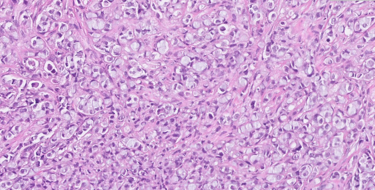
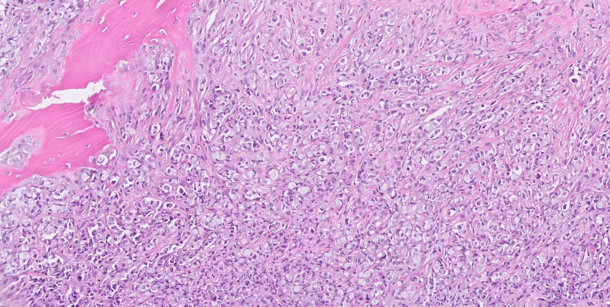
Invasive urothelial carcinoma with lipoid and micropapillary features — a frequent association worth recognizing. Lipid-filled cytoplasmic vacuoles alongside micropapillary clusters/retraction spaces make for a striking diagnostic image. #GUPath#UroPath