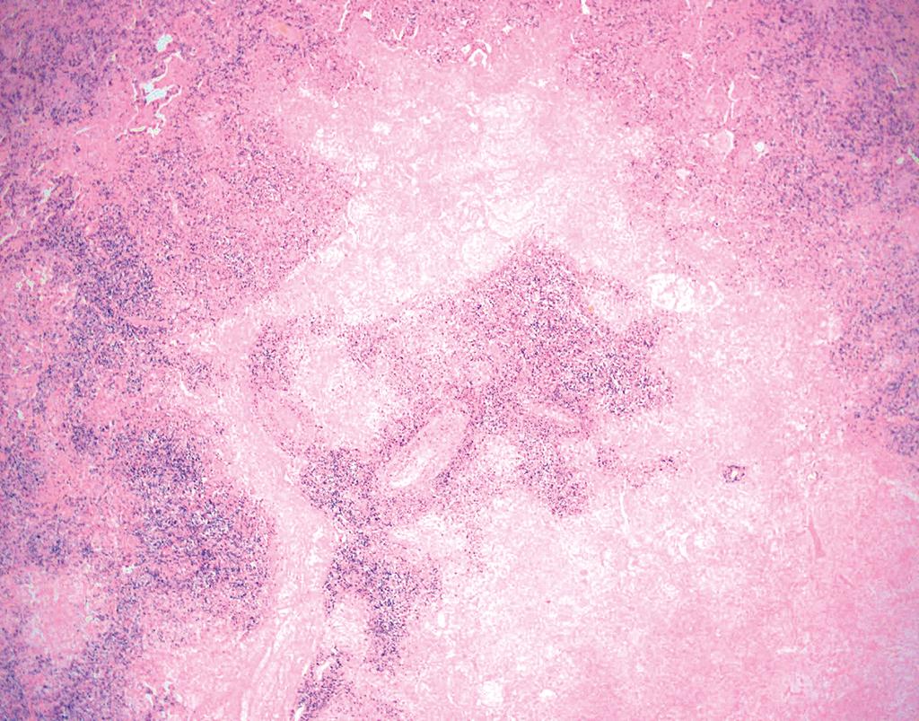
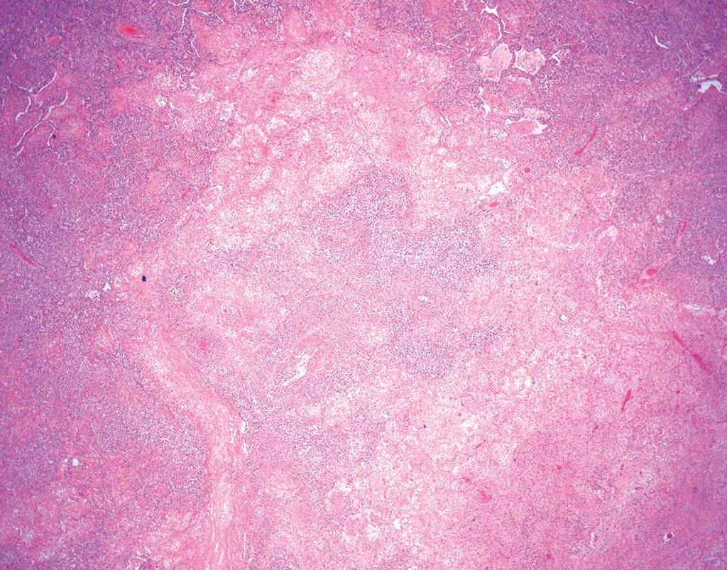
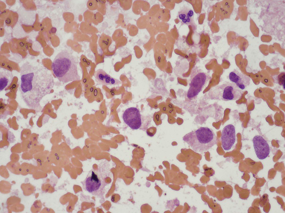
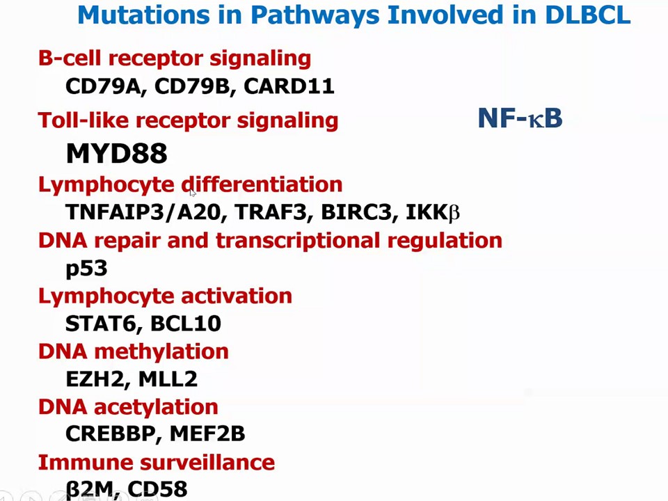
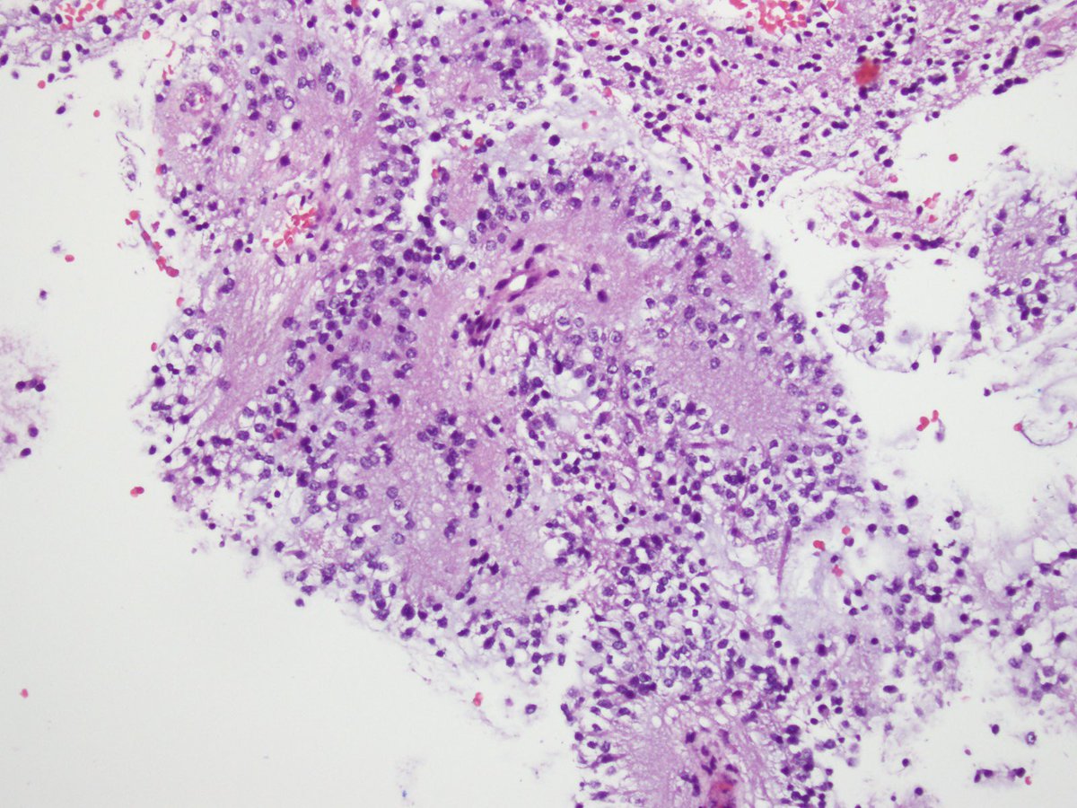
✅ Lymphomatoid Granulomatosis 🎯🫁
• Angiodestructive polymorphous infiltrate with geographic necrosis
• Driven by EBV-infected B cells in a background of reactive T cells
• Graded (1-3) based on the absolute density of large atypical cells
• Despite the name, well-formed granulomas are characteristically absent
#PathQuiz 🔬🫁 #ThoracicPath
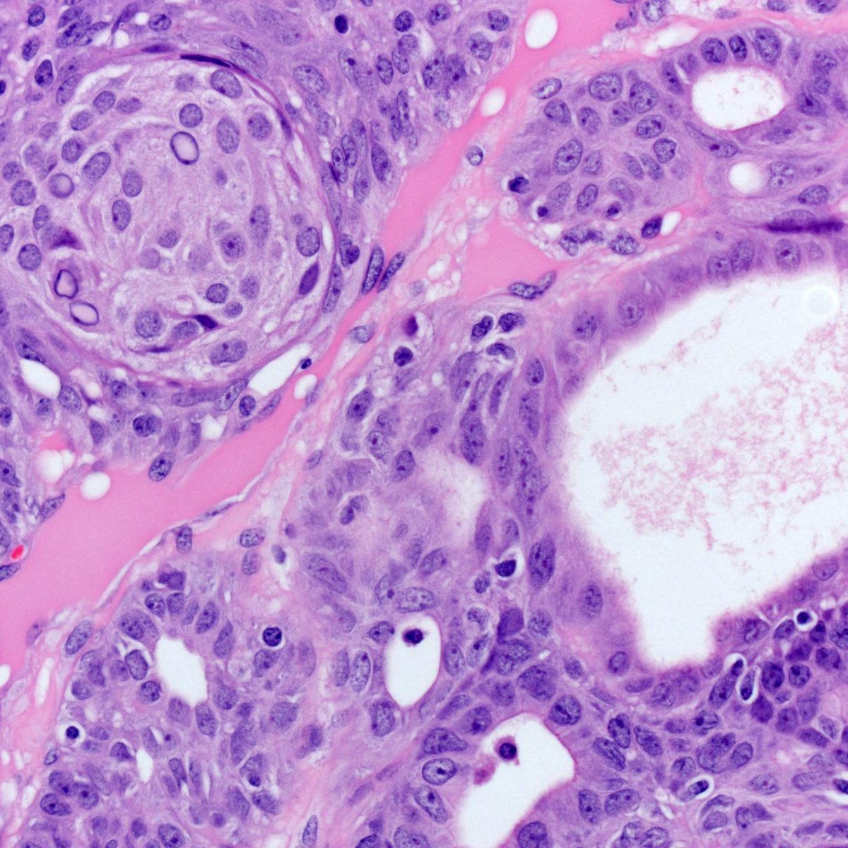
A) Mucoepidermoid Carcinoma
B) Papillary Adenocarcinoma
C) Sclerosing Pneumocytoma
D) Papillary Adenoma
#Pathology
Clue in caption 🧐
#PathQuiz 🔬
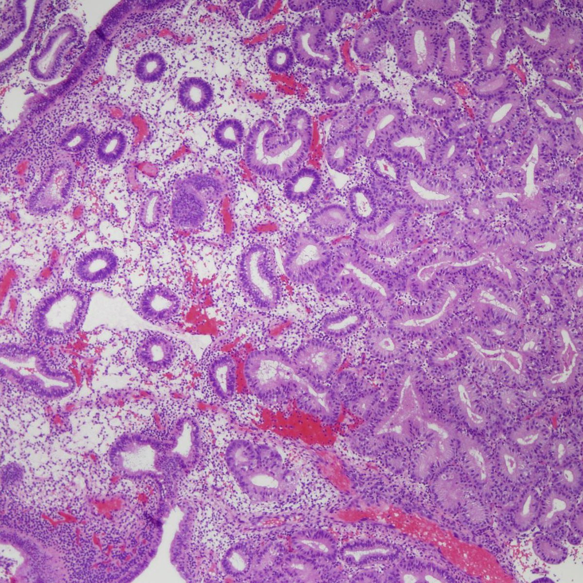
A) Hyperplasia without atypia
B) Hyperplasia with atypia
C) Endometrial polyp
D) Atypical polypoid adenomyoma
#Pathology#GYNPath#Endometrium
Clue in caption 🧐
Patchy moderate chronic active colitis
Dr. Krasinskas recomendation for this case:
- If I receive some fragments of colonic mucosa showing chronic active colitis, and others that appear "normal", I use the term "patchy," as in this case.
On initial presentation, IBD/UC tends to be diffuse and involves the rectum, distal colorectum or the entire colon (pancolitis).
Uninvolved "skip" lesions are uncommon in untreated UC but can be seen in treated UC and in segmental colitis associated diverticulosis (SCAD)
This patient had diverticulosis, making the histologic findings c/w SCAD
Dr. Krasinskas - Tips and Hints in Surgical Pathology, Thoracic pathology #USCAP #pathology #PathX
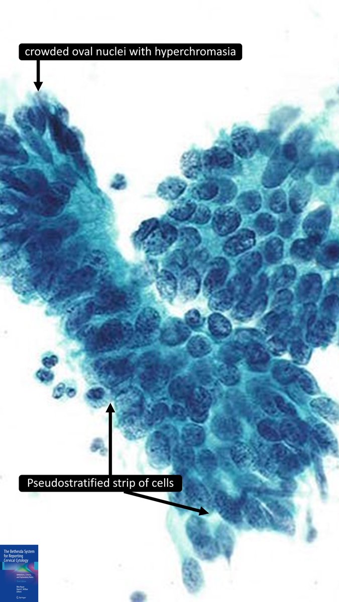
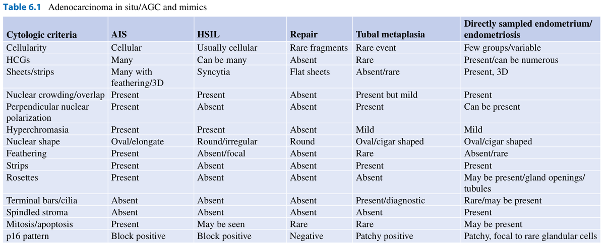
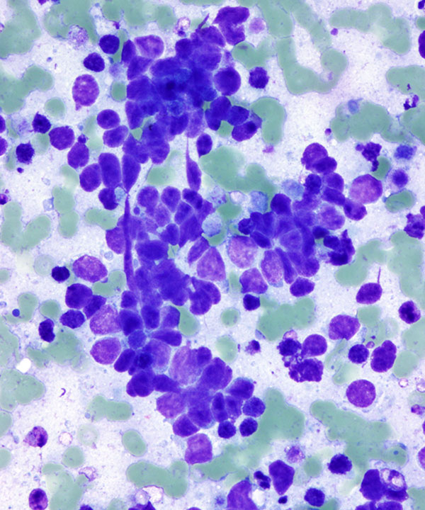
Answer for #2: Atypical endocervical cells
Cells: sheets, strips, pseudostratified
Nuclei: enlarged, elongated, hyperchromatic with heterogeneous & coarse chromatin
Hyperchromasia that is more monotonous than simple metaplasia
Cytoplasm: may be abundant, but N:C is increased; distinct cell borders
#CytoPath #PathSky #PathTwitter #Pathology
AP case of the month: Woman in her 6th decade with progressive gait issues & paresthesia. MRI showed an enhancing intradural extramedullary thoracic spinal cord mass. Previously resected and recurred after 3 yrs. Histology: pleomorphic cells, EMA+, vimentin+, Ki-67 ~20–25%.
#Cytopathology is always helpful!
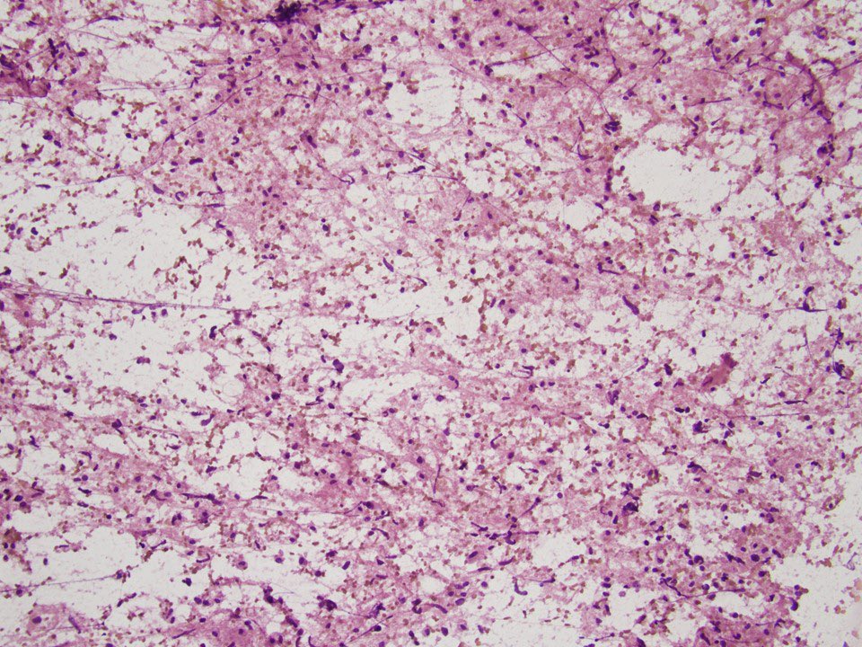
A squash prep during a frozen section for a neurological tumor. Pleomorphic cells with cytoplasmic extensions, suggestive of a high grade astrocytoma. IHC is +ve for GFAP. NGS is +ve for IDH1 mutation. The final Dx is glioblastoma. @SaeedAsiry
Diffuse astrocytoma, MYB or MYBL1-altered. An infiltrating, but grade 1 glioma! (Note the trapped neurons—evidence of infiltration) Tumor cells rel. monomorphic, round to ovoid or spindly. It is strongly associated with epilepsy. #neuropath#neuropathology#pathx#PathTwitter
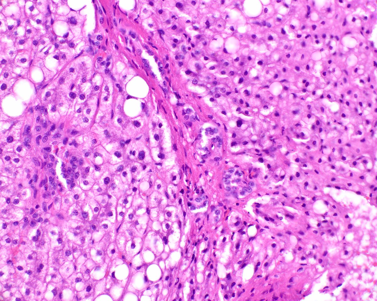
Such a perfect example of hepatic focal nodular hyperplasia with the classic "map-like" glutamine synthetase immunohistochemical staining pattern. Note that the adjoining normal liver tissue lacks that pattern. #UMiamiPath
Posterior fossa 4th ventricular brain mass in a young patient, what molecular alterations are likely detected in this lesion? 🤔
#pedipath#neuropath#pathology
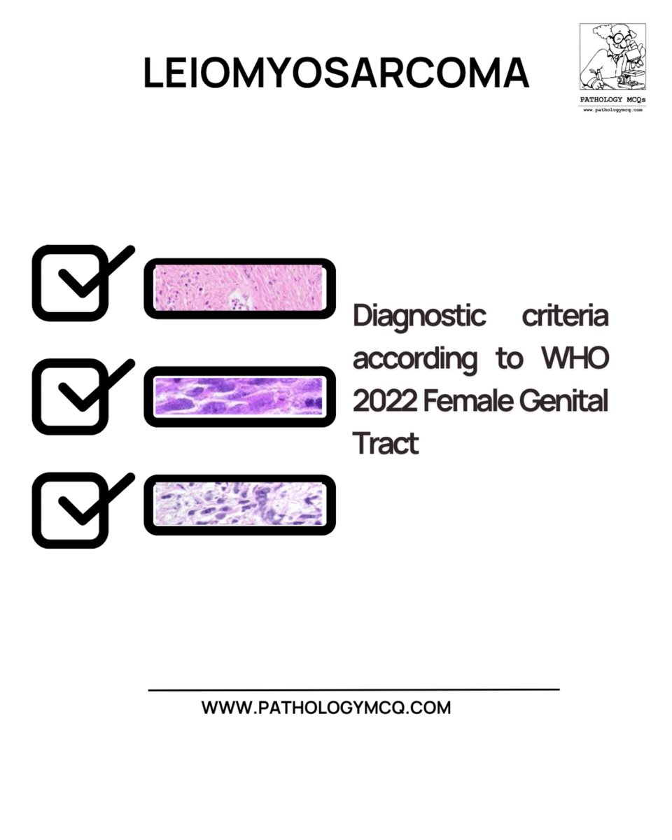
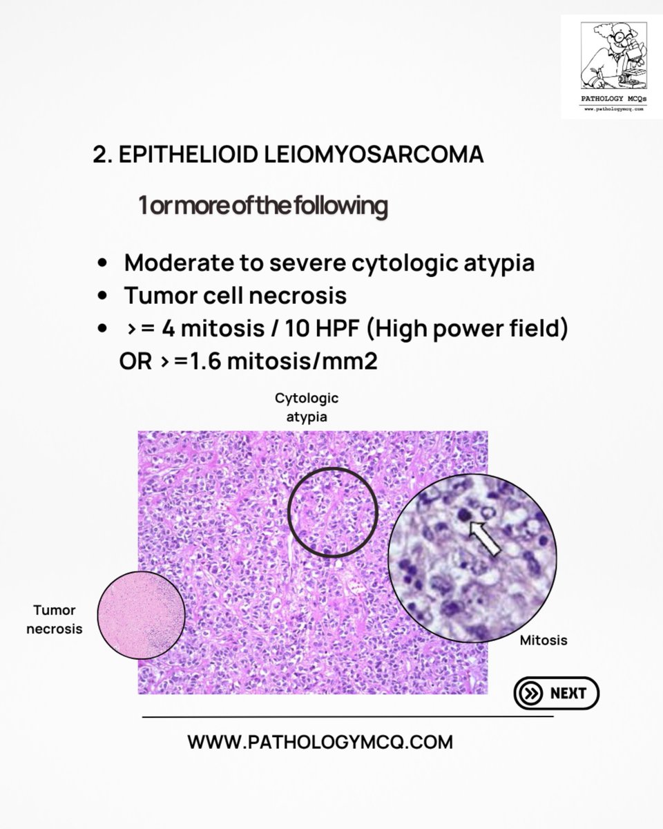
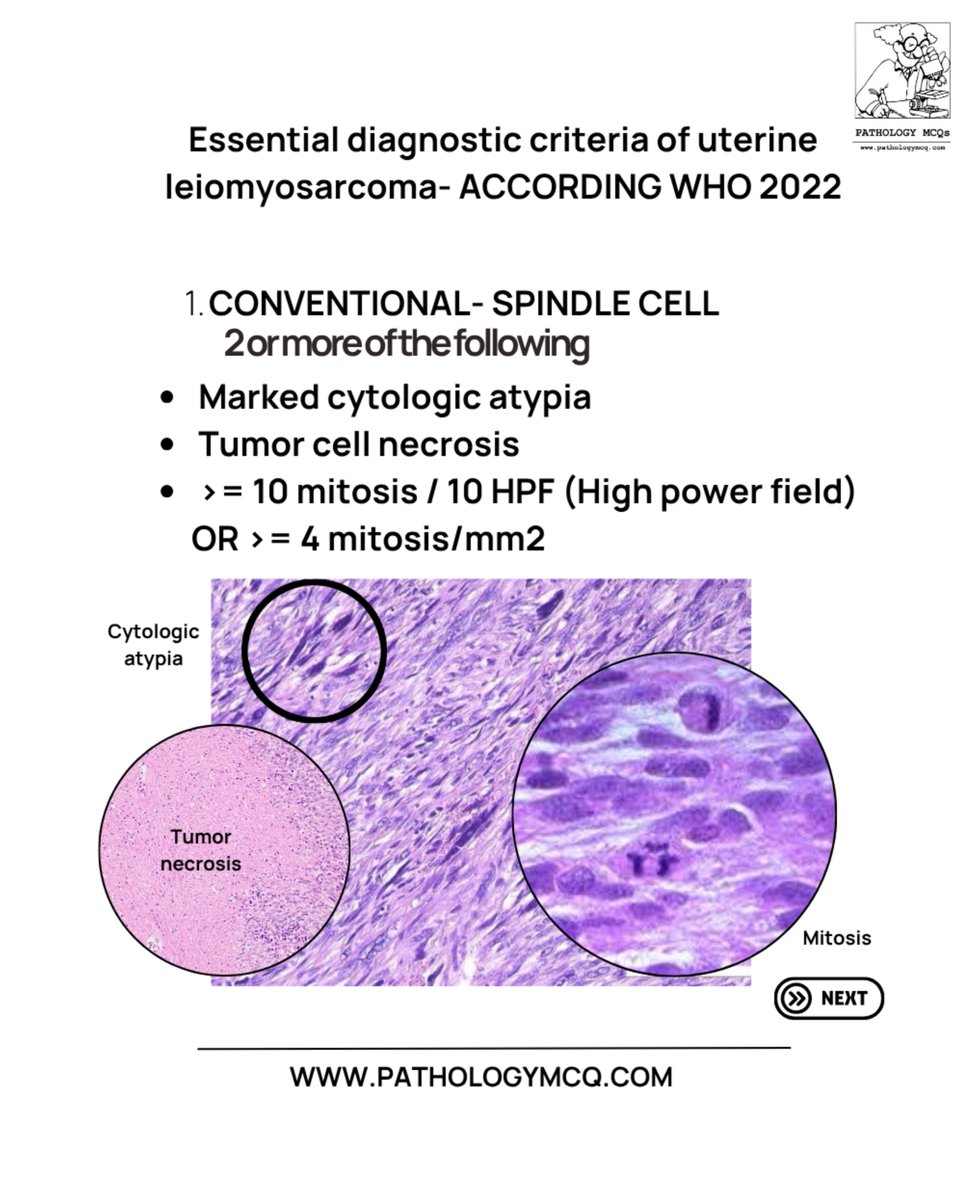
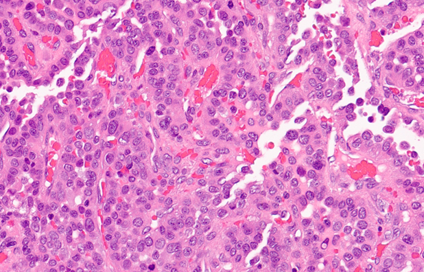
💥 LEIOMYOSARCOMA simplified!
WHO 2022 👉 Spindle, Epithelioid & Myxoid types with key histologic clues 🔬
🎉 Don’t miss the Pathology Day 2025 offer — 20% OFF on WHO-based modules! 🧠
#PathologyMCQ#WHO2022#Leiomyosarcoma#PathologyDay
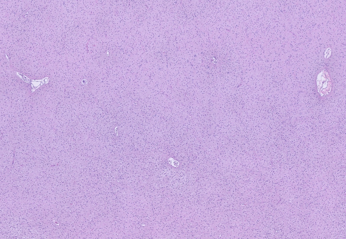
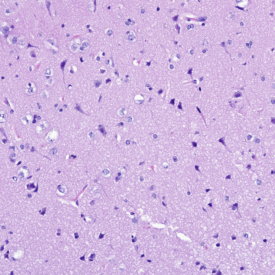
This might LOOK like unremarkable cortex, but an IDH1 R132H immunostain highlights a few scattered oligodendroglioma cells lurking in the background. Tricksy!!! #pathology#neuropath#PathTwitter