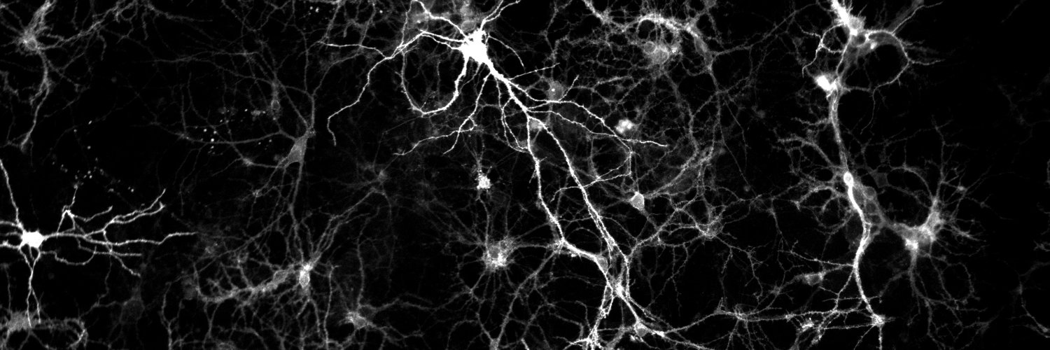
Happy to introduce our cell-tracking software for large-scale imaging datasets! https://t.co/YiT2sF38EO At Tsinghua University (Prof. Qionghai Dai & @JiaminWu7 ), we develop large-scale imaging systems and computational algorithms for neuroscience and immunology.
Directional elements: the preprints are out! Congratulations to our amazing team linked here:
https://t.co/SxNw2isNcd
https://t.co/f48w42FKcJ
https://t.co/o5NXdGcViw
We screened for principles governing global brain dynamics by developing a set of new methods: 1) conformal immersion microscopy for recording high-speed/high-resolution neural activity across dorsal cortex; 2) unbiased computational screening of brain-spanning activity for fast directionally-propagating spatiotemporal elements; and 3) novel genetically-encoded voltage sensing integrated with designed spectrally-compatible opsins (derived from our channelrhodopsin structure work) for systematic causal testing.
This unbiased screening/testing approach (which we show is applicable either to voltage or calcium imaging) allowed discovery and functional validation of a surprisingly well-defined set of directional elements that generalized across cell types and frequencies. The ability to work over long timescales at high speeds and with broad scope, anchored in optogenetic causal testing, unveiled rich spatiotemporal structure that was remarkably tractable.
From the perspective of natural brain function, the directional elements were found to be behaviorally relevant and robust to diverse perturbations; however, we also found specific conditions allowing elemental incidence and boundaries to be selectively modulated, which may provide translational as well as basic-science insight...
I'm so grateful to all our collaborators, and honored to work with all the outstanding students, postdocs, and staff who worked together to develop and apply this approach...
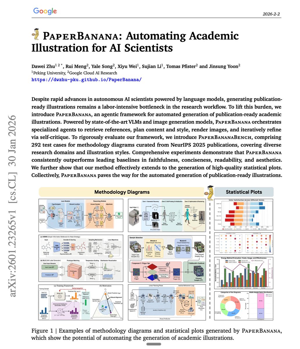
Google just dropped another banger!
the figures in this paper were drawn by the system described in the paper.
PaperBanana is an agentic framework that generates publication-ready academic illustrations from methodology descriptions.
no manual design, no Figma, just your method section and a caption.
here's how it works:
five specialized agents collaborate in sequence:
> Retriever: finds relevant reference diagrams from a curated set of NeurIPS papers. matches by visual structure, not topic.
> Planner: translates your methodology text into a detailed visual description using in-context learning.
> Stylist: applies aesthetic guidelines (color palettes, typography, layout) auto-summarized from hundreds of top-tier papers.
> Visualizer + Critic loop: generates the image, critiques it against source text, and refines. repeats for 3 rounds.
one surprising finding: randomly selected examples work nearly as well as semantically matched ones. what matters is showing the model what good diagrams look like, not finding the topically perfect reference.
in blind evaluations, humans preferred PaperBanana outputs nearly 3 out of 4 times.
it also extends to statistical plots using code-based generation for numerical precision.
link in the next tweet.
Our November issue is now live! 🎉
https://t.co/PBA3WcH2Wh
On the cover, cell tracking in a developing zebrafish embryo visualized using Ultrack. Cover by Alexandre Dizieux, Jordão Bragantini, Loic Royer, Merlin Lange, CZ Biohub. Paper here: https://t.co/SuOLwJUfl7
In our November issue we feature several papers on the theme of tools for cell segmentation and tracking. For a roadmap to the issue, read our Editorial.
https://t.co/Fusdt0Z2oB
Clearance of intracranial debris by ultrasound reduces inflammation and improves outcomes in hemorrhagic stroke models - @StanfordRad https://t.co/BludN4qtBd
😁👾Excited to introduce 𝙎𝙥𝘼𝘿 - enabling scalable label-free images-based cell profiling for chemical/genetic screen via continuous spinning + ultrafast imaging
🔄 𝙎𝙥𝙞𝙣𝙣𝙞𝙣𝙜 𝘼𝙧𝙧𝙖𝙮𝙚𝙙 𝘿𝙞𝙨𝙠 (𝙎𝙥𝘼𝘿)
- Optofluidic 96-chamber circular array design supporting continuous live-cell imaging
- Ultrafast quantitative phase imaging (QPI) at 1000’s rpm → sub-cellular-resolution label-free imaging across 100 cm2
- Disk design is fully compatible with standard protocols
📊 𝙈𝙖𝙨𝙨𝙞𝙫𝙚-𝙎𝙘𝙖𝙡𝙚 𝘼𝙣𝙖𝙡𝙮𝙩𝙞𝙘𝙨:
- 10 GB/sec real-time processing
- *InMorph* profiling strategy→ Rich biophysical signatures from label-free data
- Easily scalable to 100s of TBs for CRISPR/drug screens
✅ 𝙈𝙪𝙡𝙩𝙞-𝙛𝙖𝙘𝙚𝙩𝙚𝙙 𝙑𝙖𝙡𝙞𝙙𝙖𝙩𝙞𝙤𝙣s
- Mechanical stability testing
- Cell health assays
- Transcriptomic analysis
We demonstrated that SpAD enables sensitive, large-scale screening of drug responses and CRISPR gene knockouts without labeling - allowing unbiased morphological phenotyping at a new scale.
Years in the making - A great work (fine art) executed by our postdoc @DicksonSiu1 and Alan Wong from @hkusbms@hkumed
👉 Preprint: https://t.co/r42qS2wyqU
#LabelFree #CRISPR #Drugscreen #QPI #Biophysics #SpAD #microscopy #omics
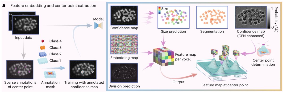
CELLECT: Contrastive embeddings for real-time, large-scale cell tracking
Modern microscopes can film living tissues in 3D for hours, producing terabytes of data with cells that divide, drift, and deform. Traditional trackers either demand dense annotations or expensive global optimization—and often break under frequent mitosis or strong motion.
Hongyu Zhou and coauthors introduce CELLECT, a contrastive embedding approach that learns a latent notion of “cell-ness” and tracks in that space rather than raw intensity. A 3D U-Net predicts confidence maps, compact feature embeddings, and division probabilities; a lightweight center-enhancement branch sharpens peaks; and two tiny MLPs clean up duplicates within a frame and link cells across frames—including splits—using only sparse center labels.
Trained once on a single public C. elegans dataset, the same model generalizes across modalities (confocal, light sheet, two-photon, light-field) and species without retraining, delivering state-of-the-art accuracy at real-time throughput.
The numbers are compelling: ~56× faster than linajea on benchmark embryos (~2 s per 512×512×41 frame) and higher long-track fidelity (46% vs 22% over ~250 min). It scales to terabyte videos on a single GPU and remains robust in dense, dividing tissues.
What this unlocks: continuous 3D tracking of B cells through germinal center formation, quantitative readouts of neutrophil–bacterium–macrophage encounters in vivo, and stable neural signal extraction under strong nonrigid brain motion—all from the same pretrained model.
This points to a practical future for quantitative biology where we keep the microscopes we have, keep the photons we capture, and recover usable, lineage-aware cell trajectories in real time—without drowning in annotations.
Paper: https://t.co/vOyvWpQLQw
It was my honor to work with the excellent researcher Hongyu Zhou (non-X user).
We hope CELLECT helps other researchers working on large-scale imaging or immune cell migration studies!
Happy to introduce our cell-tracking software for large-scale imaging datasets! https://t.co/YiT2sF38EO At Tsinghua University (Prof. Qionghai Dai & @JiaminWu7 ), we develop large-scale imaging systems and computational algorithms for neuroscience and immunology.
Our centimeter-scale mesoscale imaging system enables long-term, single-cell–resolution imaging with minimal phototoxicity. To tackle the massive data challenge, we built CELLECT — a contrastive learning–based tool for automated cell tracking, segmentation, and event detection.
Endomucin immunostaining of a cleared young mouse skull reveals a continuous vascular network in the parietal bone, enabling high-resolution 3D visualization of cranial blood vessels.
In a recent preprint at bioRxiv (Yang et al., https://t.co/MblqlBKo92), a group of authors led by Dr Anjali Kusumbe challenges an article from my lab published in 2024 (Koh et al. 2024, PMID: 39537918). The new preprint refers to Extended Data Figures and Supplementary videos that are unfortunately not provided at bioRxiv. Nevertheless, the manuscript text and the 5 main figures plus a proposed model contain a couple of major issues that I will cover here.
1/