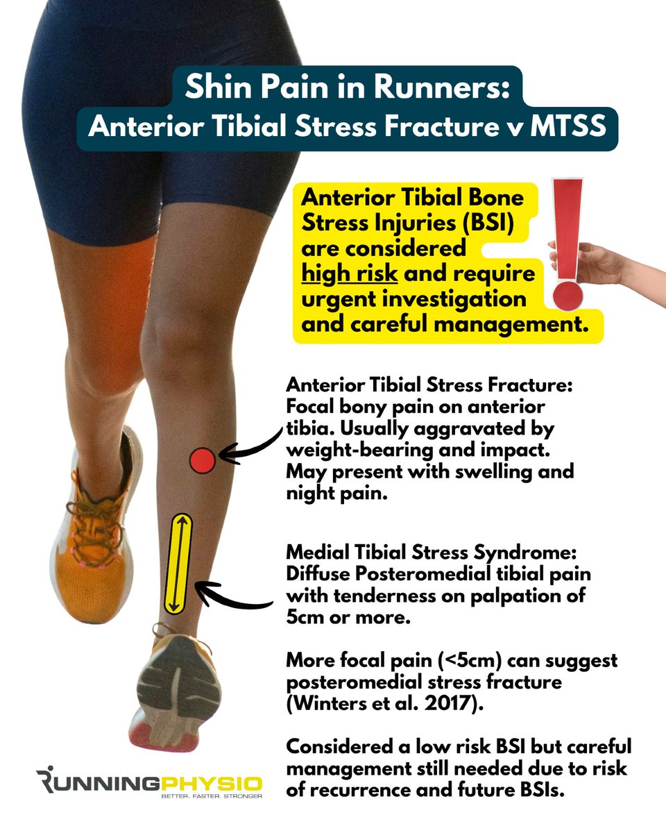
‼️Anterior Tibial Stress Fractures are high risk.
They present with more focal anterior tibial pain than Medial Tibial Stress Syndrome (MTSS) so be specific about pain location and behaviour when taking a history.
Anterior Tibial BSIs can lead to a true fracture of the tibia and require surgical intervention. They’re associated with prolonged periods away from sport and a high complication rate so need urgent investigation and careful management are needed including exploring energy availability and bone health.
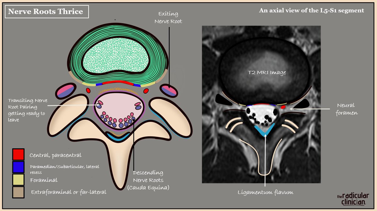
Nerve Roots 'Thrice'
Clinical anatomy applied
Lumbosacral nerve roots can be described in three geographical locations on axial T2 MRI, each with important clinical implications if they are compressed or inflamed. The picture illustrates the L5-S1 level, so we are looking at the L5 disc. Focus on the colour codes in order to appreciate disc location in relation to each geographical nerve root location.
Descending (bilateral S2, S3, S4 and S5 roots as part of the cauda equina).
An acute central or paracentral disc herniation here is rare but can cause cauda equina syndrome if compressed. Also relevant with crowding as seen with LSS.
Transiting/Traversing (here bilateral S1 roots).
As the bilateral transiting nerve roots leave the thecal sac centrally, they remain enveloped by its outer connective tissue layer, referred to as the nerve root sleeve. The lateral recess is the most common site of disc related compression because of its anatomical position and the root being fixed in place. This is why radicular pain is more commonly noted in one leg. Note that the nerve roots do not lie flat when you do.
Exiting (here L5).
As the nerve root enters its respective neural foramen. This is the second most common site of compression, typically from a far lateral disc herniation or from crowding within the foramen.
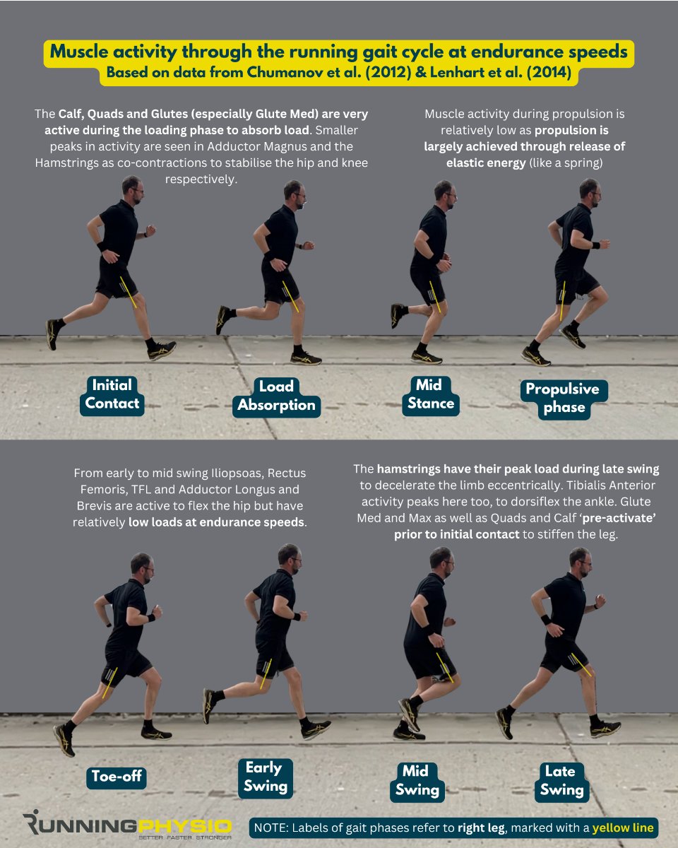
Peak forces on the calf, quads, glutes and hamstrings are high during running which is why it’s important to assess and strengthen these muscles after injury. We cover how to do this and lots more in our free Achilles video series. Access it now at https://t.co/KEji9T24t3
This graphic is a nice illustration of how muscle forces shift at different running speeds.
This can help guide return to sport or return to run protocols, especially if an athlete is coming back from an injury in one of these areas.
It also gives you a clearer picture of
INFOGRAPHIC: Muscle activity during running 🏃♀️
Peak loads on the calf, hamstrings and hip flexors will increase significantly with faster running and sprinting.
Differential diagnosis is important in shin pain as *high risk* stress fractures can occur in this region and are often missed. We cover the top 3 causes of shin pain and their management in our free video series which is available now at https://t.co/uIo2StrBOU