Chuchú en retiro. Dentro del museo ferroviario
Ambiente y calidad de los platos 10/10
Servicio malo. No llegan a cubrir todo. Se puede mejorar recién arrancan
A very impressive study for how we could prevent lung cancer more than 5 years before it is diagnosed. Using machine learning, discovery of a 14-plasma protein signature of risk that predicts responsiveness to an antibody therapy to interleukin, IL-1β
Validated across 8 cohorts
@CellCellPress@CharlesSwanton
https://t.co/qpPtgs1dH0
@paisa_ar@TheotokosLover Aprovecho para consultar. Lei por ahi quebhaybque curar los de madera con grasa (manteca x ej) yo lo estoy curando con yerba humeda y por ahora viene bien. Es probable q se pudra?
🧵 Seems like chronic pulmonary edema cases are following me lately 👀
I haven't found a pathognomonic CT sign described in the literature for this entity, but after seeing a few of these, I started calling it "nutmeg lung" 🫁
Has anyone else noticed this pattern? Is there a better name for it in your practice? 🤔
#Radres #FOAMed #RadTwitter
In addition, the other day I came across a compelling case from the folks @AbdominalCase : HALT-D (Heart-After-Liver Transplasnt with Domino).
Mindblowing surgical choreography 🤯😅
Thanks for sharing such educational and interesting cases for all! @YashantAswani
https://t.co/cCvMAl5Vey
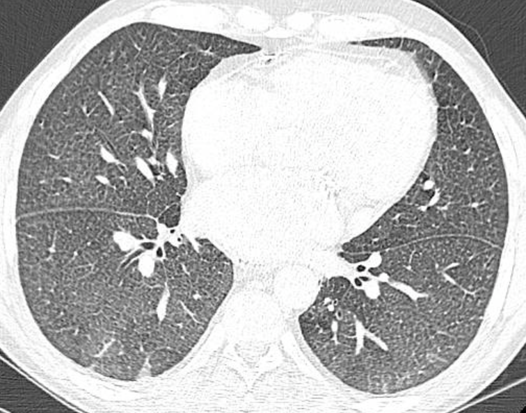
Yesterday a colleague shared this fascinating case 🫀
20F with history of Fontan surgery (TGA) Chest CT showing diffuse bilateral solid nodules.
Now take a look at the CT slices of the abdomen 🚨 Heterogeneous liver appearance + suspicious focal hepatic lesion
We’re probably looking at FALD-associated HCC with pulmonary metastases!
A reminder that Fontan patients need long-term hepatic surveillance 👁️
#Radiology #FOAMed #RadTwitter #RadRes
Another case to add! 📚
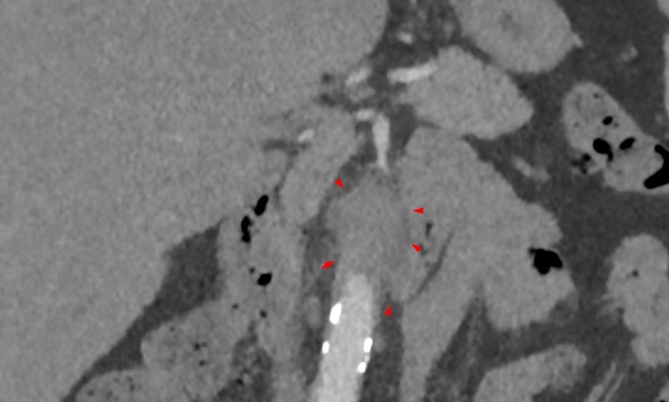
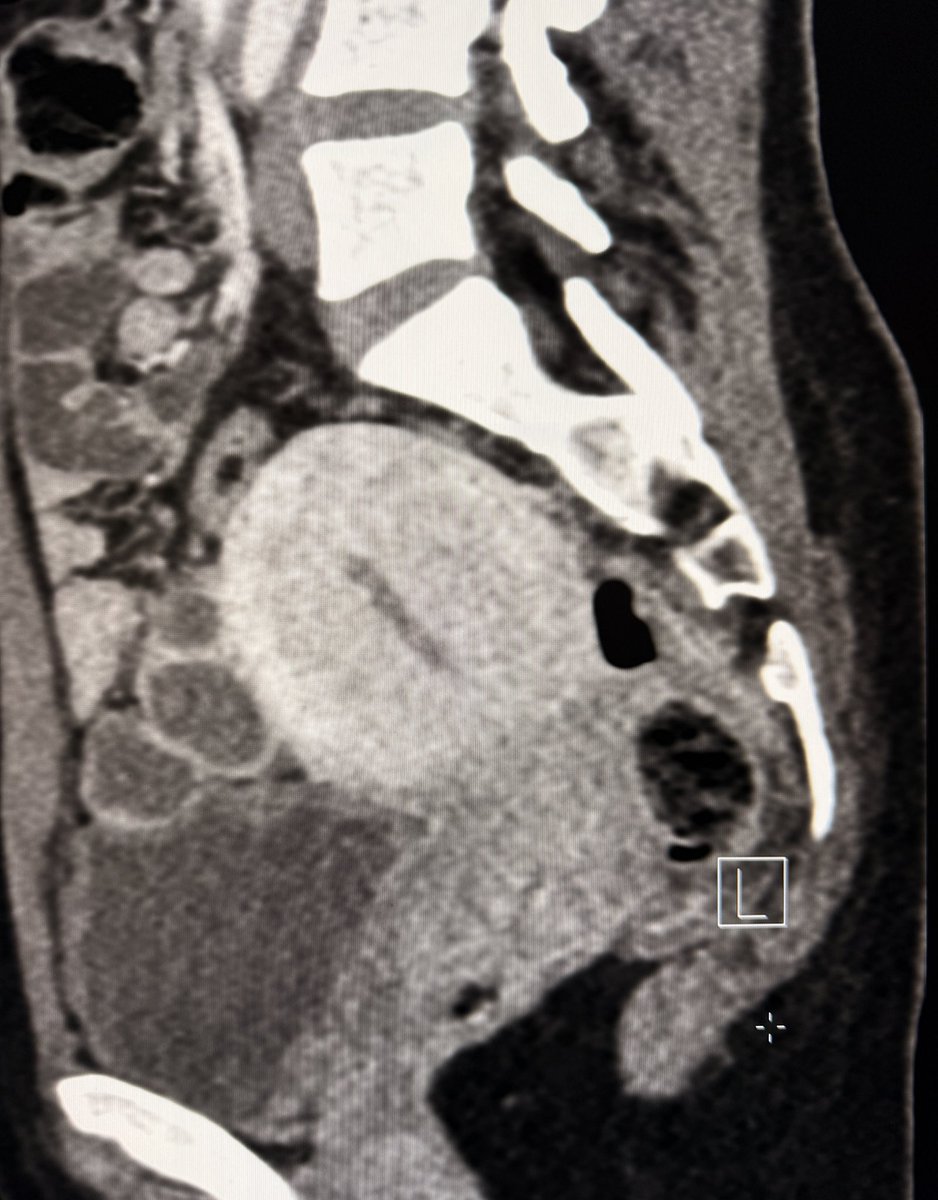
16F with Myasthenia Gravis — whole-body 18F-FDG PET/CT showing periaortic soft tissue thickening with increased metabolic activity surrounding the abdominal aorta.
No aortic dilatation. Posterior sparing. Same pattern 👀 👀
Extra findings: FDG-avid lymphadenopathies.
IgG4 also pending on this one 😅
I wonder if there's any association between aortitis and MG 🤔
#Radres #FOAMed #PETCT #vascular
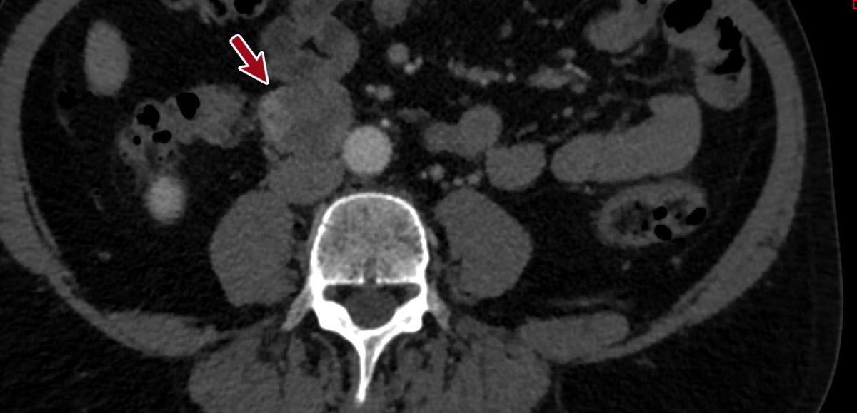
50F with abdominal pain. What’s surrounding the aorta? 👀
(Periaortic soft tissue thickening, non-circumferential, with posterior sparing and no aortic dilatation)
👉 Inflammatory periaortitis? IgG4-related disease high on the list.
IgG4 pending… 🥶
#Radiology#FOAMed #RadTwitter
@Kdog1980YNWA Absolutely! PET catches metabolic response earlier and is the gold standard in many protocols.
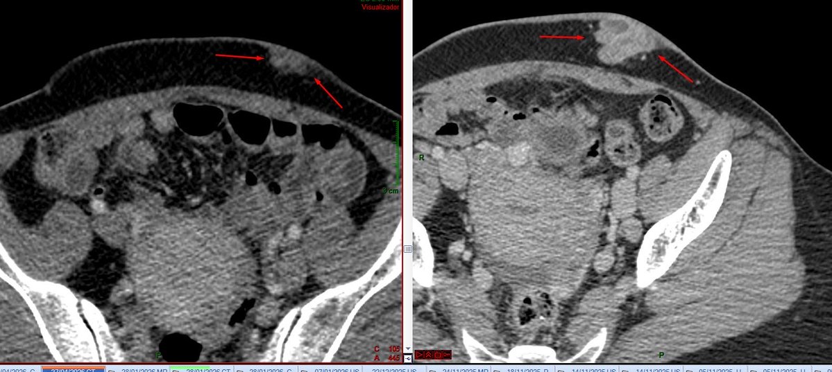
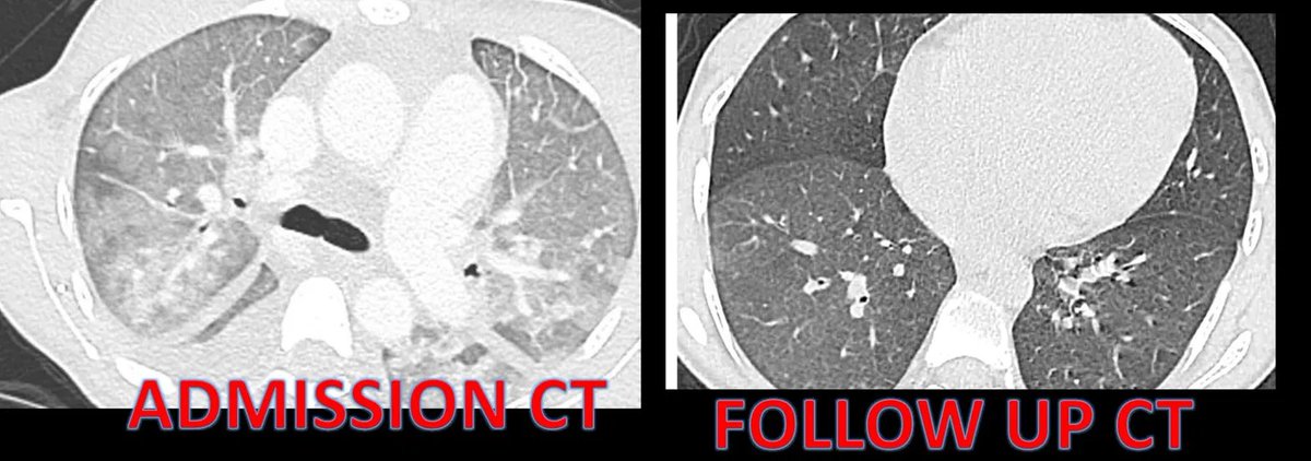
Here the CT change was so striking it was hard not to share 😄
No conflict of interest — just a radiologist amazed by science!
BRAF+MEK inhibition in metastatic melanoma.
Only 3 months between these CTs. The difference speaks for itself. 🎯
#Radres#Oncology#TargetedTherapy#Melanoma#Radiology
@peedrobenedetti@danyscht@jmtelechea No sólo eso, sino que Claude está dando respuestas erróneas! Volviendo a gpt 5.4 después de un mes de cambiarlo por Claude!
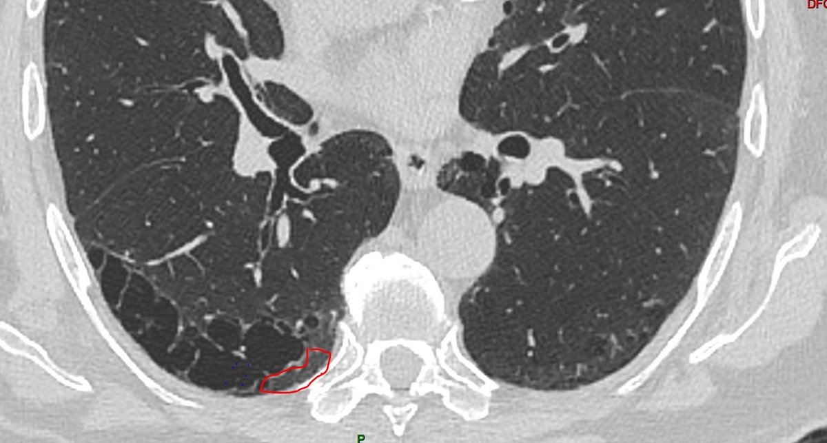
Digging into 2024 radiology gold ⛏️✨
These key points on SRIF are worth revisiting.
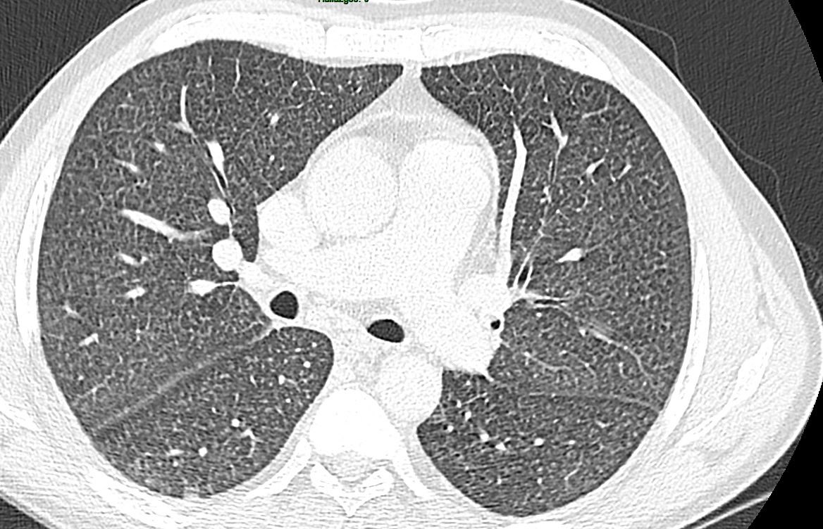
Applying them to this 78-year-old smoker:
– Mild emphysema
– Cysts within reticulation & large irregular cysts
– No subpleural predominance
– No honeycombing or traction bronchiectasis
Pattern favors SRIF ✅
To answer the question in this post, HRCT readers should be aware of two things.
First, they need to understand the features of smoking-related interstitial fibrosis (SRIF). SRIF manifests in three main appearances:
1.Involvement of existing centrilobular and paraseptal emphysema by developing dense, definable walls and causing irregular, heterogeneous shapes and sizes, thereby disfiguring their usual appearance.
2.Cysts within reticulation.
3.Large irregular cysts.
It’s important to note that the second and third appearances do not abut the pleura. SRIF typically occurs without other fibrotic features such as traction bronchiectasis, bronchiolectasis, irregular reticulation, or honeycombing.
The second question to address is whether SRIF contributes to combined pulmonary fibrosis and emphysema (CPFE). The answer depends on the presence of fibrotic features such as traction bronchiectasis, bronchiolectasis, irregular reticulation, or honeycombing. If these features are present, SRIF is not the contributor to CPFE.
Based on these guidelines, you can determine which of the four cases are CPFE due to SRIF.
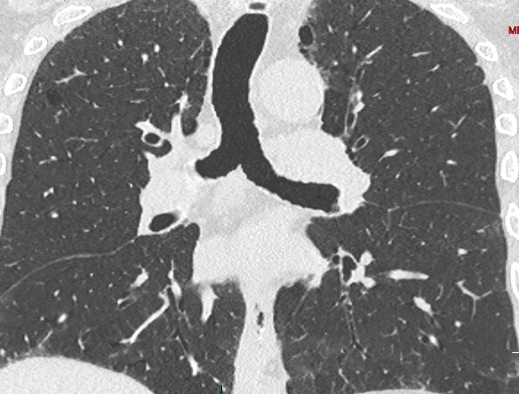
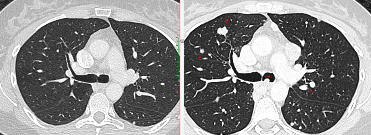
🫁 When heart failure mimics interstitial lung disease
A patient with congestive heart failure showed an indeterminate reticular lung pattern on chest CT.
Was this interstitial lung disease… or something else? 👇
#Radiology#ChestRadiology#MedTwitter
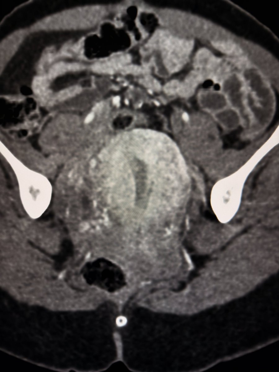
On portal venous phase CT, the uterus enhances more than the cervix. This differential enhancement is common and physiological. Yet it is often mistaken for cervical pathology.
The relatively lower attenuation of the cervix is often overcalled a cervicitis or a cervical mass.
Understanding normal enhancement patterns helps avoid these errors and keeps us from creating pathology where none exists.
—Pearls, Pitfalls, and Wisdom from my reporting list