Can you get the diagnosis when it comes to carotid stenosis?
Having some confusion about the strokes you see w/ICA occlusion?
Why are there so many different patterns of infarcts w/ICA occlusion/stenosis?
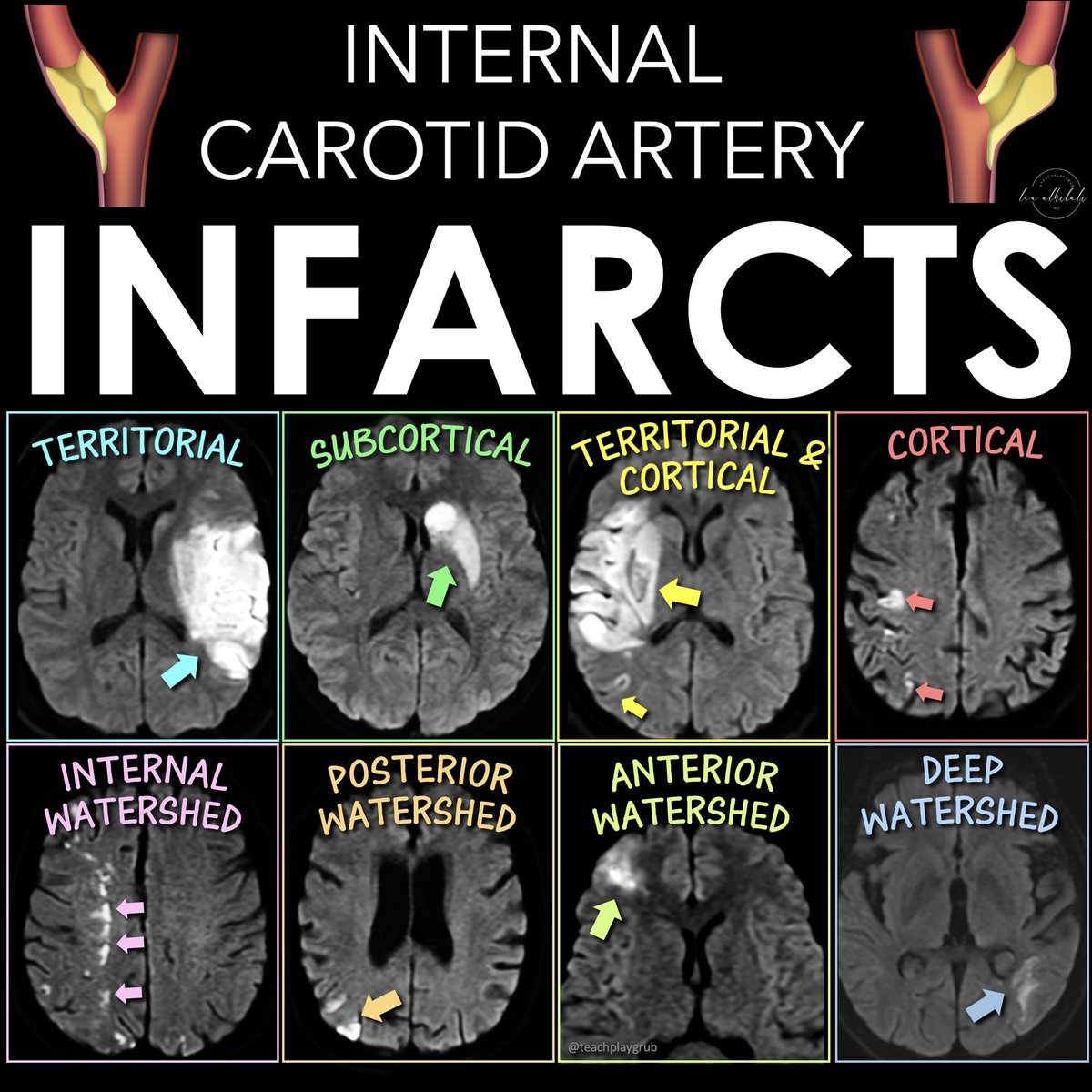
There are 5 main infarct patterns—and they reflect the different types of disruption to flow you get w/carotid disease: artery to artery emboli, embolic shower, & hypoperfusion
Here’s what you see:
1. Territorial infarction w/cortical or subcortical involvement
--From artery-to-artery or distal embolism.
--More than one occlusion indicates more proximal arterial dz
2. Subcortical infarction
--When ICA, MCA, & ACA perforating deep arteries are occluded
--Can be from MCA occlusion w/good collaterals or emboli directly to deep perforating arteries
3. Combined territorial & smaller cortical and/or subcortical infarcts
--From partial embolism fragmentation or emboli shower of various sizes
4. Multiple small infarcts in the distal territories of the MCA & ACA
--If in more than one arterial territory, it’s suggestive of cardio-embolism
--If single arterial territory, it’s from a parent artery steno-occlusive lesion
5. Watershed infarctions.
Two types:
--External border-zone (located between the cortical territories of ACA, MCA, & PCA)
--Internal borderzone (between the deep & superficial networks of MCA & ACA)
--Traditionally thought from low-flow, but emboli are often a cause
--Hemodynamic mechanism may be more important in internal borderzone infarcts & embolism in external borderzone infarcts
Now you can draw the right conclusions with ICA occlusions!
1/Want to TRI to learn something new about the TRIGEMINAL nerve?
If you’re only looking at the skullbase, you are missing a significant part of the trigeminal nucleus!
Let my help you TRI to up your game when it comes to TRIGEMINAL anatomy
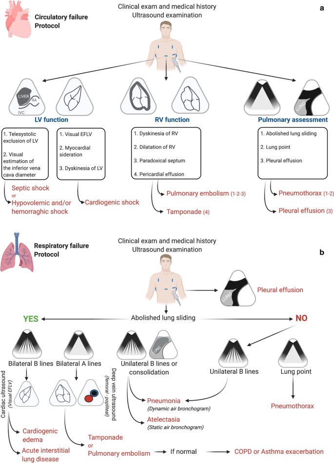
Bedside #POCUS during ward emergencies is associated with improved diagnosis and outcome.
#FOAMed#FOAMcc
🔗 https://t.co/R0S532nbHH
(*lung point should correspond to abolished lung sliding arm of the figure)
Thanks to the AMAZING @TheSonoGraphics team for these Advanced Echo Cards! Available to download FREE on POCUS 101!
👉🔗https://t.co/yynidLrxZI
✅TEE
✅Cardiac Output
✅Tamponade
✅Diastology
✅EF Calcs
✅Right 💔 Failure
✅Wall Motion Abnl
✅And MORE!
See 👀 Cards 👇 #POCUS
Rationale and Design for the BLOCK-SAH Study (Pterygopalatine Fossa Block as an Opioid-Sparing Treatment for Acute Headache in Aneurysmal Subarachnoid Hemorrhage) https://t.co/UMY8S59zgr
Have MULTIPLE questions about MULTIPLE sclerosis?
Having trouble seeing neuromyelitis optica?
In a fog about MOG?
Here’s the cheat sheet you NEED to distinguish the demyelinating diseases!
Demyelinating diseases predominantly involve the optic nerves, brain, & spine.
The three main chronic demyelinating diseases are Multiple sclerosis (most common), neuromyelitis optica (NMO), & myelin oligodendrocyte glycoprotein (MOG) antibody associated disease or MOGAD
Each has its own features in the optic nerve, brain, & spine. Here’s how to remember them!
▶️MS
🔸Optic nerve:
--MS only has 2 letters, so MS involvement of the optic nerve tends to be short segment
🔸Brain:
--Letter M makes the shape of the perivascular distribution of lesions along the ventricles (Dawson’s fingers)
--Letter S makes the shape of the subcortical U fiber involvement
🔸Spine:
--MS is only 2 letters, so lesions are usually less than 2 vertebral bodies in length
▶️NMO
🔸Optic nerve:
--NMO is a longer abbreviation, three letters, so longer involvement
--NMO can stand for Near My Occiput. Occiput is posterior, so more posterior nerve involvement
🔸Brain:
--NMO can stand for Near My Ocean. What is your brain’s ocean? The ventricles. NMO lesions are all periventricular
🔸Spine:
--NMO is 3 letters, so lesions usually more than 3 vertebral bodies in length
▶️MOGAD
🔸Optic Nerve:
--Remember MO’ GAD-olinium. So things that cause more regions of enhancement. MOGAD lesions are commonly bilateral & long segment & enhancement can extend perineural
🔸Brain:
--Remember LO’ GAD. MOGAD typically involves the lower areas of the brain
🔸Spine:
--Remember MO’ PLAID. MOGAD can give a plaid-like H shape in the cord from predominantly gray matter involvement
Hopefully, this cheat sheet will help you remember how to distinguish the demyelinating diseases!
It ain’t lyin’ about diseases of myelin!
Sink or swim!
Brain is swimming in CSF!
CSF is key for protection, buoyancy, & hormone/waste transport
Most know basic ventricular anatomy well, but not subarachnoid anatomy.
How many cisterns do YOU know?
Here’s a quick guide to the key midline cisterns & what to know for each
▶️Cisterna Magna:
🔸Largest of the cisterns
🔸Contains CN 9-11 & vertebral arteries
🔸Remember this bc the LARGEST cistern contains the LARGEST cranial nerve (vagus) and the two cranial nerves around it
▶️Prepontine cistern:
🔸Contains CN 6 & basilar artery
🔸Remember this bc the shape of the number 6 mirrors the curvature of the anterior pons, where this cistern lies
▶️Chiasmatic cistern
🔸Contains CN 2 & pituitary stalk
🔸Easy to remember bc the CHIASM is CN 2
▶️Interpeduncular cistern:
🔸Contains CN 3 & mammillary bodies
🔸Remember this bc if you lay the number 3 on its side it looks both like the two cerebral peduncles (where this cistern lies) & well, two mamillary bodies
▶️Quadrigeminal cistern
🔸Contains CN4 & pineal gland
🔸East to remember bc QUAD is the prefix for FOUR
▶️Lamina terminalis:
🔸Contains the ACOMM
🔸Remember this bc the shape of subarachnoid hemorrhage after ACOMM rupture follows this cistern anteriorly
Now when you look at cisternal anatomy, you will definitely be able to go with the flow!
ICU stories (another common/"boring" one):
Middle-aged pt w history of head/neck cancer, hypertension, hyperlipidemia, chronic kidney dz (baseline creat 2.0 mg/dl), & diabetes 2 presented to the ED at noon time complaining of extreme weakness (leading to falls), thirst/hunger