New paper from the lab! Many animal cells exhibit chiral cell surface dynamics, which are proposed to drive left–right asymmetric patterning. However, the molecular mechanisms linking these events remain unclear. https://t.co/yY0aiEMhEB
New paper from the lab! Many animal cells exhibit chiral cell surface dynamics, which are proposed to drive left–right asymmetric patterning. However, the molecular mechanisms linking these events remain unclear. https://t.co/yY0aiEMhEB
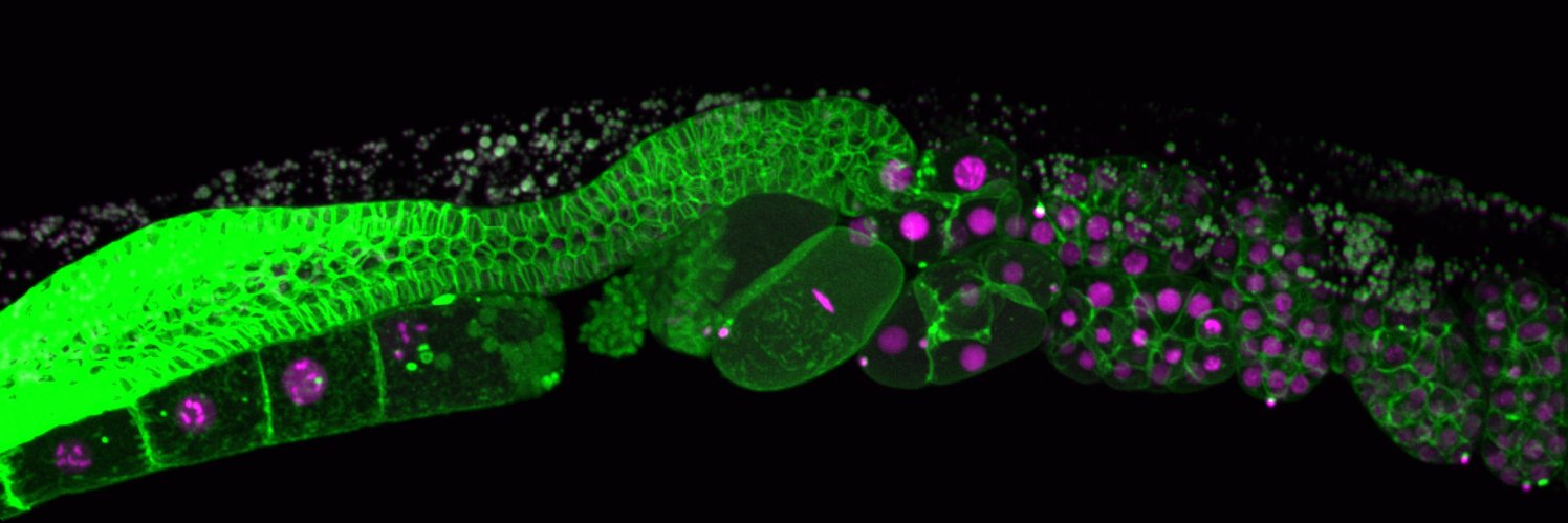
The established chiral cadherin patch interacts asymmetrically with the contractile ring, likely driving its rightward closure, the first detectable left–right asymmetry in C. elegans.
Only 2 weeks left to apply for the Assistant Professor position in Cell & Developmental Biology in Zoology, UBC!
Open to all areas and systems. This is a fantastic department with supportive colleagues https://t.co/6Vi4nGs6G4
Check out our latest paper, out now in @NatureComms
In epithelial cells and animal zygotes, the cytokinetic contractile ring closure is often asymmetric and plays critical roles in tissue integrity. How is it regulated? (1/8)
https://t.co/9RaxXlRjRB
Our analysis revealed that a previously proposed, cortical flow-dependent positive feedback in ring assembly (ref: Reymann et al., eLife 2016, Khaliullin et al., eLife 2018), is responsible for mechanosensation. (6/8)
Our lab is interested in cytokinesis regulation and function in multicellular contexts, and the tools and findings from this simple unicellular model are useful for that purpose. Now we are working on the later developmental stages. More to come (8/8)
Lastly, we found that anillin is required for this mechanosensation and that anillin is required for the maintenance of this positive feedback loop. How? We don't know at this point. (7/8)
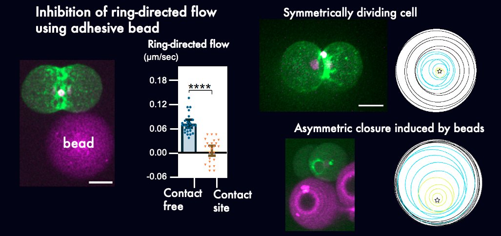
We found that at the lagging edge, ring-directed flow is inhibited due to cortical compression. We also found that artificial inhibition of ring-directed flow using adhesive beads is sufficient to induce asymmetric ring closure in symmetrically dividing cells. (5/8)
Ring-directed cortical flow during cytokinesis has been known for decades, but we were able to track it from the cell surface into the division site, probably for the first time. And yes, it delivers cell cortex materials to the ring, promoting cytokinesis. (4/8)
We used asymmetric ring closure in C. elegans zygotes as a model (ref Maddox et al., Dev Cell 2007) and found that the cell cortex moves differently between the leading and lagging edges. At the leading edge, there is a ring-directed flow. (Movie: myosin) (3/8)