Emerging MRI biomarkers such as the central vein sign and paramagnetic rim lesions show promise for differentiating multiple sclerosis from mimics and reducing misdiagnosis. @radiologyto https://t.co/AZHvMrrQSX
Adopting semiautomated coregistration-fusion into clinical practice can improve detection of radiologic disease activity in #MultipleSclerosis.
Study from @StMichaelsFdn@UofT ▶️ https://t.co/25tJQDocTH
Follow-up MS MRI report “stable”
But could MRI disease activity have been missed? — In this study 20% of F/U MRI reported “stable” using routine comparison had at least one missed lesion. These missed lesions were associated with future disease activity.
https://t.co/Crdbjb7R0R
Latest NAIMS consensus paper out!Imaging chronic active lesions. PRL SEL PET. Tremendous work by Francesca Bagnato @VanderbiltU with a v special mention in acknowledgments. Definition is 1st step in dx biomarker @danielsaloreich@PascalSati@takishinohara
https://t.co/b6MUZpslia
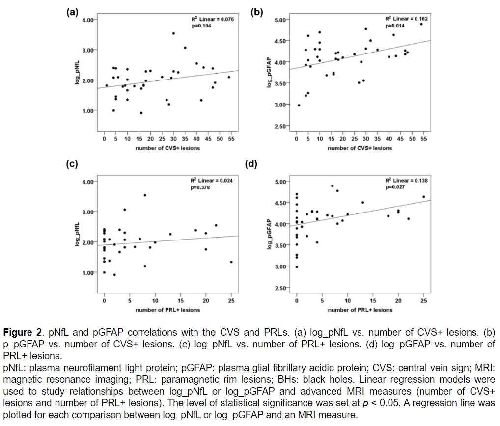
Plasma glial fibrillary acidic protein #GFAP levels were associated with the number of white matter lesions with central vein sign and with paramagnetic rim in people with radiologically isolated syndrome #RIS.
Study from @Raph_Schneider @UnityHealthTO ▶️ https://t.co/CYcevHj50F
‘Evolution of brain MRI lesions in paediatric myelin-oligodendrocyte glycoprotein antibody-associated disease (#MOGAD) and its relevance to disease course!’
Congratulations to Dr @drmankad, Dr Jackie P & Dr Yael H on our Medical Advisory Board and collaborative team for this excellent and very detailed paper! Check it out
Link: 👉🏻 https://t.co/qGfXTnNyoy
In this multicenter study, the central vein sign #CVS provided high specificity to #MultipleSclerosis diagnostic process, especially when identifying up to six CVS+ lesions.
Study from @DanOntaneda @CleClinicLCM @NIH_NINDS ▶️ https://t.co/LC0gRHbowK
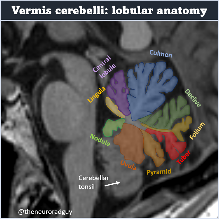
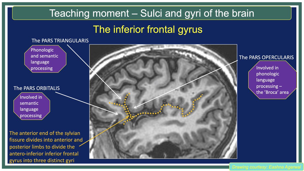
Small but beautiful: lobular anatomy of the vermis. A step-by-step tweetorial with many useful mnemonics can be found on the page of
@teachplaygrub; nice illustrations of the same anatomy can be found on the page of the excellent
@drharunyildiz. #FOAMrad#FOAMed#neurorad
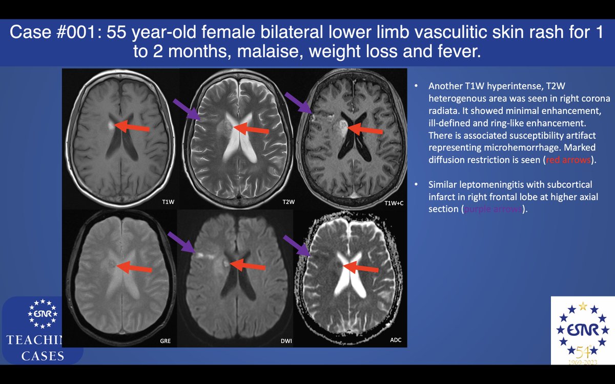
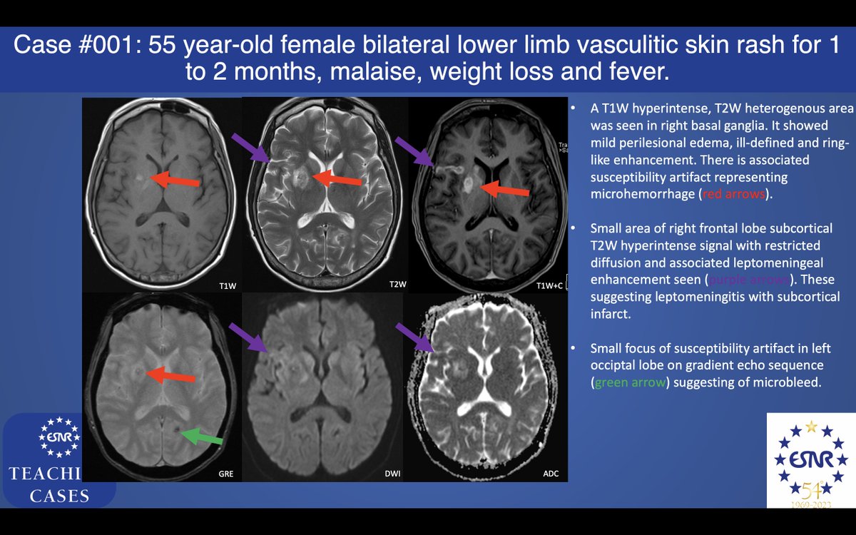
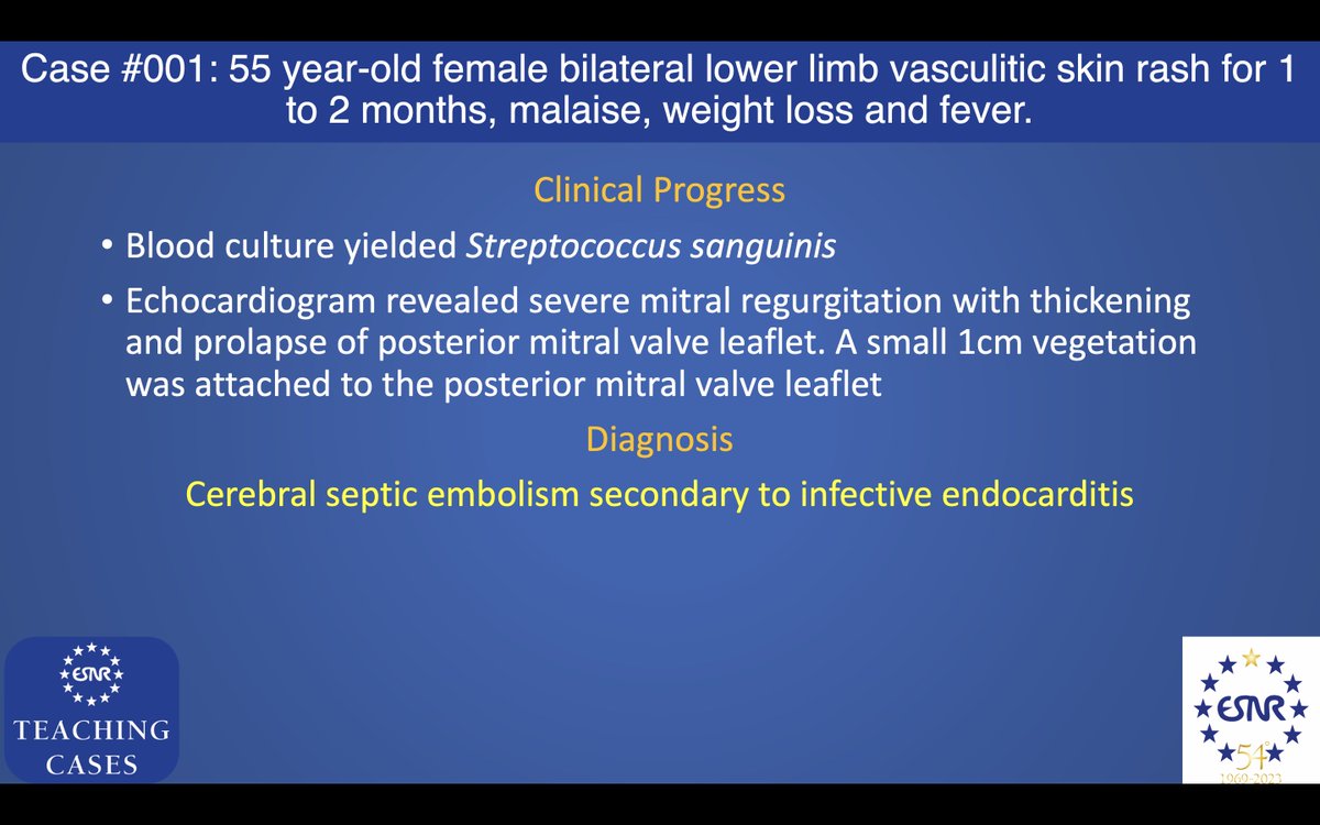
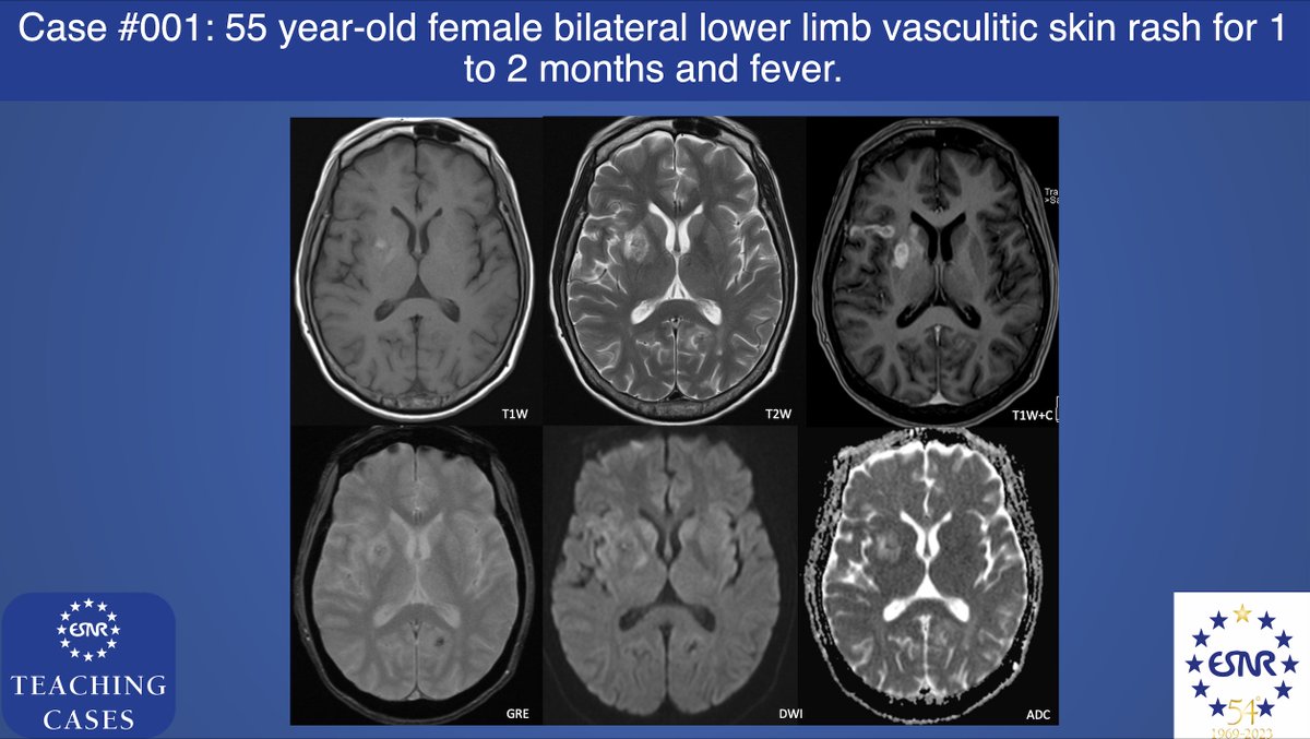
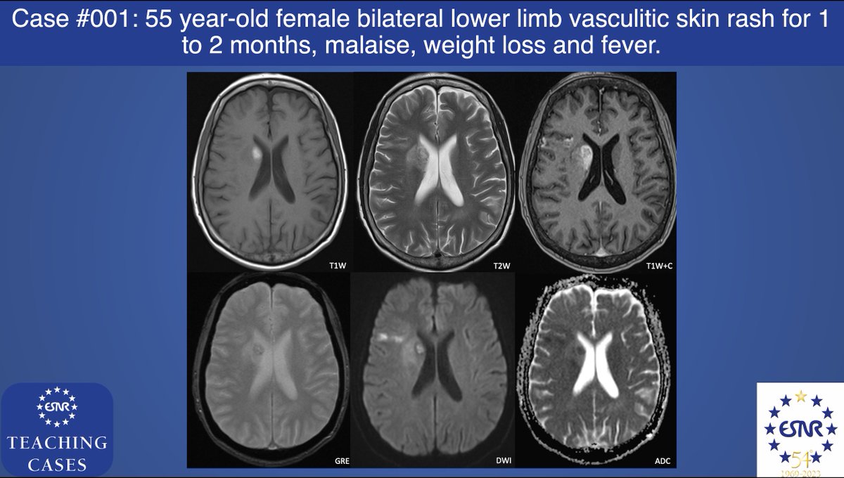
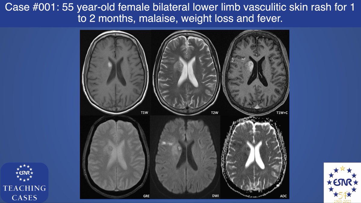
#ESNRTC case 1: 55 year-old female with good past health
•Bilateral lower limb vasculitic skin rash for 1- to 2- months
•Fever, malaise and weight loss
•white cell count (14.0 x 10⁹/L), raised erythrocyte sedimentation rate (107mm/hr)
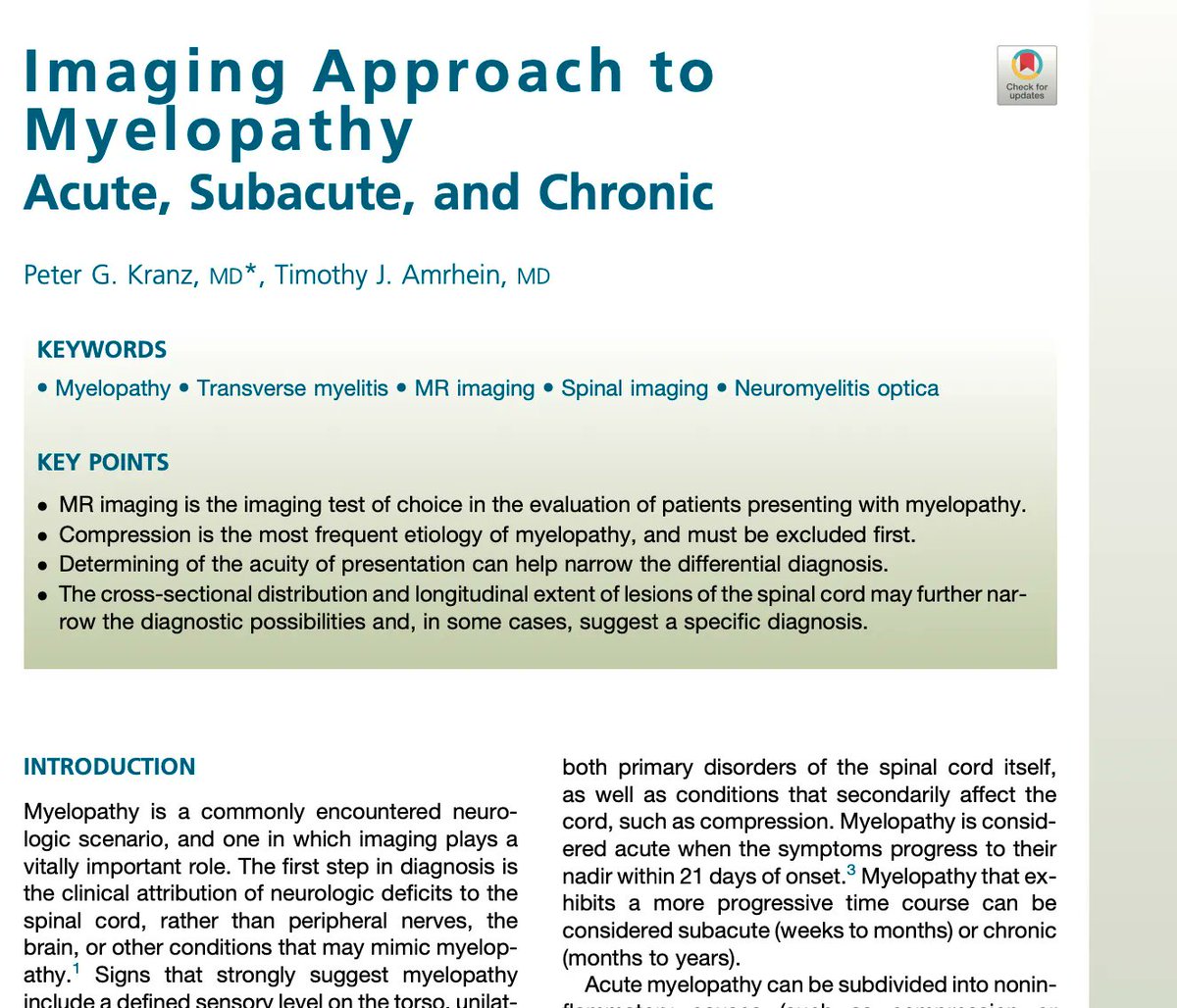
Happy Holidays, from ASSR. Please spend time doing what matters most to you. Once you're done with that, remember this tweet so that you can go read @Miriam_Peckham's fav spine article on approach to myelopathy by @PeterGKranz & @TimAmrheinMD https://t.co/MtiZ3mJp49