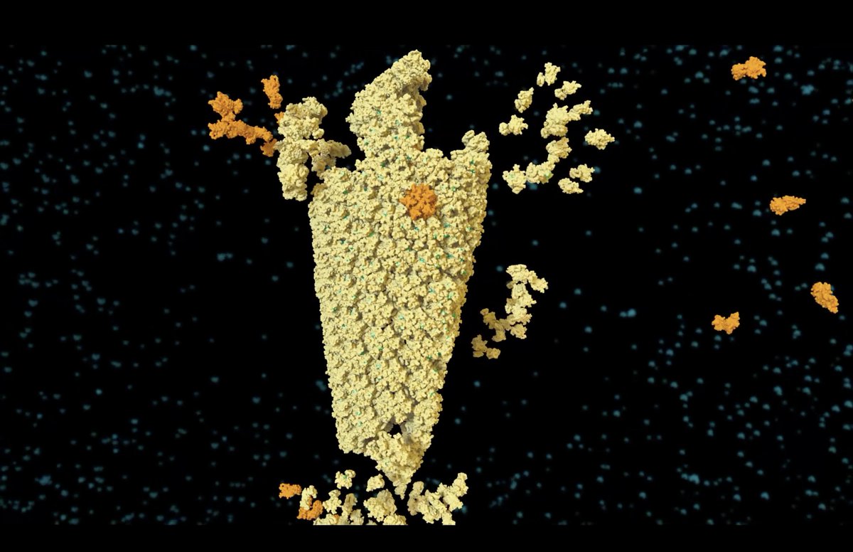
Researchers found that treatment of intact HIV capsids with lenacapavir causes capsids to rupture, with ruptures occurring first in areas of the capsid with the highest level of curvature. #ASBMB2026#ASBMB26
Watch in 3D at PDB-101: https://t.co/JiUkpxoadf
Spent this past summer designing & teaching my very first undergraduate class on AI and the Evolution of Structural Biology!
Check out the animation I created to teach my students about protein purification (https://t.co/yDYT5G7wbV)
Meet Dr. Laura Dassama, an Assistant Professor at Stanford University. This illustration by @torrez_rach showcases how the Dassama Group uses tools of chemistry and physics to provide molecular insights into complex biological processes.
Founded in 2018, the Animation Lab (@janetiwasa) may be the only institution of its kind. Here, a squad of dedicated postdoctoral scholars may spend months translating the smallest molecular phenomena into a visible, comprehensible spectacle. https://t.co/FZGOe7tQcv #scicomm
The Animation Lab is hiring! We're looking for a software developer for an exciting new project creating intuitive tools for molecular animation. Experience with Python req'd and Blender preferred. Please forward to any interested folks! https://t.co/Xck1B2lkkt
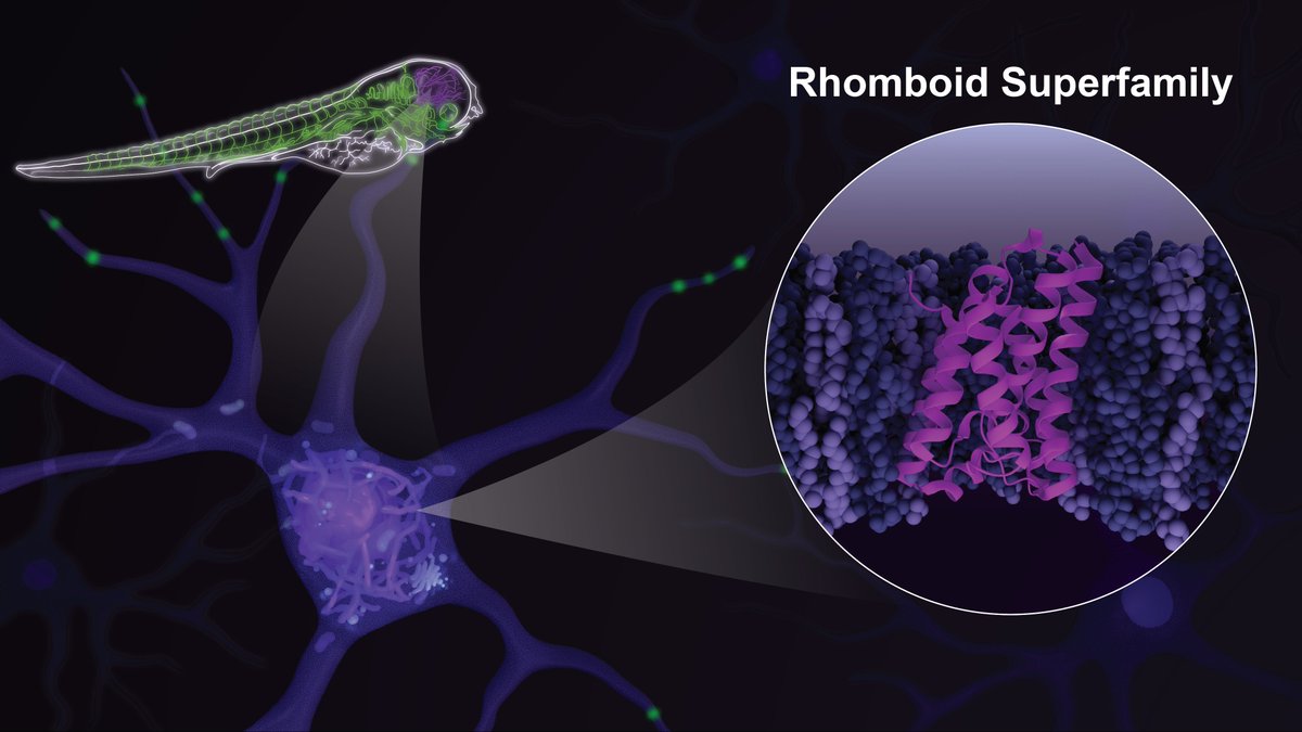
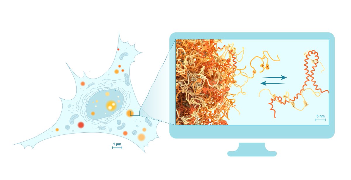
For #SeeingDiversity, we’re featuring Dr. Sonya Neal @Neal_Lab! Sonya’s lab explores the role of the rhomboid superfamily in vast membrane-related processes and how their dysregulation leads to disease. This illustration by @torrez_rach shows the multiscale means her lab uses.
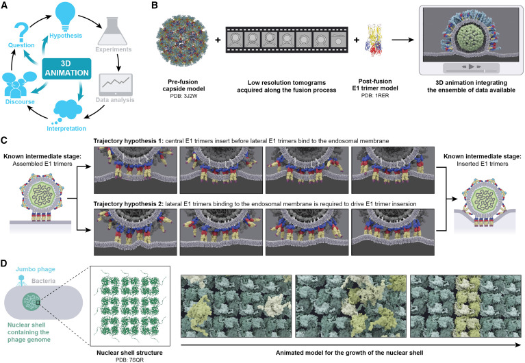
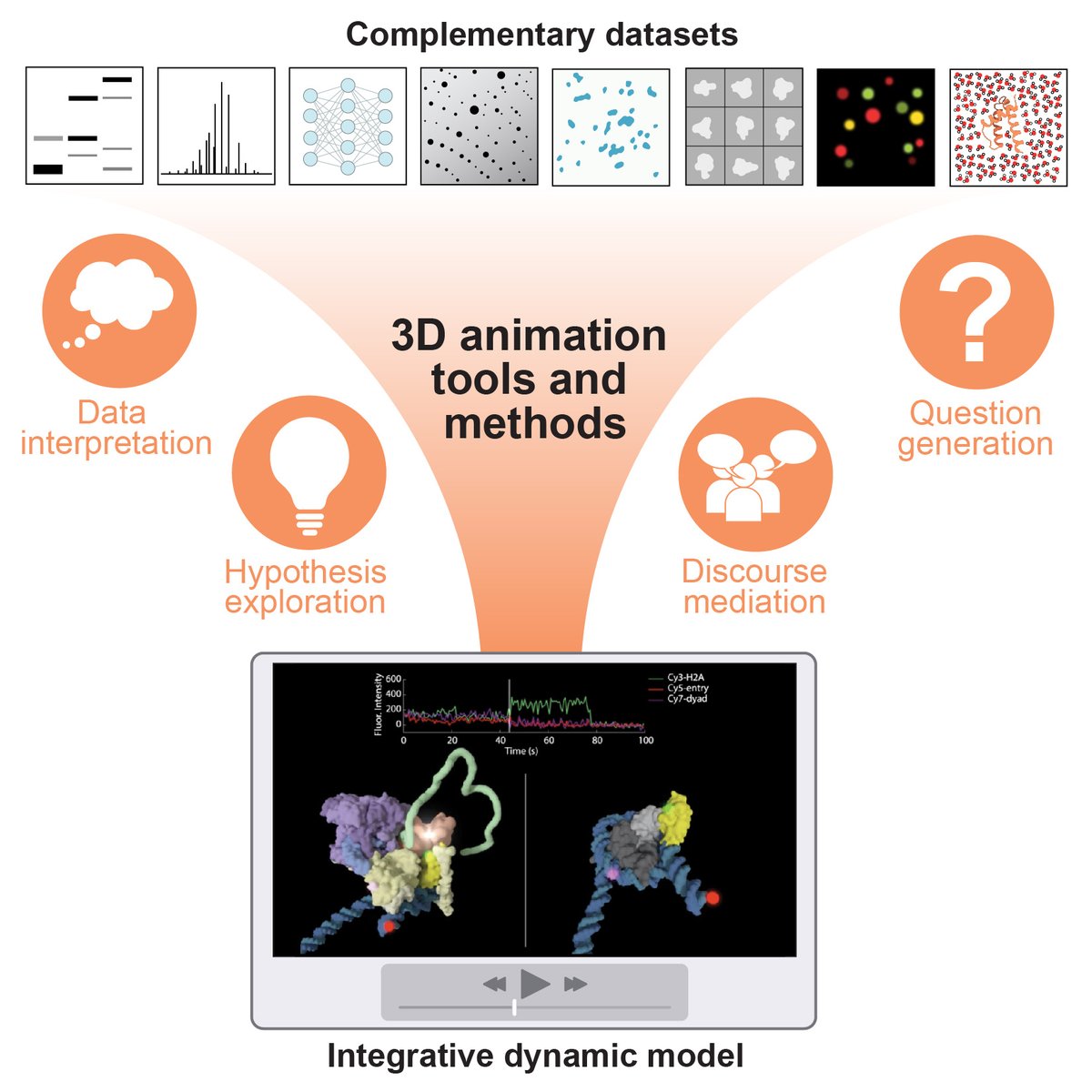
Check out the new Perspective from Margot Riggi, Rachel Torrez & Janet Iwasa “3D animation as a tool for integrative modeling of dynamic molecular mechanisms” @janetiwasa@MargotRiggi@torrez_rach
https://t.co/ySxlI11w0N
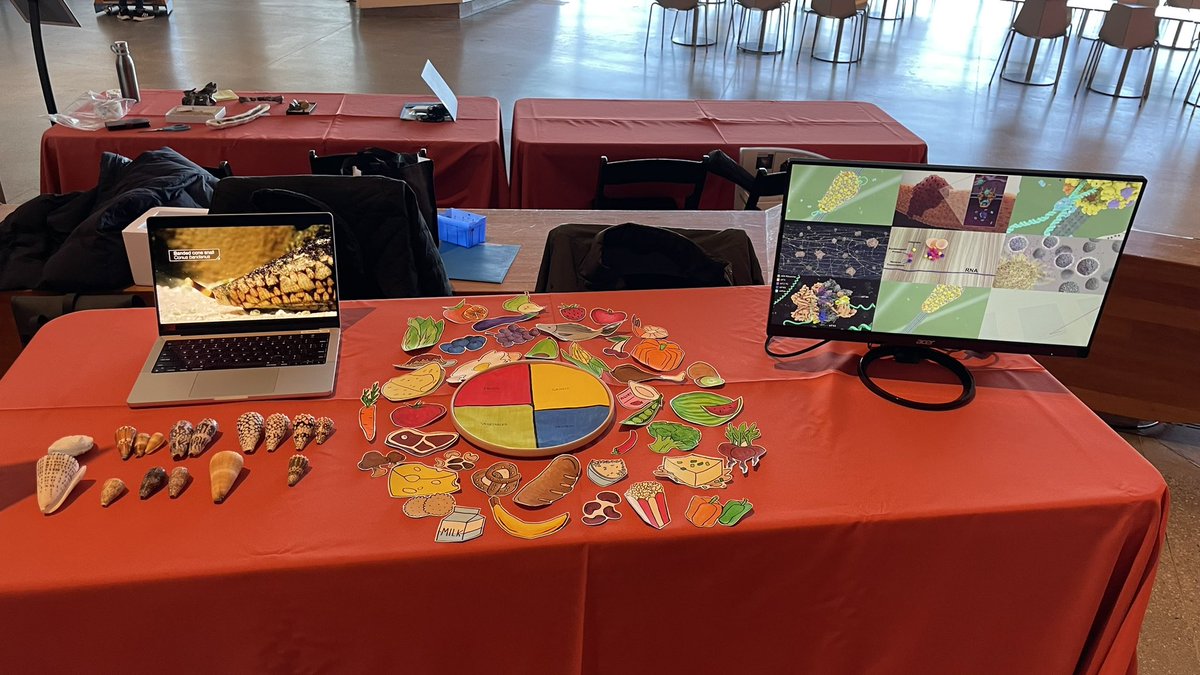
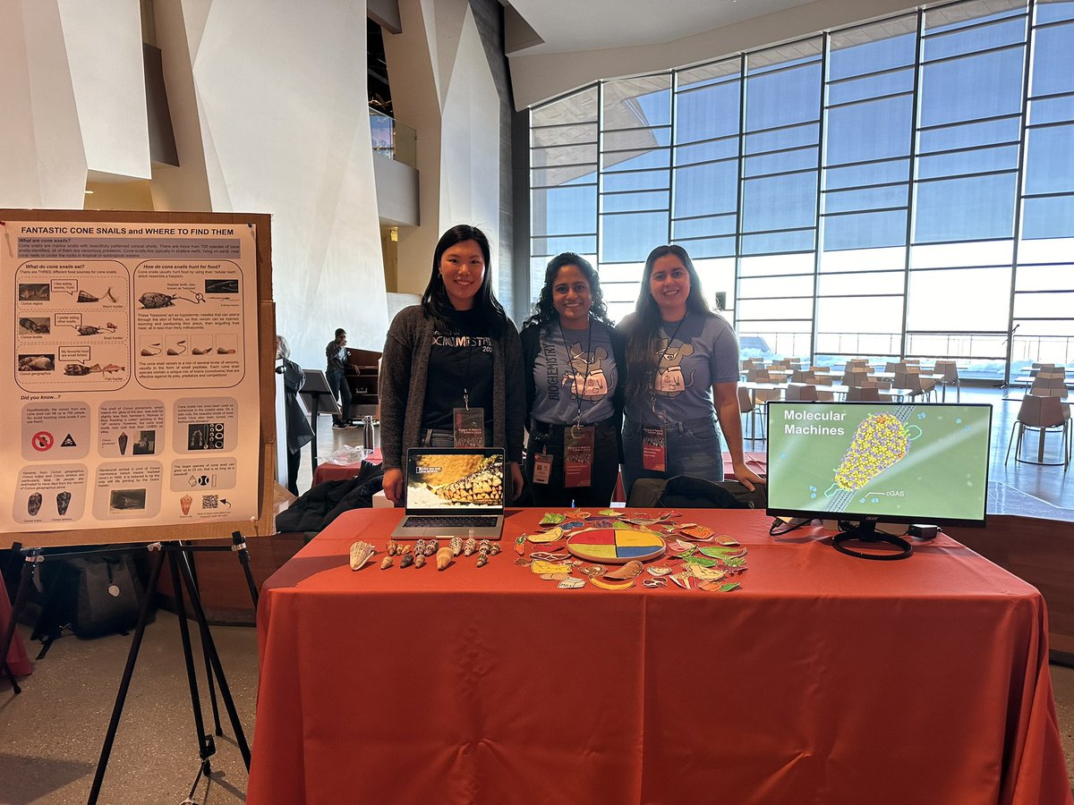
Representing @UofUBiochem at the @NHMU Women in Nature + Science event with @torrez_rach and @yeung_hyw. Come learn about molecular animations, cone snails and diets! 🐚🐠🧬🎬 🍝🥦
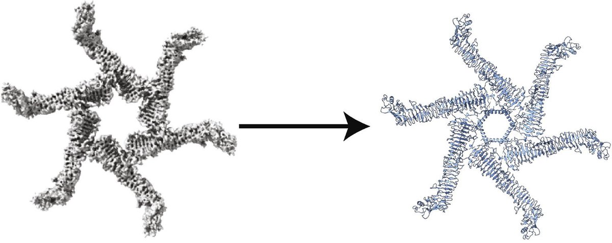
By coupling the power of single particle #cryoEM and #cryoET, LSI researchers have revealed a model for how H. pylori’s pore-forming toxin VacA reconfigures to interact with cell membranes, an essential step for its contribution to gastric disease. @JMolBiol
https://t.co/dHT3j8wLad
Check out the Animation Lab's latest paper by @MargotRiggi and @torrez_rach that describes how animation tools can be used to model dynamic molecular mechanisms! https://t.co/SabVoL9ekm
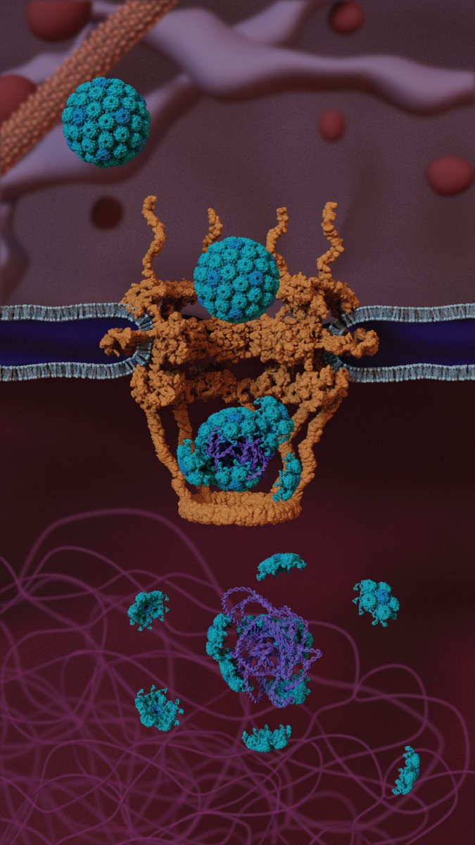
For #SeeingDiversity, meet Dr. Chelsey Spriggs @Dr_Sprggs. Chelsey is an Assistant Professor @UMCDB and her lab (@SpriggsLab) studies how viruses with DNA genomes reach the nucleus to cause infection (illustration by @torrez_rach).
Do you want to learn more about critical parameters to optimize your cryo-ET data collection? @janetiwasa@shen_utah and I released Chapter 4! https://t.co/aWJBCTZfUp
Featuring lamella and montage tomography details thanks to @erwright73, @JYang22638056, @Pangolininator!#CryoET
For #SeeingDiversity, meet Dr. Jerelle Joseph @jerelleaj, an Assistant Professor @EPrinceton! This illustration by @MargotRiggi highlights how Jerelle’s lab uses physics-based simulations as a “computational microscope” to study membraneless biomolecular condensates.
1/ Our newest work on Lis1 with the @aleschziner lab and co-first authors @eva_karasmanis, @janicereimer3, and @aga_kendrick suggests a mechanism for Lis1’s relief of dynein autoinhibition: https://t.co/eNrgQ4W3HD. @janetiwasa’s animation of our model:
The UMich Cryo-EM Biosciences Initiative has an open faculty position in CLEM and cryo-ET. Applications reviewed on rolling basis so don't wait to apply: https://t.co/Tu97fkzJd9
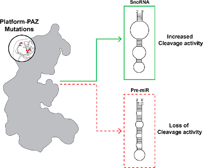
Dicer plays a role in RNA maturation, and dysregulation can lead to disease. Ohi, Garner and colleagues explore the role of residues distal to the active site, finding disease-related mutations with unique impacts.
@UMichPharmacy@UMLifeSciences
https://t.co/9iNgWer4oD
#RNA