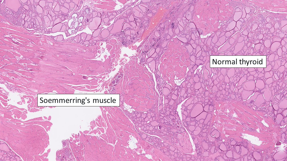
In thyroid cancer, skeletal muscle invasion represents extrathyroidal spread, but this is only reliable in the lateral lobes and not the isthmus. This is due to Soemmerring's muscle arising from the hyoid bone and inserting on the isthmus. Be careful when assessing tumors here!
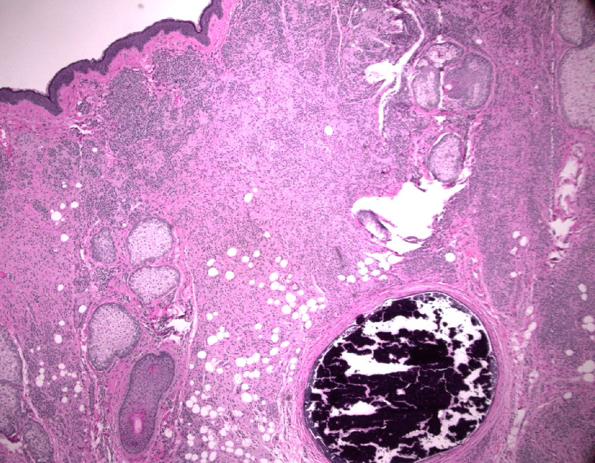
Nodal nevus in the capsule vs Melanoma Metastasis (subcapsular). Same patient. Nice contrast in cellular morphology and IHC #dermpath#pathology#IHCpath
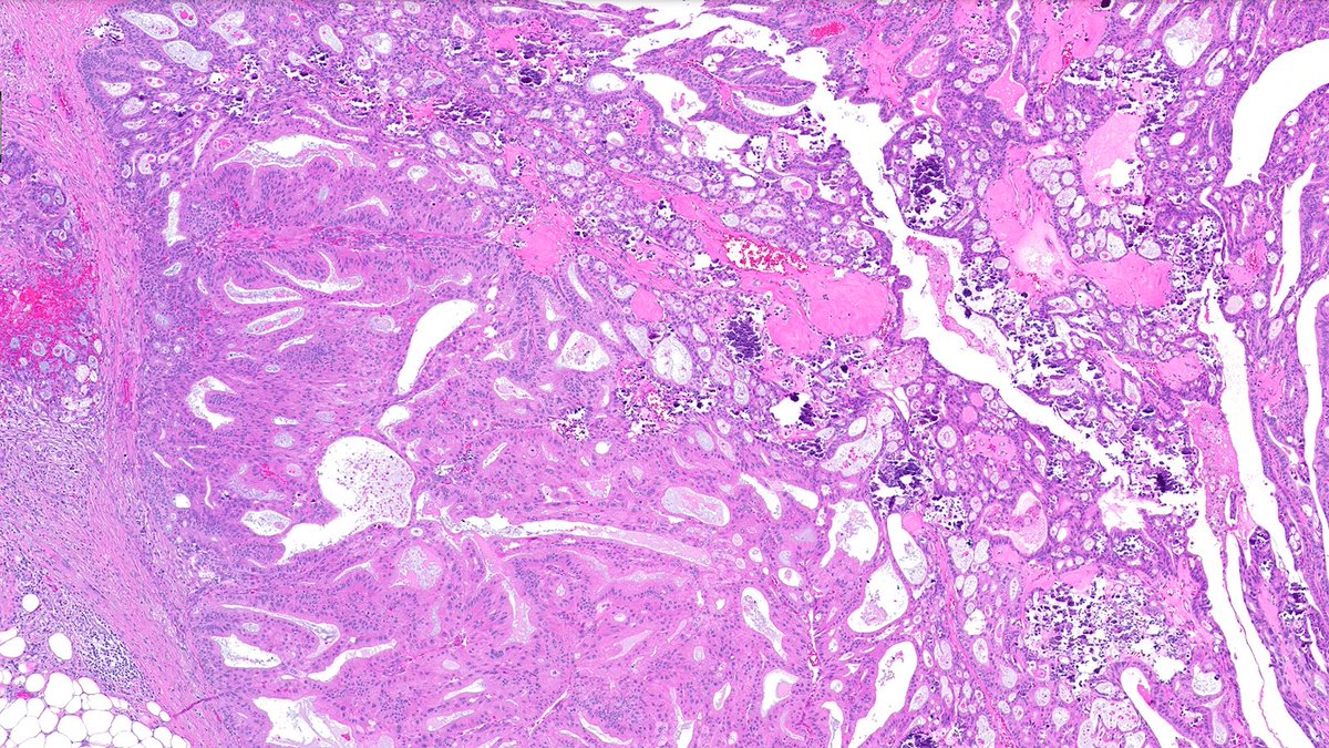
Case of Apocrine Encapsulated Papillary Carcinoma (EPC)🔬
A vanishingly rare entity—fewer than 10 cases reported. This apocrine EPC lacks myoepithelial cells and is ER-negative, yet remains staged as pTis.
#PathX#PathTwitter#breastpath
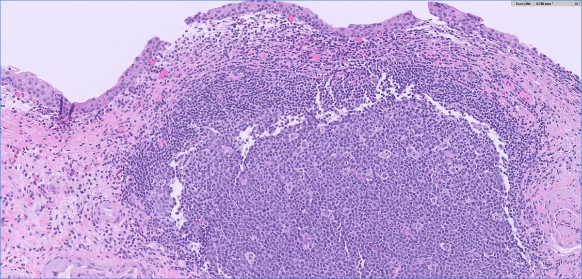
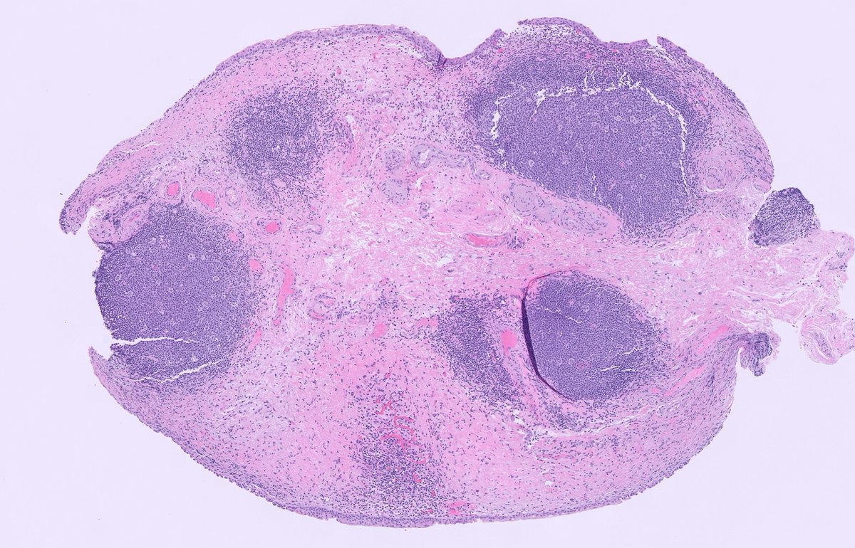
A young adult presented with multiple erythematous to violaceous plaques on the extremities.
💡 Take a look at the histopathology — what is your diagnosis?🔮🔬
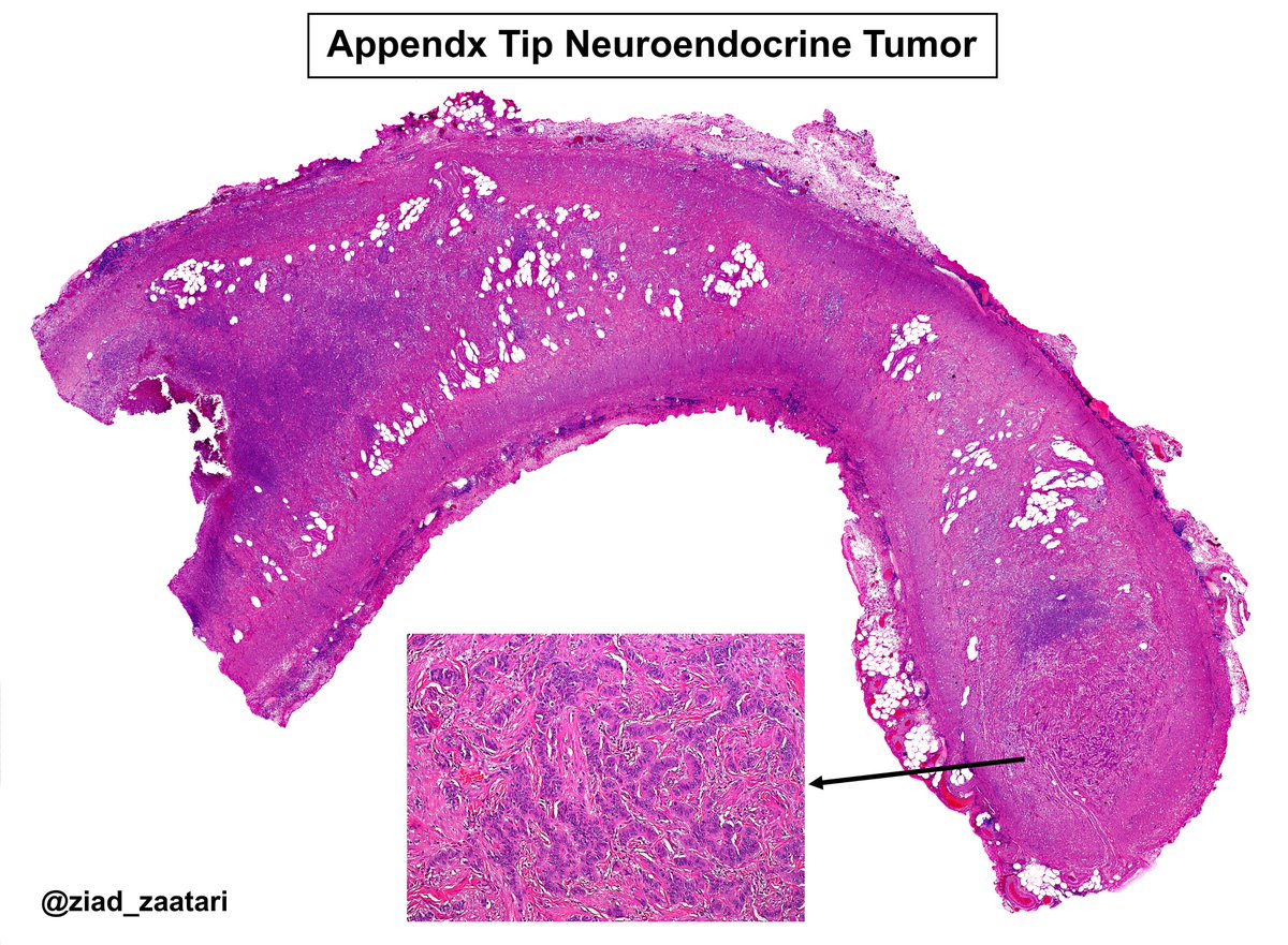
🔬🧑🏫 Lessons in Pathology: The reason why we submit the appendix tip in appendectomy specimens... Sometimes you will find a neuroendocrine tumor, like in this case!
So always look carefully and don't forget!
#GIpath#Pathology#PathologyBasics
A blob of dystrophic calcification within a nevus. If Nanta can do it with bone, I think I do it with this calcification. I will call it Nevus of Kolker
BAP1-inactivated melanocytoma:
- Large epithelioid melanocytes with distinct cytoplasmic borders
- Symmetrical and tends to show maturation with depth
- Loss of nuclear BAP1 staining
Whole slide image and more info at: https://t.co/toL8thD0go
Pleomorphic adenoma-the ultimate borderland 1/2
Avoid missing a myoepithelial carcinoma/myoepithelial carcinoma ex PA
Clues (for myoepithelial carcinoma)
-Uniform myoepithelial proliferation (pic 2)
-Expansile lobulated growth (pic 3)
-Zonal distribution of hypercellular and hypocellular regions
Dr. Sajed #USCAP25 #pathology #PathX
Case of Carcinoma with Apocrine Differentiation 🔬
This special type shows tumor cells with prominent nucleoli and abundant granular eosinophilic cytoplasm. Molecularly, this carcinoma is characterized by AR upregulation without ER activation.
#PathX#PathTwitter#breastpath
Let’s review HER2 immunohistochemistry this week!
Case of HER2 Null 🔬
HER2 Null cases show complete absence of membranous staining.
Be sure to check out my @captodayonline HER2 webinar Tuesday June 10th. Register here: https://t.co/MZVZSBRDjT
#PathX#PathTwitter#breastpath
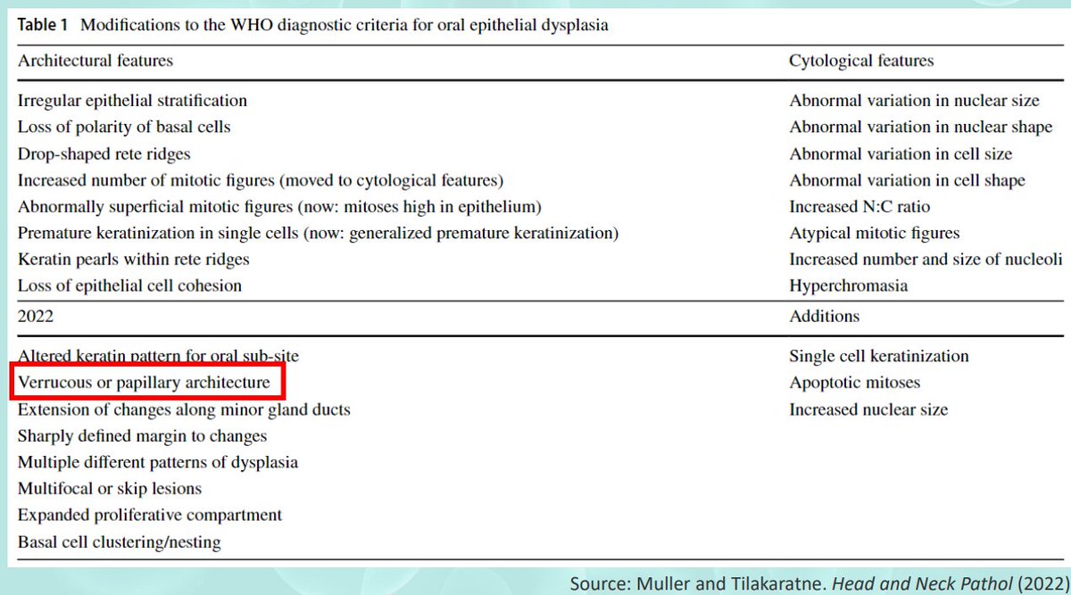
Oral epithelial dysplasia
WHO-2022: 3-tiered grading system
Defining dysplasia grade only in this manner oversimplifies the complexity of grading
Cytological atypia confined to the basal third may be sufficient for a diagnosis of severe dysplasia, depending on individual features present, particularly bulbous rete processes, budding and disorganization of basal cells, and marked pleomorphism
Pics Credit: Dr. Hernandez-Prera #USCAP25 #pathology #PathX
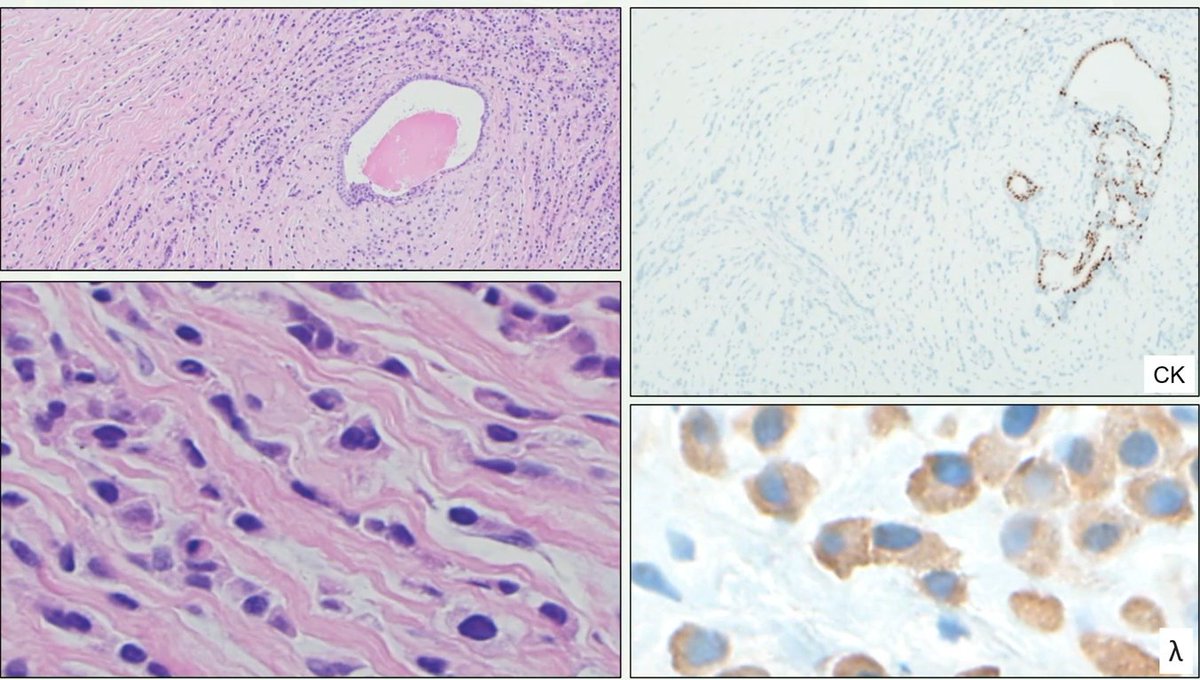
Breast-Challenges in diagnosis
Case diagnosed as invasive lobular carcinoma due to negativity for ecadherin
This lesion was also triple negative and CK negative
Patient had history of plasma cell neoplasm
Lambda stains were positive
Final diagnosis: metastasis of a plasma cell neoplasm
Dr. Sahin #USCAP25 #pathology #PathX
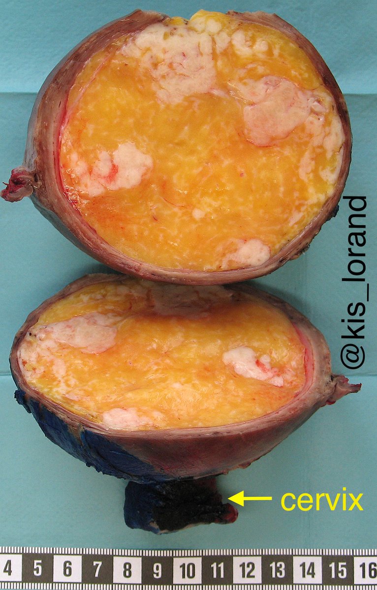
Let me contribute to the "big specimens' season" with (literally) an orange in the uterus 😀😀 The bonus questions are:
1. what was the preoperative diagnosis in the endometrial biopsy?
2. what did the pathologist do after seeing the biopsy?
#pathology#gynpath
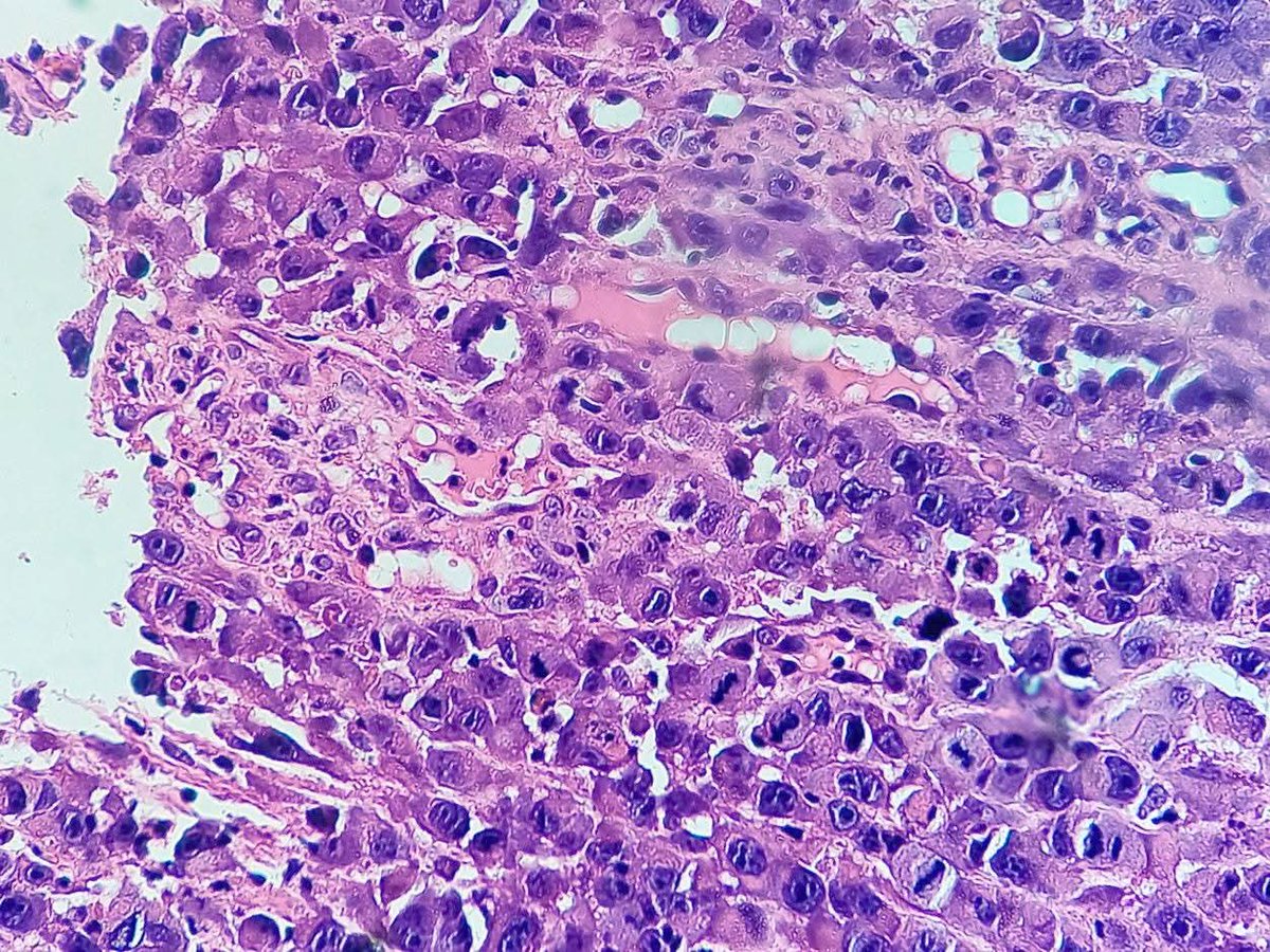
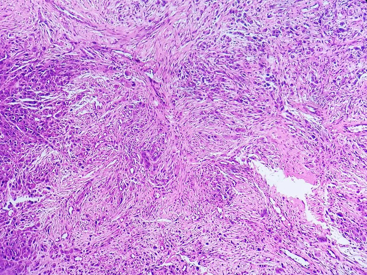
80 years old man with ulcerated lesion on hard palate. H&E sections reveal infiltrating pleomorphic tumor cells with rhabdoid morphology and sarcomatous appearanc.IHC came out positive staining for Pan CK, CK 5/6, p40, INI 1 and negative staining for desmin, myogenin and S 100.