✅Myeloid sarcoma presenting as an isolated intracranial relapse of AML.
💡It may closely mimic a meningioma. In patients with a hematologic malignancy in remission, marked diffusion restriction and disproportionate edema should raise suspicion for extramedullary relapse.
https://t.co/MdgJbz1NZB
✅Powassan virus encephalitis.
💡This rare tick-borne flaviviral infection is endemic in parts of northeastern North America, including Connecticut. In a febrile patient with recent tick exposure, bilateral deep gray matter involvement should raise suspicion.
https://t.co/oLS3iyFLkO
🩻 Case Study of the Week!
A middle-aged patient with diabetes presented with complaints of general malaise, dull abdominal pain, and weight loss of 12 pounds during the prior 3 months.
🔗 See all the case details, more images, and diagnosis: https://t.co/LKlT4FsGOt
#Radiology #RadEd #MedEd #RadiologyEducation #COW #CaseOfTheWeek
✅Synchronous pineal and suprasellar pineoblastoma, mimicking bifocal germinoma.
💡Marked diffusion restriction may favor a highly cellular embryonal tumor, but pineoblastoma and germinoma can overlap on MRI. Histologic confirmation is essential.
https://t.co/soW3cfHhe3
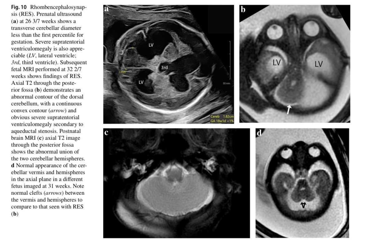
La rombencefalosinapsis es una anomalía crítica de la organización prenatal que suele pasar desapercibida.
💡 Hallazgos clave: Fusión de hemisferios cerebelosos, agenesia del vermis y 4to ventrículo en "diamante".
RM fetal en 3er trimestre es el estándar para su confirmación.
✅The upper lesion is a dural cavernous hemangioma; the lower one is a parasagittal meningioma.
💡Dural cavernous hemangiomas can enhance intensely and may even show an apparent dural tail, closely mimicking meningioma. The dural tail sign is helpful—but not specific.
https://t.co/NrMKr5R8cg
@daniel_gewolb@Radiopaedia Talk of the shrimp & it appears.
⛔️ ASSYMETRY of Shrimp is PARAMOUNT for PML.
Also appreciate the associated cerebellar atrophy. ⬇️
https://t.co/IKUKf5q2YN
✅Intracranial histiocytic sarcoma.
💡A rare malignant histiocytic neoplasm that may mimic a high-grade meningioma, even with a dural tail. Rapid systemic progression should raise suspicion; diagnosis relies on histiocytic markers such as CD68 and CD163.
https://t.co/aKqa5NDVVw
Las guardias de 24h se diseñaron para una época en la que "estar de guardia" significaba literalmente estar disponible en el hospital por si surgía una emergencia real mientras dormías o descansabas.
Hoy son 24 horas de trabajo a destajo sin parar. Ya no funcionan.
✅Rabies encephalitis
💉The patient had received vaccine doses but no rabies immunoglobulin. Survival from clinical rabies is exceedingly rare: he required 5 months of intensive care and was discharged with severe neurologic sequelae.
https://t.co/weenZDyRIa
⭐️ ~17% of patients with classic trigeminal neuralgia have aberrant nerve anatomy which may cause or contribute to their symptoms and is associated with worse surgical outcomes! ⚡️
⭐️ In patients with classic trigeminal neuralgia: Check the angle of the trigeminal nerve as it crosses through the porus trigeminus into Meckel's cave!
🔷Case example in 🧵
#Neurology #Neurosurgery #radres #futureradres #Medicine #MRI #FOAMed @ASNRographics@TheAJNR@Radiopaedia
⭐️ Answer: Sinonasal Chondrosarcoma
▶️Sinonasal chondrosarcomas are rare tumors that arise from cartilage remnants and typically occur in middle age and older adults w/o strong sex predilection
▶️This particular case is difficult to distinguish from a chordoma since there is involvement of the skull base with midline location (arising from the nasal septum). However, the mass is centered more anterior in the posterior nasal cavity/sphenoid sinus, the enhancement pattern is "arabesque"-like rather than “honey-comb” pattern enhancing septa and although calcifications can be seen with chordomas it is considered rare and they are more typical in chondrosarcoma with the rings and arcs pattern
Case study: Sylvian Fissure Lipoma with Associated Focal Cortical Dysplasia and Venous Malformation
Sylvian fissure lipomas are rare congenital lesions, asymptomatic in most cases, and therefore discovered incidentally. Association with other coexisting abnormalities, such as focal cortical dysplasia or venous malformations, is even rarer, as in this case.
👀 Do you know what to look for? https://t.co/9nwIf19S6Z
#Radiology #RadEd #MedEd #RadiologyEducation #CaseStudy #NeuroRad
Queen Square Teaching Images #10
Leptomeningeal enhancement in ATTR (hereditary transthyretin amyloidosis).
Brain often symptomatic later than peripheral nerves. Amyloid deposition in the meninges, or an amyloid angiopathy of smaller vessels.
#NeuroRad
✅MOGAD-related encephalitis.
💡In children, MOGAD often presents with encephalopathy rather than isolated optic neuritis or myelitis. Large, bilateral but asymmetric, poorly defined lesions involving both white matter and deep gray nuclei should raise suspicion.
https://t.co/M4NCGaZ6Wi
💡Acute necrotizing encephalopathy (ANE). The combination of recent influenza infection, bilateral thalamic lesions, diffusion restriction, petechial hemorrhage, and the trilaminar ADC pattern is highly suggestive.