Athlete’s Heart (Syndrome) refers to physiological cardiac changes that occur as a result of the hemodynamic stress of regular strenuous exercise.
Watch our video, “HCM versus Athlete’s Heart,” from our Microlesson Series on HCM! https://t.co/e8XH6luFOV #HCM#athletesheart
Understanding the Phases of Left Ventricular Diastole
Diastole begins with aortic valve closure (end-systole) and ends with mitral valve closure (end-diastole). During this time, the left ventricle relaxes and fills with blood through four distinct phases:
1. Isovolumic relaxation
- Begins immediately after the aortic valve closes.
- Both the aortic and mitral valves are closed, so LV volume does not change.
- As LV pressure falls below left atrial pressure, the mitral valve opens.
2. Early rapid filling
- Blood flows quickly from the left atrium into the relaxing left ventricle.
- This produces the E wave on transmitral Doppler.
- Most ventricular filling occurs during this phase in healthy adults.
3. Diastasis
- As atrial and ventricular pressures equalize, blood flow slows markedly.
- There is little or no transmitral flow, and the mitral valve remains partially open.
- Diastasis is longer at slow heart rates and may disappear at faster heart rates.
4. Atrial contraction
- Atrial systole raises left atrial pressure, causing a second phase of ventricular filling.
- This generates the A wave on transmitral Doppler.
- In normal individuals, atrial contraction contributes about 20% of total LV filling.
The transmitral Doppler tracing mirrors these filling phases:
🟦E wave = early rapid ventricular filling
🟩Low-flow interval = diastasis
⬜A wave = atrial contraction
The Doppler velocity curve is essentially the first derivative of the LV volume curve, meaning it shows how quickly ventricular volume changes during diastole.
The right ventricle follows the same sequence of diastolic filling, although its diastolic period is usually slightly shorter because RV systolic ejection lasts a little longer.
Reference: Catherine M. Otto, The Practice of Clinical Echocardiography.
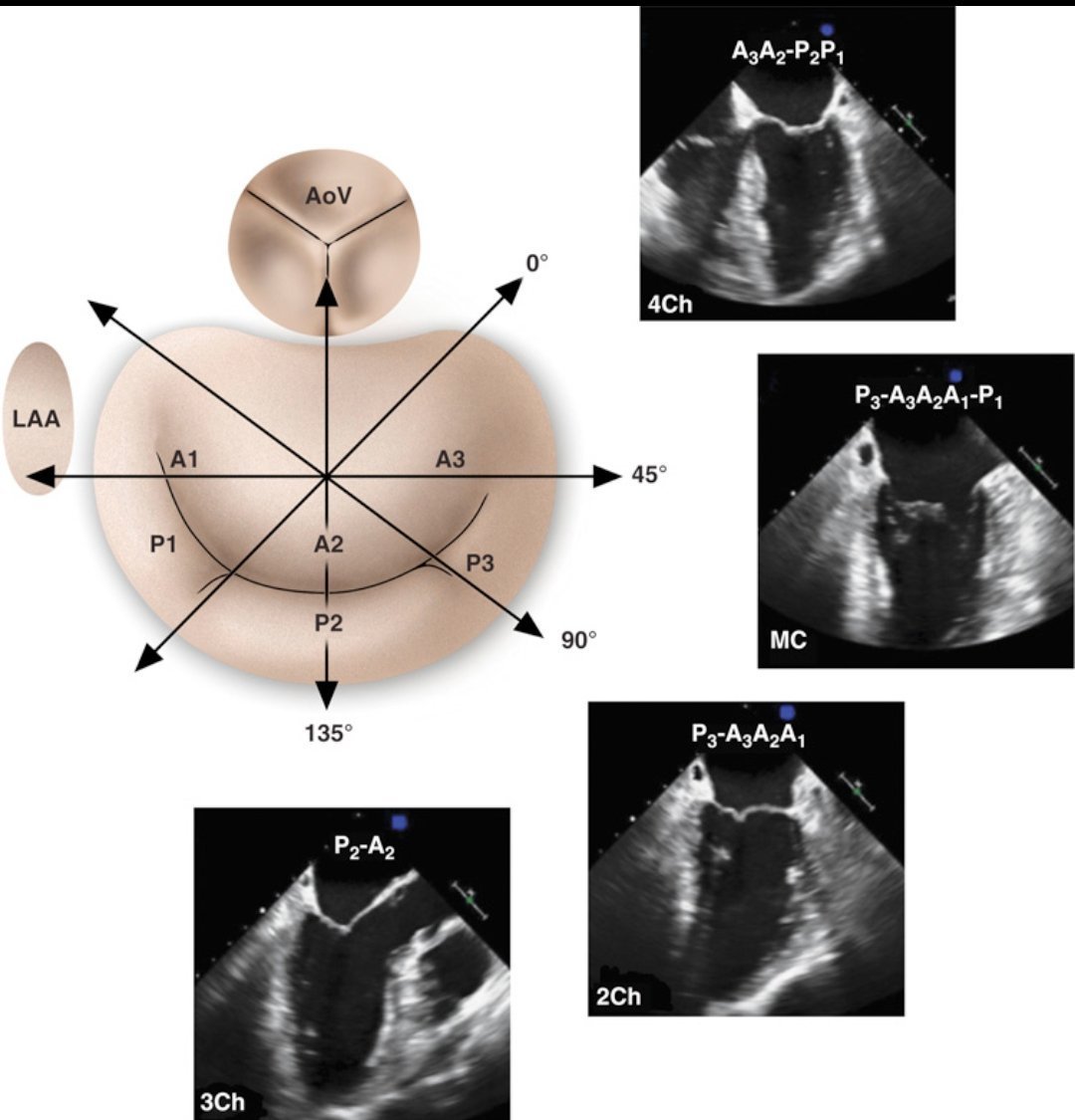
Mitral Valve Anatomy in TEE!
This schematic shows the mitral valve's labeled segments with corresponding transesophageal echocardiography (TEE) views at different angles. Understanding probe positioning helps optimize imaging for structural assessment
NBTE nonbacterial thrombotic endocarditis - look for sterile “vegetations” (platelet fibrin deposits) along the valve leaflet’s closing edges that appear to "kiss" in this characteristic pattern
#echofirst
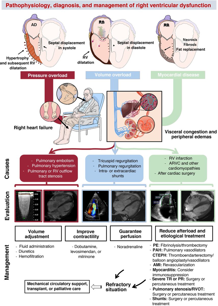
🚨 New #VExUS post on https://t.co/hqYw2WlFqR
Confused by abnormal hepatic vein Doppler waveforms? This post breaks them down using physiology, making it easier to connect waveform changes to the underlying hemodynamics.
🔗https://t.co/smNhInCSx9
#POCUS#echofirst#Nephpearls
If a component of VExUS feels difficult, the answer is to understand it better, not to eliminate it altogether. Simplification can be helpful to some extent, but oversimplification often comes at the cost of losing valuable physiological information.
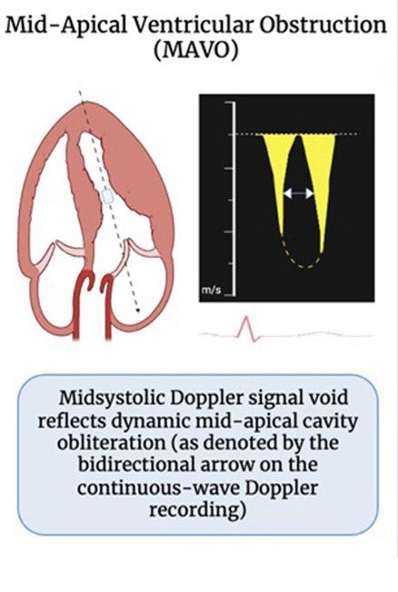
Have you seen the “void”?
in midapical HCM with midventricular cavity obliteration, CW Doppler shows midsystolic interruption of forward flow -the “void”
MAVO HCM ⬆️ prevalence of apical aneurysm, can miss Dx if use peak gradient only #echofirst
🔗 https://t.co/wtNKtom3r3
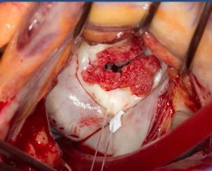
Just gave a presentation in STS2025, introducing an innovative way of doing septal myectomy to treat hypertrophic obstructive cardiomyopathy(HOCM). I call it MESM - Minimally Invasive Electro Septal Myectomy. Thanks Dr. Badhwar for his kindly comments.
🔴Simple tools to locate origin of PVCs⤵️
1.QRS Morphology
🔹RBBB: Left Ventricular (LV)
🔹LBBB: Right Ventricular (RV)
2. Inferior leads (II, III, aVF)
🔹Positive: Base/Superior
🔹Negative: Apex/Inferior
3. Leads I, aVL
🔹Positive: Right side
🔹Negative: Left side
Many patients with HOCM are finding relief with the trans-axillary minimally invasive Modified Morrow procedure. We successfully operated on four cases in a single day, all with excellent outcomes. Each patient was extubated right at the table immediately after skin closure!
For the surgery of apical hypertrophic cardiomyopathy, can it only be done through median sternotomy and a left ventricular apex incision? Is there a minimally invasive option available?
The mechanism of left ventricular outflow tract obstruction (LVOTO) is complex in hypertrophic cardiomyopathy (HCM).
We aimed to evaluate the impact of mitral valve geometry on LVOTO by echocardiography. Read our @JournalASEcho article: https://t.co/fSjQ9fZFkO
Our August 2024 @JournalASEcho is online! https://t.co/iDs8kfrLjR
This issue features:
🔷Progression of Mild Mitral Annulus Calcification to Mitral Valve Dysfunction and Impact on Mortality
🔷Mitral Geometry on the Mechanism of LV Outflow Tract Obstruction in HCM
🔷And more!