Worried that hydrocephalus is hiding in plain sight?
Normal pressure hydrocephalus (NPH) can mimic volume loss
How can you tell the difference?
Many findings can be seen with NPH—you may use 1 or more. But do you know how well your sign performs?
Here are how common NPH signs perform:
Evans index
SENS: 100
SPEC: 74
Cingulate sulcus sign:
SENS: 63
SPEC: 61
Callosal angle:
SENS: 49
SPEC: 78
Disproportionately enlarged subarachnoid spaces (DESH):
SENS: 94
SPEC: 41
Anteroposterior diameter of the lateral ventricle index:
SENS: 98
SPEC: 100
For the SINPHONI trial, a combination of Evans index > 0.3 & DESH had a 70-80% PPV for shunt responsiveness.
Now you know the signs of NPH & how good they may or may not be. Hopefully, you can now set the bar high when it comes to hydrocephalus!
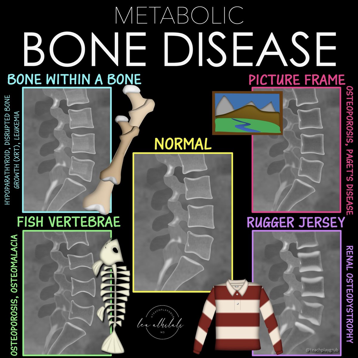
Can’t metabolize all the metabolic bone diseases?
When you see weird bones, do you just say, “suggestive of metabolic bone disease” and hope no one asks questions??
Is trying to memorize all their patterns disrupting your homeostasis?
Metabolic disease are complex but there are a few that are a slam dunk
Bookmark this figure for the for the 4 classic xray patterns you NEED to know!
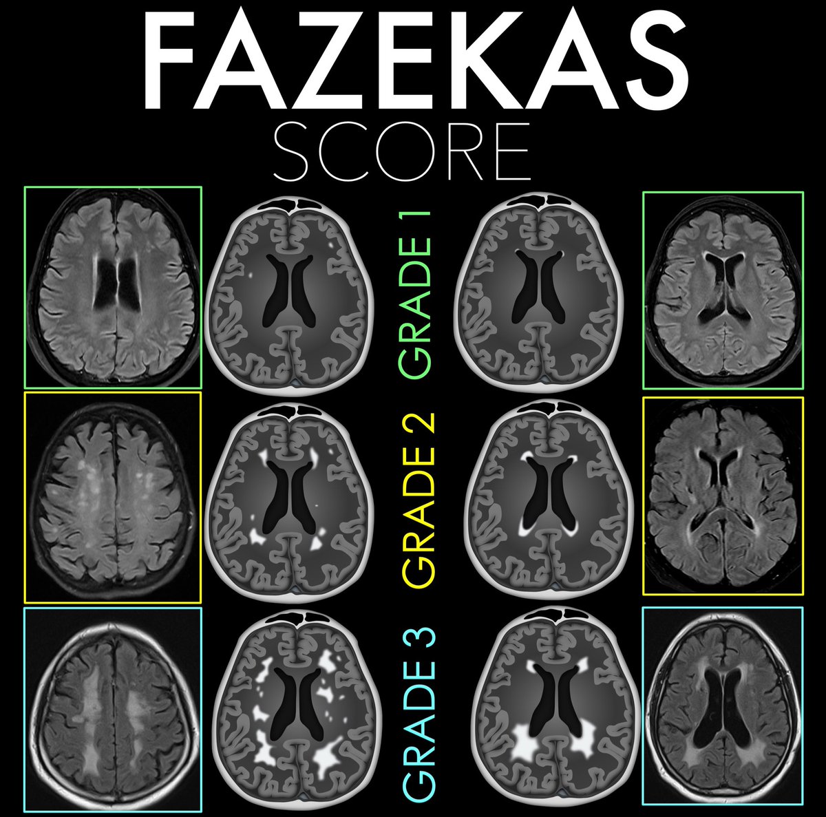
Feel like waving the white flag over white matter disease?
Do you grade the degree of small vessel disease, or just mention & hope no one asks for more?
Degree of small vessel disease is important & the Fazekas scale is an easy way to semi-quantitate disease burden
Here’s how to remember it:
Think about how the number looks or feels — it grows in intensity!
Deep White Matter Fazekas:
1 → “1-hit wonder” Punctate foci — just tiny spots, like little dots.
2 → “2 = two-gether” Beginning confluence of foci — two dots coming together, mild merging.
3 → “3 is a crowd” Large, confluent areas — everything blending into a mess, too much white.
Periventricular Fazekas:
1 → “1 is a pencil” ✏️ Pencil looks a number 1 (pencil-thin “cap” or rim along ventricle).
2 → “2 curves into a halo” ⭕ Smooth halo of hyperintensity — picture the roundness of 2 encircling the ventricle.
3 → “3 branches out” 🌿 Irregular fingers of white matter reaching into the deep — the edge of the number 3 look like irregular finger
Now you know the Fazekas score for small vessel disease!
Now your knowledge of small vessel disease will be anything but small!
Having trouble remembering what to look for on scans for Alzheimer’s dementia?
Want the key findings to stay in your memory?
Medial temporal atrophy (MTA) score quantifies hippocampal volume loss
It rates the amount of CSF around the hippocampus as a marker of volume loss
CSF surrounds the hippocampus like a snake surrounding its prey—how far the snake has gone in getting its prey determines the MTA score:
MTA-1: Choroidal fissure opens, looks like the snake has started to slither in
Remember: the number one looks flat like a slightly opened choroidal fissure
MTA-2: Temporal horn widens, looks like the snake has surround the hippocampus
Remember: the number two look like the CSF curving around as the temporal horn opens
MTA-3: Moderate hippocampal atrophy, looks like the snake has begun to squeeze the hippocampus
Remember: the number three has a tight curve, so it looks like it is crushing the hippocampus & it looks small (atrophied)
MTA-4: Severe hippocampal atrophy, looks like the snake has crushed the hippocampus
Remember: Number four has only a tiny hole in it, so the hippocampus has to be super tiny & atrophied to fit inside!
Hopefully, this will help you commit this rating system to memory so you’ll never forget what you need to say for Alzheimer’s!
Olivary hypertrophy is an exceptional consequence of brainstem insults. Usually occurs after a latency period of few months. @atabisneuro et al reported a case of bilateral olivary degeneration following Scrub typhus rhombencephalitis @neuro_ian Link: 10.4103/aian.aian_931_23
1/Do you know all the aspects of, well, ASPECTS?
Many know the anterior circulation stroke scoring system—but posterior circulation (pc) ASPECTS is often left behind
25% of infarcts are posterior circulation
Do you know pc-ASPECTS?!
Here’s how to remember pc-ASPECTS!
Tired of always speculating about MR spectroscopy?
If you've ever looked at an MR spectroscopy & thought: "I have no idea what I’m looking at!"--then this cheat sheet is for you!
Here are the 4 basic rules you need so you can understand the spectrum of basic spectroscopy!
First you need to know the peaks.
3 main peaks: Choline, Creatine, NAA
Remember the order bc a spectrum looks like mountain peaks & it is cold in the mountains. And CHOld CREATures NAp or hibernate in the mountains
Choline
Marker of membrane turnover
Remember: membranes coat or “CHOat” the cell
Choline = ChoLEAN, choline LEANS into the creatine peak, it’s right next to it
Creatine
Marker of energy, basically an internal control
Remember, everyone takes Creatine powder for energy!
Creatine is at ~3—creatine almost rhymes with 3
NAA Marker of neuronal health
N = Neuron
NAA has double As so it is at ~2!
Four rules:
1. Hunter’s angle:
—Most people know that the peaks of the spectrum should go up at you move lateral, called Hunter’s angle
—Most bad things reverse Hunter’s angle
—Ask yourself: Is my arrow pointed up to shoot into the air at the enemy (good) or is point to the ground where it will hit the dirt (bad)
2. TE & spectrum length are inversely related
—Spectroscopy follows the rule: speak softly & carry a big stick.
—Short TE = long spectrum, lots of extra peaks for glutamate/glycine, myoinsitol
—Long TE = short spectrum, mainly the basic 3 peaks
3. Each region has its own unique signature
—Each brain region has its own unique composition of compounds that might alter Hunter’s angle a bit, but not reverse it
—Need a control in contralateral normal brain so compare apples to apples
4. Lactate peak goes like a sine wave
—Lactate peak represents anerobic metabolism—sign of cells in trouble
It’s at 1.3ppm. Remember this bc 13 is an unlucky number & lactate is an unlucky sign!
—It’s like a sine wave: up at short TE (35), down at intermediate TE (144), and up again at long TE (244)
—You can use this flipping to better visualize the lactate peak
—You can remember it’s down in the middle TE bc when you’re caught in the middle, you’re down & out
Just remember these tricks & you will be spectacular at basic spectroscopy!
Sink or swim!
Brain is swimming in CSF!
CSF is key for protection, buoyancy, & hormone/waste transport
Most know basic ventricular anatomy well, but not subarachnoid anatomy.
How many cisterns do YOU know?
Here’s a quick guide to the key midline cisterns & what to know for each
—Cisterna Magna:
Largest of the cisterns
Contains CN 9-11 & vertebral arteries
Remember this bc the LARGEST cistern contains the LARGEST cranial nerve (vagus) and the two cranial nerves around it
—Prepontine cistern:
Contains CN 6 & basilar artery
Remember this bc the shape of the number 6 mirrors the curvature of the anterior pons, where this cistern lies
—Chiasmatic cistern
Contains CN 2 & pituitary stalk
Easy to remember bc the CHIASM is CN 2
—Interpeduncular cistern:
Contains CN 3 & mammillary bodies
Remember this bc if you lay the number 3 on its side it looks both like the two cerebral peduncles (where this cistern lies) & well, two mamillary bodies
—Quadrigeminal cistern
Contains CN4 & pineal gland
East to remember bc QUAD is the prefix for FOUR
—Lamina terminalis:
Contains the ACOMM
Remember this bc the shape of subarachnoid hemorrhage after ACOMM rupture follows this cistern anteriorly
Now when you look at cisternal anatomy, you will definitely be able to go with the flow!
1/Time to go with the flow!
Hoping no one notices you don’t know the anatomy of internal carotid (ICA)?
Do you say “carotid siphon” & hope no one asks for more detail?
Here’s a thread to help you siphon off some information about ICA anatomy!
Feeling unarmed when it comes to evaluating cervical radiculopathy & foraminal narrowing on MR?
Think of the nerve root like a hot dog, sitting between the two buns of the disc/uncovertebral joint & facet.
The more you put in your hot dog, the more the hot dog itself is squished. Same w/the nerve root.
Spurring & degenerative change are like the extra topping that push on the hot dog inside the buns.
A small amount of toppings/degenerative change, leaves the hot dog space. But if you pile on fixings, then the hot dog is taken over.
Ask yourself--how is my hot dog doing?
Mild stenosis is like just a little ketchup & mustard on the bun but hot dog still has space.
Moderate stenosis is when you aren’t just putting on sauce, you are adding things that take up space, like relish.
But there’s only so much relish one can put on, so it doesn’t take up more than half the bun.
Severe stenosis is like a chili cheese dog, where the hot dog is smothered & it has no room in the bun away from the chili or cheese. Here the narrowing is greater than 50%
This is the Kim classification & has strong correlation w/symptoms I like it bc it doesn’t require calipers to estimate a >50% narrowing
So now you know how to both image and assess stenosis in the cervical neural foramen.
Now hopefully rating cervical foraminal narrowing won’t be a pain in the neck!