Mastering TEE Probe Manipulation & Positioning!
Transesophageal Echocardiography (TEE) is a powerful imaging technique, but proper probe control is essential for optimal cardiac visualization.
🔹 (1) Probe Manipulation – Understanding Movements
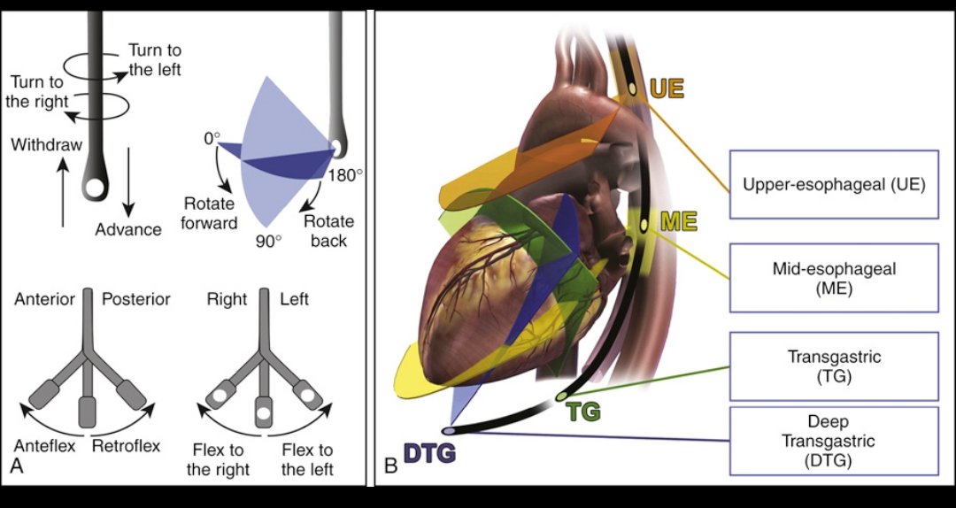
TEE probe movement is controlled via a handle with adjustable knobs:
✅ Transducer Rotation (Electronic Control):
🔄 Forward rotation (0° → 180°)
🔄 Backward rotation (180° → 0°)
This adjusts the transducer plane to obtain different imaging angles.
✅ Anteflexion & Retroflexion (Large Knob):
▶️ Anteflexion: The probe tip bends forward (toward anterior structures like the LVOT, mitral valve).
◀️ Retroflexion: The probe tip bends backward (helpful for imaging the LA, pulmonary veins, or deep transgastric views).
✅ Right & Left Flexion (Small Knob):
↪️ Right Flexion: The probe tip moves to the right, optimizing alignment with right heart structures.
↩️ Left Flexion: The probe tip moves to the left, improving views of left-sided heart structures.
✅ Advance & Withdraw:
⬆️ Advancing moves the probe deeper (toward the stomach for transgastric views).
⬇️ Withdrawing pulls the probe back (toward the upper esophagus for great vessel imaging).
🔹 (2) Esophageal Probe Positions
TEE imaging is performed from different locations along the esophagus and stomach to achieve various cardiac views:
📍 Upper Esophageal (UE):
🔹 High esophageal position (near the oropharynx)
🔹 Best for imaging the great vessels & aortic arch
📍 Mid-Esophageal (ME):
🔹 Most commonly used position
🔹 Offers multiple cross-sectional views of the atria, ventricles, valves, & aorta
📍 Transgastric (TG):
🔹 Probe advances into the stomach
🔹 Provides short-axis views of the left ventricle (LV)
📍 Deep Transgastric (DTG):
🔹 Further advancement into the stomach
🔹 Optimized Doppler alignment with the LVOT, aortic valve, & ascending aorta
TEE is both an art & science—refining probe manipulation improves diagnostic accuracy and procedural success!
Source: Catherine M. Otto, Textbook of Clinical Echocardiography
#Cardiology #TEE #Echocardiography #MedicalImaging
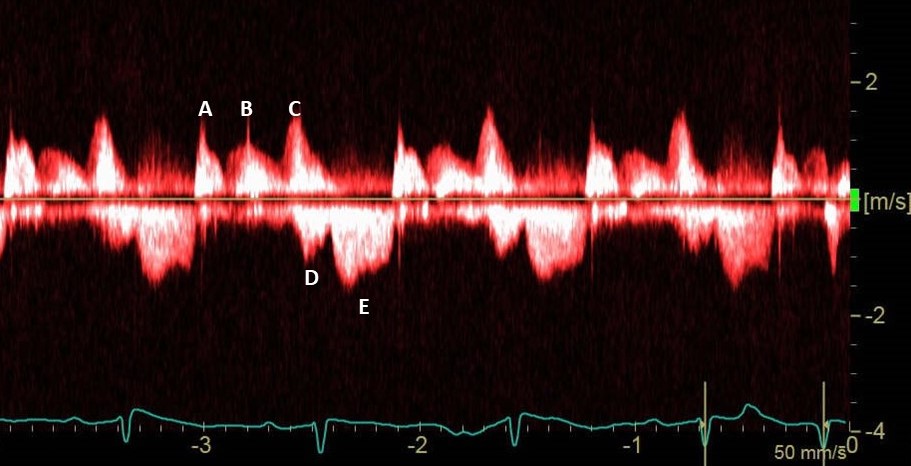
A patient in ICU. CW Doppler interrogation is performed across the mitral valve in 4-chamber view. What are the waves labeled A-E and what are the hemodynamic implications?
Every house from chautaquea on sunset to village burned down
Ralph's and gelsons gone
palisades village ALL there and stayed in tact
car wash gone
Pali elementar gone
gas stations gone
all of alphabet street gone
library gone #PalisadesFire
Did you know?
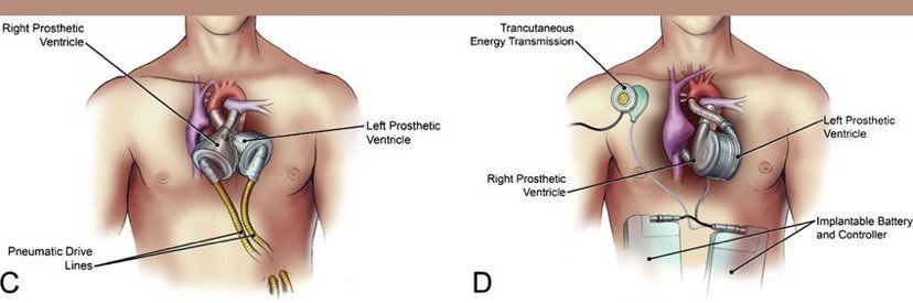
Back in 2004, the SynCardia 70cc Total Artificial Heart received FDA approval. Here's to many more years of innovation, progress, and lifesaving moments with the SynCardia Total Artificial Heart!
#SynCardia#heartfailure#CVD#TAH#MCS#artificialheart#medtech
I have a meeting this week with @HeartHackathon on all things upcoming AHF competitions.
With the recent outcomes regarding kidney and heart xenotransplant recipients… the future of (h)tx is still reliant on MCS, and I’m very proud to support that road in the interim.
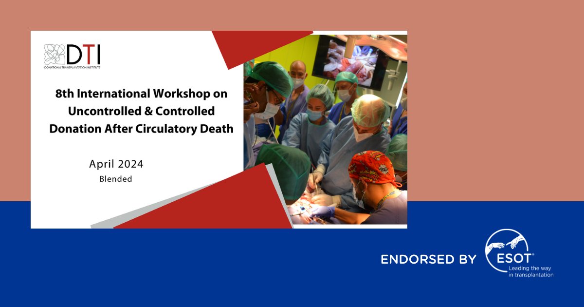
The International Workshop on Uncontrolled & Controlled Donation After Circulatory Death is addressing the growing interest in DCD donors to increase organ availability. The VitaSmart from #BridgetoLife will be there as part of the Machine Perfusion wet lab.
#MachinePerfusion
Sign up for the webinar and submit your queries for John via the Google Forms link: https://t.co/uTkxhm9KFc
Date: 29th February, 2024
Time: 1pm-2pm AEDT
#artificalheart#hearthackathon#webinar
From an amazing person and good friend - Lance White. Lance spent 2.5 years on the TAH. Heart Transplant Recipient's New Lease On Life https://t.co/zbDl4gwPlH