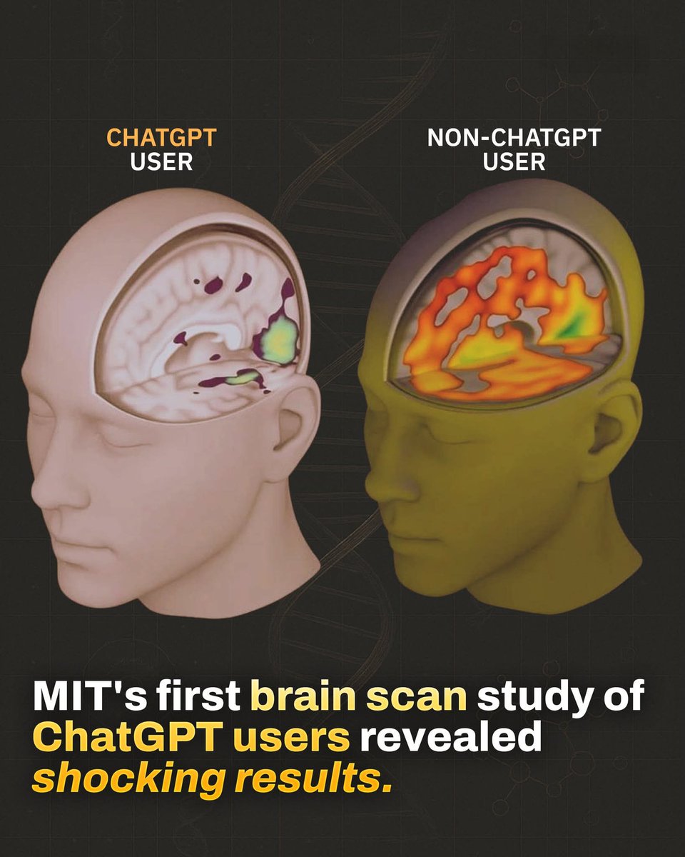
🧠 MIT recently completed the first brain-scan study on ChatGPT users—and the results are deeply revealing.
Rather than boosting brain function, prolonged AI use may be dulling it.
Over four months of cognitive data suggest we might be measuring productivity all wrong ⤵️
In MIT’s study, participants had their brains scanned while using ChatGPT.
→ 83.3% of users couldn’t recall a single sentence they’d written just minutes earlier.
→ In contrast, those writing without AI had no trouble remembering.
Brain connectivity dropped sharply—from 79 to 42 points.
→ That’s a 47% drop in neural engagement.
→ The lowest cognitive performance among all user groups.
Even after stopping ChatGPT use in later sessions, these users showed continued under-engagement.
→ Their performance remained lower than those who never used AI.
→ This suggests more than dependency—it’s cognitive weakening.
Beyond the scans, educators flagged the writing itself.
→ Essays were technically solid, but often called “robotic,” “soulless,” and “lacking depth.”
Here’s the paradox:
→ ChatGPT makes you 60% faster at completing tasks…
→ But it reduces the mental effort required for learning by 32%.
The top-performing group?
→ Those who began without AI and added it later.
→ They retained the best memory, brain activity, and overall scores.
Using ChatGPT can feel empowering—but it may quietly offload your thinking.
→ You gain speed, but lose engagement.
→ You get answers, but stop learning how to think.
The takeaway isn’t to avoid AI—but to use it intentionally.
→ Use it to assist, not replace your mind.
→ Build cognitive strength—not dependency.
MIT’s early study on AI and the brain lays out the stakes. The way we use these tools matters more than ever.
This trial has two goals: ensure the brain implant is safe; and to restore a person's ability to communicate with real-time speech
https://t.co/VyJCLX0FAw
Higher polygenic risk scores for bipolar disorder were associated with favorable treatment outcomes after lithium augmentation in antidepressant non-responders with unipolar depression. https://t.co/qDvVNk3v67
Vo et al. analysed brain imaging data from over 3,000 people with Parkinson's disease and found widespread structural brain changes, including in early stages. These patterns were shaped by both brain network connectivity and local biological features. https://t.co/eWsETyB5Qy
PhD Students - Which journal will accept your paper?
Find out using in 3 simple steps.
1. Go to https://t.co/MJXMwkIjcz
2. Upload your paper
3. Review-it will suggest 10 most relevant journals.
These journals are likely to accept your paper.
7 Sci-Hub Alternative Websites
Paper you need to ask for payment & can't use sci-hub?
You don't have to pay to read academic papers.
These are 7 sci-hub alternative websites to download papers for free.
Saccular aneurysms account for approximately 90% of lesions and typically form at arterial bifurcations in the internal carotid artery, the anterior and posterior communicating arteries, and the middle cerebral artery. In the posterior circulation, they commonly occur at the basilar artery bifurcation and cerebellar artery branch points (seen in figure). Less common types of unruptured intracranial aneurysms (UIAs) include fusiform aneurysms involving elongated segments of the artery, mycotic aneurysms associated with infections, and dissecting aneurysms resulting from arterial injury. Up to 20% of patients with UIAs have multiple intracranial aneurysms. The formation of aneurysms is thought to result from degeneration of the internal elastic lamina, endothelial dysfunction, and hemodynamic stress; inflammation, which leads to instability of the vascular wall, plays a crucial role in both their formation and rupture.
Learn more in the new Clinical Practice article, “Unruptured Intracranial Aneurysms,” by Christopher S. Ogilvy, MD (@chrisogilvyMD), from @BIDMChealth and @harvardmed: https://t.co/lAIar9rnAx
#Neurology #Cardiology
🍓 "Strawberry Gelatinous Appearance" — A Possible Face of Myxoid Liposarcoma
Myxoid liposarcoma (MLS) is one of the most morphologically distinctive soft tissue sarcomas, both microscopically and macroscopically. In some cases, it displays the so-called "strawberry gelatinous appearance" — a reddish-pink, translucent, gelatinous cut surface, sometimes with hemorrhagic areas. Although not a constant finding, this pattern reflects the tumor’s rich myxoid matrix and delicate vasculature, and is appreciated by pathologists as a suggestive clue, though not pathognomonic.
🔬 Overview
Accounts for approximately 30–40% of liposarcomas in adults
Peak incidence: 3rd to 5th decade
Most common sites: deep soft tissues of the thigh and retroperitoneum
🧠 Classic Histology
Abundant myxoid stroma rich in hyaluronic acid
Delicate branching capillaries in a “chicken-wire” pattern
Presence of immature lipoblasts with cytoplasmic vacuoles indenting the nucleus
May show hypercellular areas with round cells, indicating higher grade
🔎 Gross Pathology
Usually soft, gelatinous, and translucent on cut surface
In some cases, a reddish or hemorrhagic, strawberry-like appearance
While not universal, this appearance is recognized as a possible characteristic pattern
🧬 Genetics and Molecular Diagnosis
Characteristic translocation: FUS::DDIT3 (most common) or EWSR1::DDIT3
Detected by FISH, RT-PCR, or sequencing
🧪 Immunohistochemistry
S100: may be positive in lipoblasts
CD34 and MDM2: generally negative
Note: Diagnosis is primarily based on morphology and molecular findings
📉 Prognosis and Risk Factors
A round cell component >5% is associated with worse prognosis
Metastases more commonly occur in soft tissues, bones, and retroperitoneum (less commonly in the lungs)
Purely myxoid tumors tend to have a more favorable clinical outcome
📚 References
Potterveld S, Clay MR. Myxoid liposarcoma. https://t.co/yteSms2T6w website. Available at: https://t.co/CoCgpiSCMS. Accessed June 24th, 2025.
WHO Classification of Tumours Editorial Board. Soft Tissue and Bone Tumours. 5th ed. Lyon: IARC; 2020.
Antonescu CR. The role of genetics in the diagnosis of soft tissue tumors. Mod Pathol. 2014;27(Suppl 1):S36–S43.
⚠️ Disclaimer: This content is intended for educational purposes only and should not be considered a substitute for professional medical advice, diagnosis, or treatment.
#MedicalEducation #NotasDePatologia #SoftTissuePathology #MyxoidLiposarcoma
Case report: an atypical postvaccinal encephalitis in a 60-year-old man, with radial perivascular enhancement consistent with inflammation of glial fibrillary acidic protein–rich white matter loci. https://t.co/AyEzugwpPn
Poor sleep may double the risk of depression, impair toxin clearance from the brain by up to 60%, and fuel insomnia, which affects 10% of adults.
Sleep isn’t just rest—it’s a cornerstone of mental health and neuroprotection.
Here’s what clinicians need to know about sleep stages, insomnia, and evidence-based interventions for improving patient outcomes 🧵👇