Grateful to receive a Certificate of Scientific Contribution at #AORTIC. This recognition reflects our team’s commitment to strengthening cancer care and research in #Africa 🎉
May we also #PrayTogether for #Tanzania, where, following the recent elections, violent clashes have broken out, leaving many victims. I urge everyone to avoid all forms of violence and to follow the path of dialogue.
I���m delighted to share our recent publication on services available for CNS tumors in Sub-Saharan Africa. This collaborative work highlights the current capacities, challenges, and opportunities to strengthen neuro-oncology care across the region.
https://t.co/ijavwZNnCJ
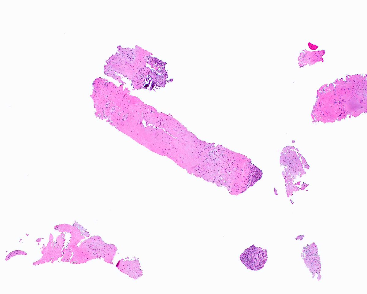
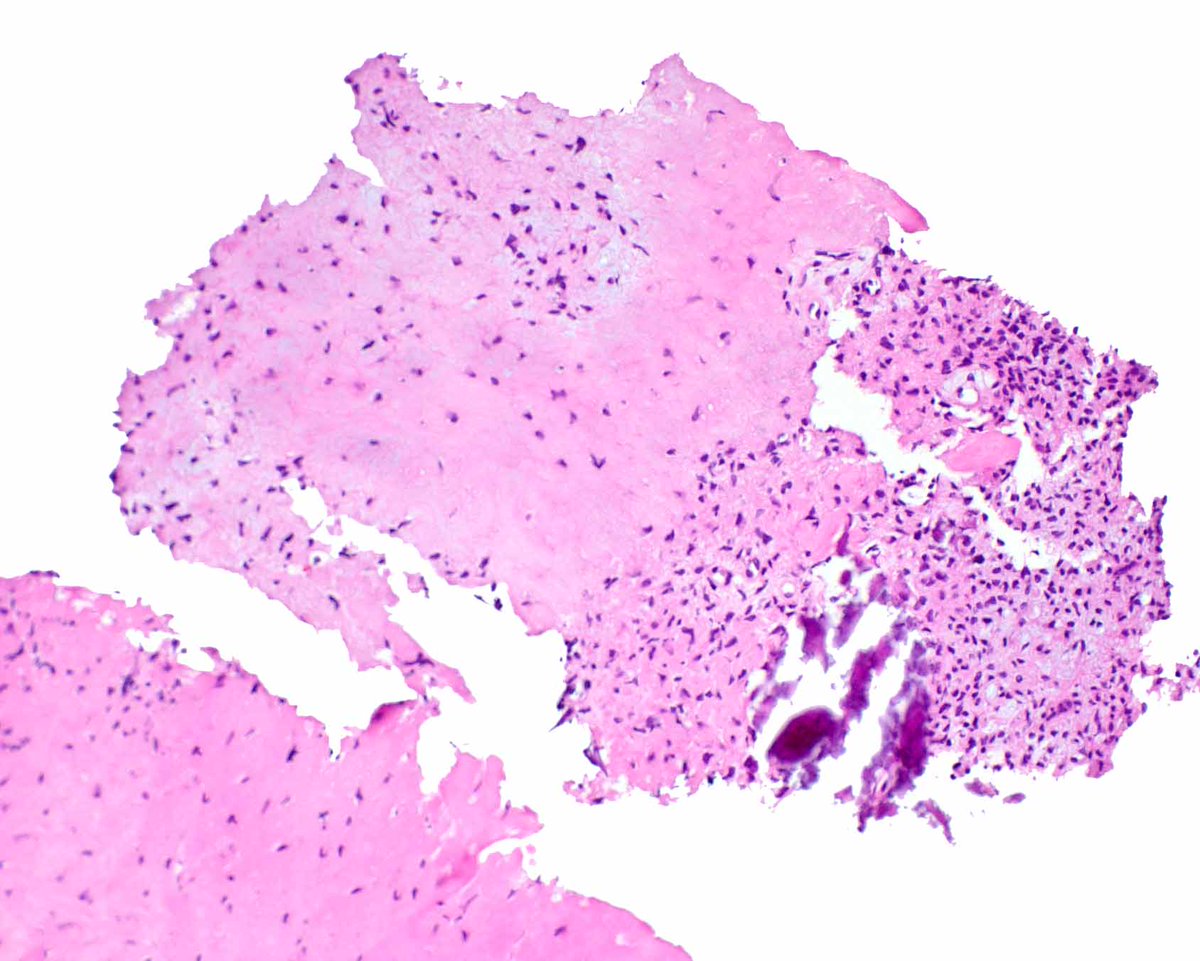
Modern immunohistochemistry can be so reassuring on tiny needle biopsies, like the ones for this chondromyxoid fibroma.
1PMIDs: 10349990. PMID: 35650682; PMCID: PMC9481662.
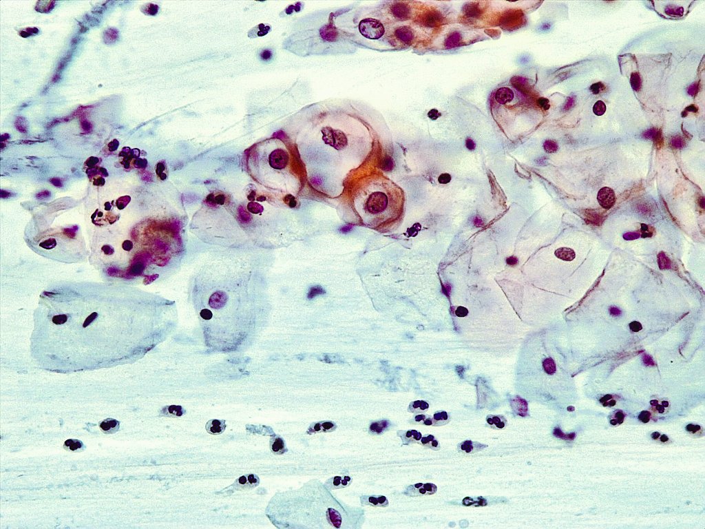
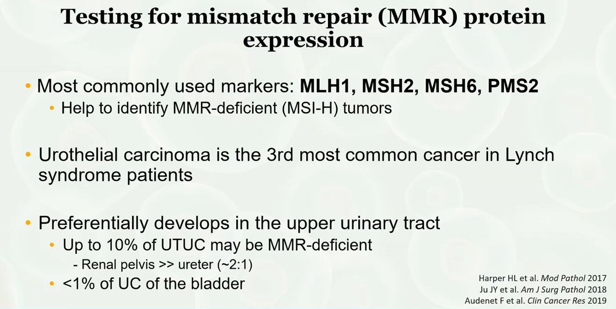
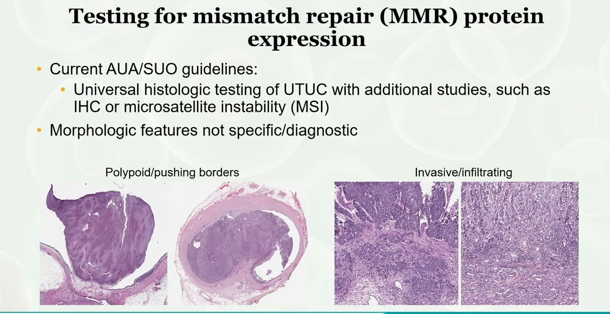
Urothelial carcinoma and MMR testing:
-Urothelial carcinoma is the 3rd most common cancer in Lynch Syndrome patients
-Preferentially develops in upper urinary tract
Dr. Al-Ahmadie #USCAP25#pathology#PathX
The Genitourinary Pathology Society (GUPS) and the International Society of Urologic Pathology (ISUP), collaborated on several important projects where publishing joint standardized guidelines is critical for ensuring uniform practice.
Continued in comments..
🧬 Vaněk Revisited: A Practical Guide to Diagnosing Inflammatory Fibroid Polyp
The inflammatory fibroid polyp (IFP) is a benign, subepithelial mesenchymal lesion most commonly found in the gastrointestinal tract — particularly in the gastric antrum and small intestine.
🔬 Historical context:
Originally described by Jan Vaněk in 1949 as a “submucosal gastric granuloma with eosinophilic infiltration” [Am J Pathol 1949;25:397], the lesion was long referred to as “eosinophilic granuloma” or “Vaněk’s polyp.” These terms are now considered outdated and imprecise. The current terminology, inflammatory fibroid polyp, better reflects its fibrovascular and inflammatory nature.
🧫 Classic histologic features:
• Proliferation of spindle-shaped fibroblastic/myofibroblastic cells
• Prominent small vessels with an arborizing pattern
• Mixed inflammatory infiltrate rich in eosinophils, lymphocytes, and plasma cells
• Poorly circumscribed submucosal growth
🧬 Genetic profile and differential diagnosis:
Activating PDGFRA mutations are frequently found, supporting a diagnosis of benign neoplasia and helping to distinguish IFPs from other GI mesenchymal lesions (e.g., GIST).
🧪 Special stains:
Although not essential for diagnosis, PAS and Alcian Blue may be used to demonstrate mucin in residual epithelial areas, but their role in the primary diagnosis is limited.
📌 Pathologist tips:
❌ Avoid outdated terms like “eosinophilic granuloma” or “Vaněk’s polyp”
✅ Prefer: “Inflammatory fibroid polyp with typical histologic features”
✅ Consider immunohistochemistry (CD34, vimentin) or molecular analysis in atypical cases
📚 References:
Vaněk J. Gastric submucosal granuloma with eosinophilic infiltration. Am J Pathol. 1949;25(3):397–411.
Assarzadegan N, Gonzalez RS. Inflammatory fibroid polyp. https://t.co/yteSms2T6w. https://t.co/LsPZnYMtO6.
⚠️ Disclaimer: This content is intended for educational purposes only and should not be considered a substitute for professional medical advice, diagnosis, or treatment.
#Vanek #GIPath #MedicalEducation #NotasDePatologia #Histopathology #InflammatoryFibroidPolyp #PathTwitter
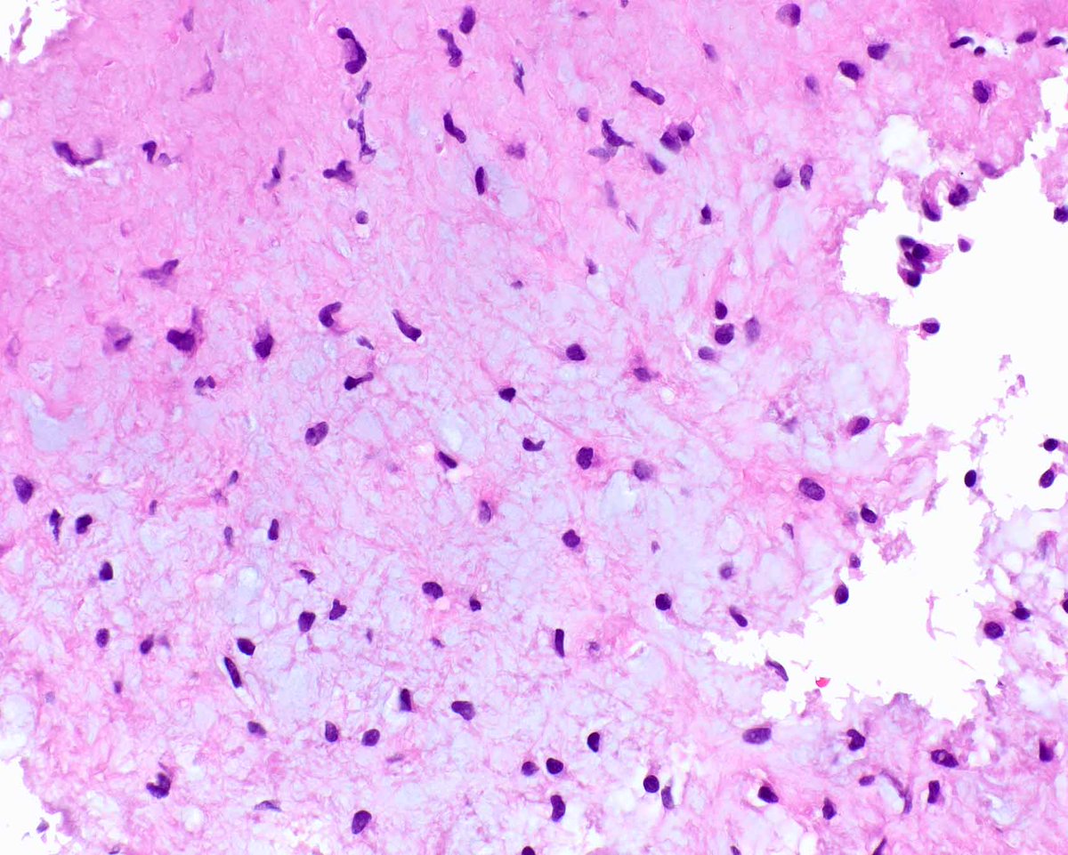
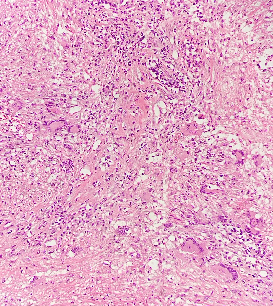
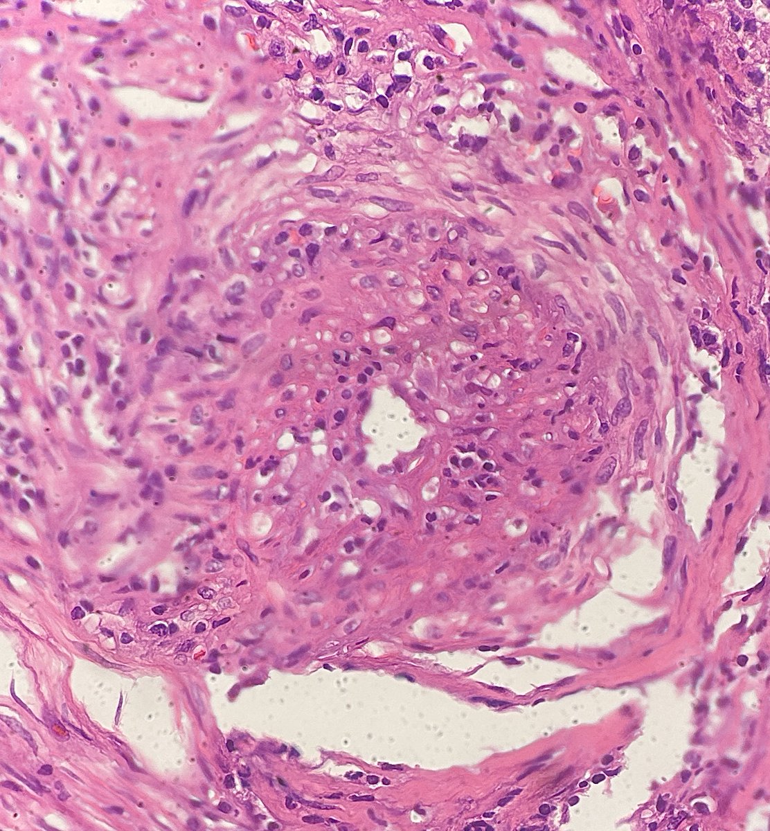
🧠 Acute Bacterial Meningitis: a neurological emergency with high mortality potential
📌 Acute bacterial meningitis is a purulent inflammation of the leptomeninges caused by bacterial infection. It often progresses rapidly and carries a significant risk of death or long-term neurological sequelae.
🔍 Etiology:
Causative organisms vary by age and immune status:
Neonates: Streptococcus agalactiae, E. coli, Listeria monocytogenes
Children and young adults: Neisseria meningitidis
Adults and elderly: Streptococcus pneumoniae, Listeria monocytogenes
Immunocompromised hosts: Listeria, Gram-negative bacilli, Mycobacterium tuberculosis
⚠️ Classic clinical triad:
Fever
Severe headache
Nuchal rigidity
Other features may include altered mental status, nausea, vomiting, photophobia, and positive Kernig or Brudzinski signs.
🧠 Morphologic features:
Grossly, there may be cerebral edema and purulent exudate over the convexities or the base of the brain, depending on the pathogen (Neisseria → base; Pneumococcus → convexities).
Microscopically:
Dense neutrophilic infiltrate in the subarachnoid space
Leptomeningeal vasculitis with thrombosis of small vessels
Possible parenchymal extension with necrosis in advanced cases
Bacteria visible with special stains (e.g., Gram)
May involve ventricles (ventriculitis) or lead to abscess formation
🩸 Cerebrospinal fluid (CSF):
Turbid or purulent appearance
Elevated WBC count (>1,000/mm³, neutrophil predominance)
Elevated protein
Decreased glucose
Gram stain and culture are critical; PCR increases sensitivity
💊 Treatment (start immediately):
Empiric antibiotic therapy based on age and clinical context
Adjunctive dexamethasone (especially in pneumococcal meningitis)
Intensive supportive care as needed
🛡️ Prevention:
Vaccination against Haemophilus influenzae type b, Streptococcus pneumoniae, and Neisseria meningitidis has significantly reduced incidence in immunized populations.
📚 Take-home message:
Prompt diagnosis and early treatment are key to improving outcomes. An integrated clinical, laboratory, and pathological approach is essential.
#MedicalEducation #NotasDePatologia #Neuropathology #infectiousdiseases
#Meningitis
⚠️ Disclaimer: This content is intended for educational purposes only and should not be considered a substitute for professional medical advice, diagnosis, or treatment.
Virchows Archiv reports on a study of basal cell adenomas (BCAs) with abundant S100 protein-positive stroma, suggesting these tumors may represent a unique triphasic subset distinct from pleomorphic adenomas (PAs).
https://t.co/FRR1AHJWGm
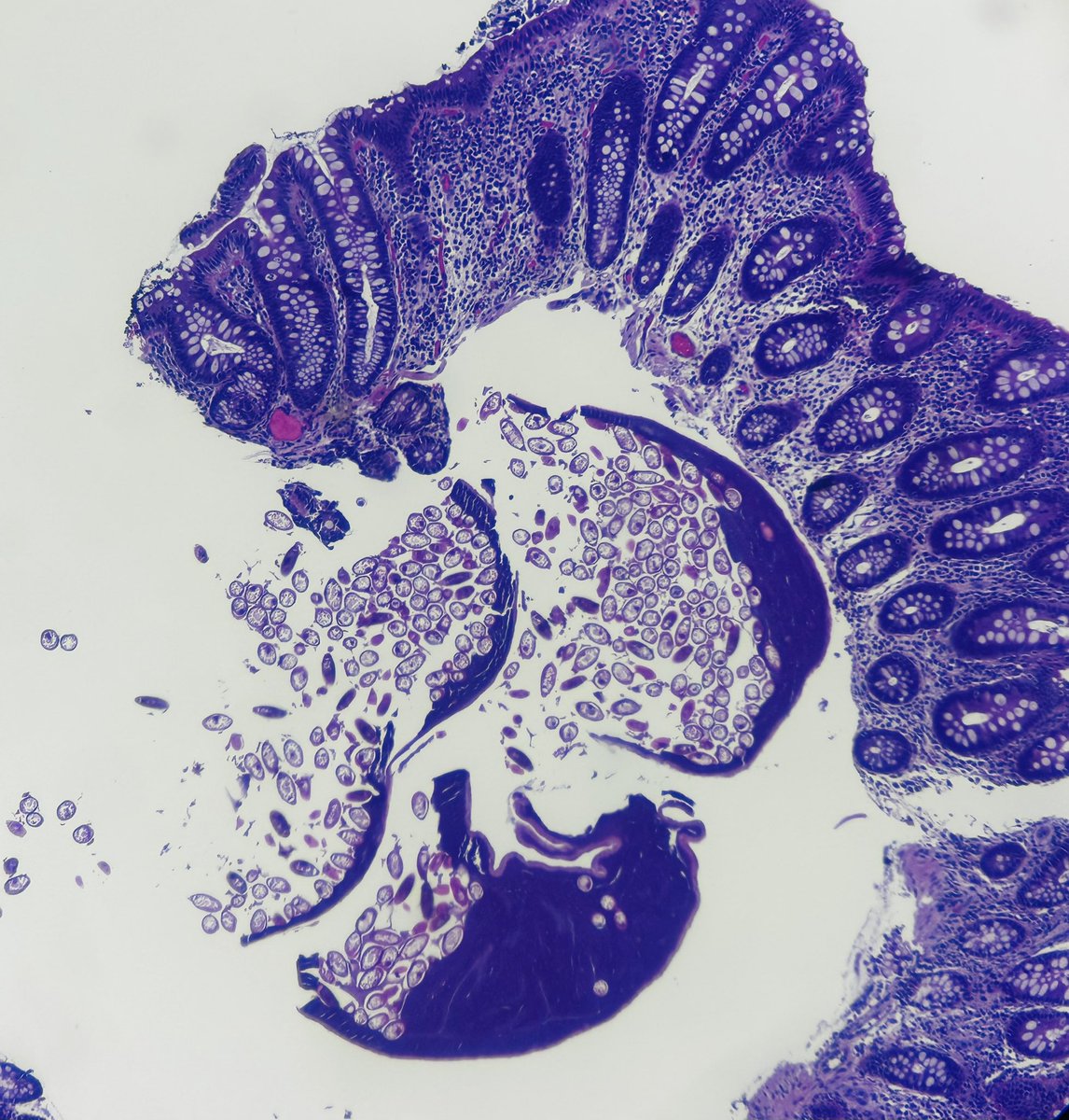
A 60-year-old cisgender man with no medical history underwent colonoscopy, revealing superficial erosions and a “foreign body.” @valensev#path#infectpath#Parasite
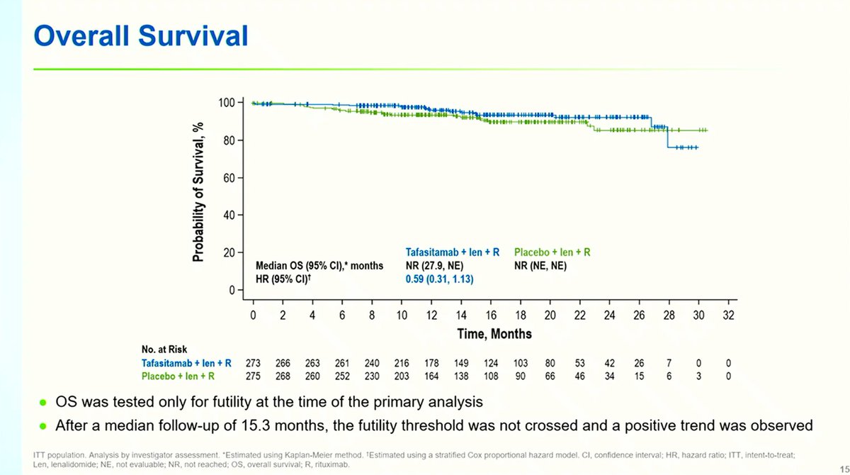
🚨 POTENTIAL NEW SOC IN R/R FOLLICULAR #LYMPHOMA!!
PFS superiority of Tafasitamab-R2 against Placebo-R2 (inMIND phase 3 trial) 📈
Besides, almost all relapses were CD19+ (thus, subsequent CAR-T access is not compromised)
Presented by Dr L Sehn at #ICML2025#18ICML@icmlugano
Proliferating trichilemmal tumour
Not malignant or benign
Just a low malignant potential tumor.
Scalp is the typical site
It is Like a pilar cyst, but with prominent epithelial infoldings
Calcification is common
#path#derm#Dermatopathology#pathology


![Notas_Patologia's tweet photo. 🧬 Vaněk Revisited: A Practical Guide to Diagnosing Inflammatory Fibroid Polyp
The inflammatory fibroid polyp (IFP) is a benign, subepithelial mesenchymal lesion most commonly found in the gastrointestinal tract — particularly in the gastric antrum and small intestine.
🔬 Historical context:
Originally described by Jan Vaněk in 1949 as a “submucosal gastric granuloma with eosinophilic infiltration” [Am J Pathol 1949;25:397], the lesion was long referred to as “eosinophilic granuloma” or “Vaněk’s polyp.” These terms are now considered outdated and imprecise. The current terminology, inflammatory fibroid polyp, better reflects its fibrovascular and inflammatory nature.
🧫 Classic histologic features:
• Proliferation of spindle-shaped fibroblastic/myofibroblastic cells
• Prominent small vessels with an arborizing pattern
• Mixed inflammatory infiltrate rich in eosinophils, lymphocytes, and plasma cells
• Poorly circumscribed submucosal growth
🧬 Genetic profile and differential diagnosis:
Activating PDGFRA mutations are frequently found, supporting a diagnosis of benign neoplasia and helping to distinguish IFPs from other GI mesenchymal lesions (e.g., GIST).
🧪 Special stains:
Although not essential for diagnosis, PAS and Alcian Blue may be used to demonstrate mucin in residual epithelial areas, but their role in the primary diagnosis is limited.
📌 Pathologist tips:
❌ Avoid outdated terms like “eosinophilic granuloma” or “Vaněk’s polyp”
✅ Prefer: “Inflammatory fibroid polyp with typical histologic features”
✅ Consider immunohistochemistry (CD34, vimentin) or molecular analysis in atypical cases
📚 References:
Vaněk J. Gastric submucosal granuloma with eosinophilic infiltration. Am J Pathol. 1949;25(3):397–411.
Assarzadegan N, Gonzalez RS. Inflammatory fibroid polyp. https://t.co/yteSms2T6w. https://t.co/LsPZnYMtO6.
⚠️ Disclaimer: This content is intended for educational purposes only and should not be considered a substitute for professional medical advice, diagnosis, or treatment.
#Vanek #GIPath #MedicalEducation #NotasDePatologia #Histopathology #InflammatoryFibroidPolyp #PathTwitter](https://pbs.twimg.com/media/GuJgoBTXoAAXs4o.jpg)