Our June issue is live! 🎉
https://t.co/AulrIZWCXv
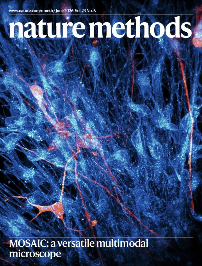
On the cover, an absolutely gorgeous image of neurites and mitochondria in a brain organoid, captured using the MOSAIC microscope.
Paper here: https://t.co/FwAYTBMnHj
@Eric_Betzig@ABCUCBerkeley One microscope, every scale. MOSAIC images life from single molecules to whole organisms: dividing cells, remodeling organelles, regenerating zebrafish, Alzheimer's tissue at the nanoscale, live mouse neural dynamics. Credit: Fu, Liu, Milkie, Ruan et al., Nature Methods (2026).
Congrats to all 49 authors on the publication of our paper on the multimodal MOSAIC microscope, to be found here: https://t.co/meZXjRMog2
A summary with movies I posted nearly a year ago can be found below.
MOSAIC: a versatile multimodal microscopy that seamlessly transitions between light-sheet, 2-photon, label-free and super-resolution modalities. @Eric_Betzig@ABCUCBerkeley@legant_lab
https://t.co/l3NtYh3OrD
🔬🔦Check out our ‘Imaging spotlight’ on Volumetric Imaging via Photochemical Sectioning (VIPS) from @Ruixuan3, Srigokul Upadhyayula and colleagues.
VIPS helps researchers scale super resolution imaging to large whole-mount tissues.
https://t.co/jhcTMYm99a
A new method called VIPS enables organ-wide nanoscale reconstruction for quantitative mapping of connectivity and molecular landscape in intact tissue.
Learn more this week in Science: https://t.co/ffOqBTWo5o
1/ Better late than never, here’s a long thread of threads summarizing our MOSAIC preprint.
If you’re curious about the process by which a new microscope is conceived and brought into the world, this is the thread for you. If you’ve come for eye candy, there’s plenty of that too below.
https://t.co/BEfAolyeR9
Finally here! Our preprint on the MOSAIC, a multimodal adaptive optical microscope enabling non‑invasive in‑vivo imaging from molecules to organisms, is out.
https://t.co/B2GHHP5b0K
@Eric_Betzig@legant_lab
The MOSAIC reconfigurable microscope by @Eric_Betzig et al is finally out!
MOSAIC integrates multiple advanced imaging techniques including light-sheet, label-free, super-resolution, and multi-photon, all equipped with adaptive optics:
https://t.co/RkwkoptVHv
Unlocking the potential to image petabyte-scale datasets 😵💫! From #CZBiohubSF Investigator Gokul Upadhyayula and co-corresponding authors & @BerkeleyMCB colleagues Xiongtao Ruan and @Eric_Betzig.
We are excited to introduce PetaKit5D, a comprehensive suite of software tools designed to tackle challenges in the handling of large datasets and make processing of petabyte-scale datasets feasible: https://t.co/P2WaWODV3F
PetaKit5D is a scalable and efficient software for processing large datasets from light sheet microscopy and other imaging modalities. It is available for use in MATLAB and Python, as well as a GUI that requires no coding. Try it and share your feedback!
https://t.co/CkCbRndwPl
Almost everyone in microscopy (especially LSFM) talks about data deluge, but the tools haven't kept apace with the problems. Check out PetaKit5D for processing and analyzing your huge datasets!