New in @BloodPortfolio 🩸
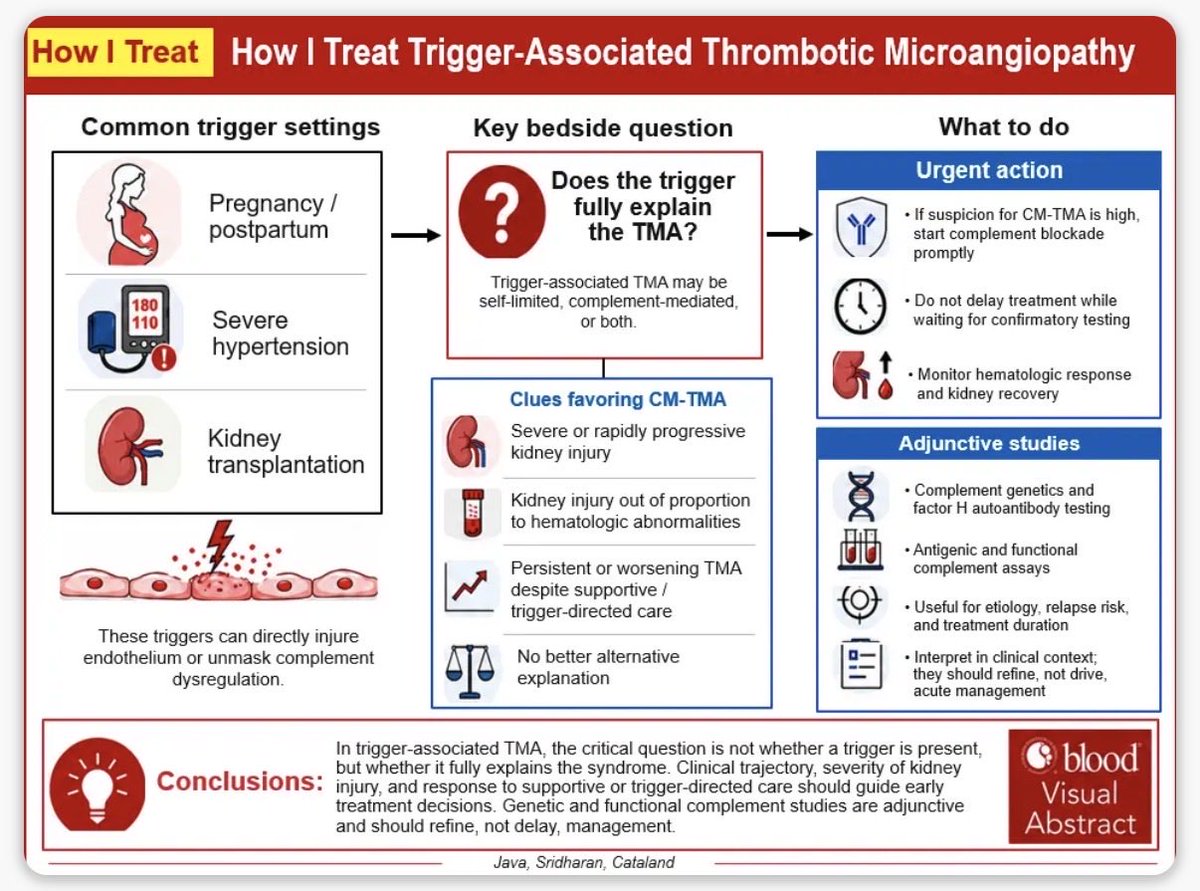
Our “How I Treat” on TMA with clinical triggers.
When pregnancy, severe HTN or kidney txp coexist with TMA, when is the trigger enough & when to suspect complement-mediated disease & how to treat? A practical framework.
https://t.co/4Tkufwccgo
Creatinine is rising. But is the kidney really to blame? Detective Nephron and Mackenzie Ula Densa tackle a puzzling case that challenges assumptions and puts your clinical reasoning to the test: https://t.co/X0x1GCn3Tf
#ASNKidneyNews@kdjhaveri
La disnea en la Insuficiencia Cardíaca Aguda (ICA) no es simplemente "líquido en los pulmones". Una reciente revisión nos demuestra que es un fenómeno neurocardiorrespiratorio multidimensional.
🧵👇
🤓Critical Mind ICU Academy is now live on Google Play📱
After months of development, clinical review, testing and refinement, I am very happy to finally make Critical Mind ICU Academy available to the critical care community.
The idea behind Critical Mind is simple: ICU education should go beyond reading guidelines and memorizing protocols. We need to learn how to think through physiology, interpret changing clinical information, make decisions and understand their consequences.
Critical Mind ICU Academy was developed from the perspective of real-life critical care practice and is designed as a structured learning environment for physicians, residents, fellows and other healthcare professionals working with critically ill patients.
The Academy combines a structured critical care curriculum with more than 100 clinical MCQs, 25 interactive ICU simulations, bedside physiology and haemodynamic training, mechanical ventilation, shock, sepsis, cardiology, neurocritical care, renal failure, antimicrobial therapy, nutrition and other core ICU domains.
It also includes a dedicated haemodynamic laboratory, an ultrasound portfolio covering cardiac, lung, vascular, FAST/eFAST and transcranial Doppler applications, progress tracking, and an offline educational AI tutor and simulation debriefing environment.
One feature that was particularly important to me was accessibility. The Academy can be used in English, Spanish, Hungarian, French, German, Portuguese, Japanese and Chinese, and its core educational environment works offline without external APIs.
This is not intended to replace textbooks, guidelines, formal fellowship training or clinical judgement. The goal is different: to create a place where trainees can repeatedly learn, reason, make decisions, make mistakes safely, and try again.
As an intensivist, I built Critical Mind around something I have increasingly come to believe in during clinical practice and teaching:
Critical care is not learned by memorizing what to do. It is learned by understanding why.
Critical Mind ICU Academy is now available on Google Play.
Launch price: USD 49.99
I would be particularly interested in feedback from ICU residents, fellows, intensivists and critical care educators as the Academy continues to evolve.
🔗 Google Play:
https://t.co/LcGJ1eQpCG
#CriticalCare #IntensiveCare #MedicalEducation #ICUEducation #FOAMcc #POCUS #Hemodynamics #MechanicalVentilation #Simulation #MedEd #DigitalHealth
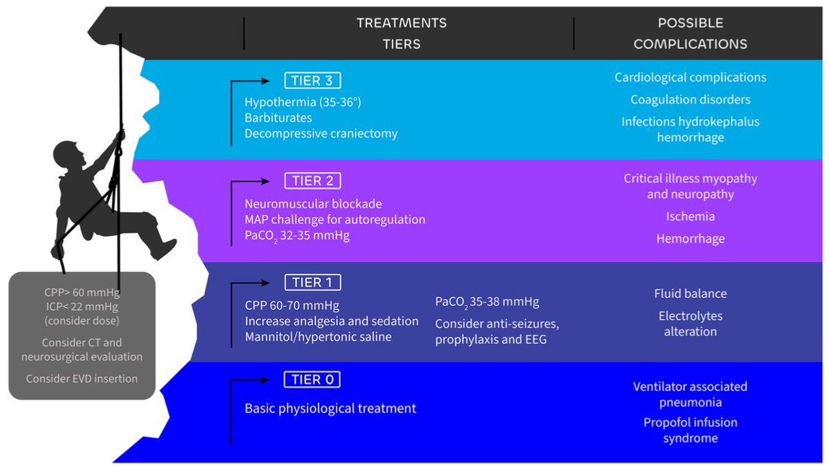
Critical Physiology Series #37
Mechanical Ventilation With Increased Intracranial Pressure: Protect the Lung Without Harming the Brain
Mechanical ventilation directly influences cerebral blood flow, intracranial pressure and cerebral perfusion pressure.
CO₂ is a cerebral vasodilator, hypercapnia increases cerebral blood volume and may increase ICP. Hypocapnia produces vasoconstriction and can rapidly decrease ICP, but excessive or prolonged hyperventilation may reduce CBF enough to produce cerebral ischemia.
For this reason, normocapnia is usually the safest starting point. Short controlled hyperventilation should be reserved mainly as a temporary rescue strategy during an acute intracranial pressure crisis while definitive treatment is established.
PEEP creates another brain lung interaction.
Increasing PEEP raises intrathoracic pressure. If this increases right atrial and cerebral venous pressure, cerebral venous drainage may deteriorate and ICP may rise. If PEEP also decreases cardiac output and MAP, cerebral perfusion pressure may fall.
But when PEEP recruits lung and improves compliance, relatively little pressure may be transmitted to the pleural space. Better oxygenation may actually benefit the injured brain. PEEP should therefore be titrated according to lung recruitability, systemic haemodynamics, ICP and CPP rather than limited by an arbitrary number.
Tidal volume, Ppl and driving pressure remain important because acute brain injury does not protect the patient from ventilator induced lung injury.
Recent evidence suggests that extremely low tidal volumes combined with aggressive PEEP may not benefit patients with severe brain injury. The objective is enough protection to avoid secondary lung injury while maintaining cerebral oxygenation and perfusion.
Mechanical power adds another dimension because respiratory rate, tidal volume, pressure and flow determine how much mechanical energy reaches the respiratory system over time.
Strong spontaneous breathing also requires caution. Excessive respiratory drive may generate large transpulmonary pressure swings while simultaneously causing fluctuations in cerebral venous pressure and ICP.
Newer strategies are becoming particularly interesting.
Fully automated ventilation can continuously adjust ventilatory support while targeting predefined PaCO₂, oxygenation and lung protective ranges. The recent BRAVE study showed that automated ventilation can maintain brain and lung protective targets more consistently, although evidence remains early and does not yet establish automated ventilation as standard care.
Modes such as NAVA or proportional assistance may help preserve diaphragm activity and improve synchrony during recovery, but evidence specifically supporting them for intracranial hypertension remains insufficient.
Recommended lectures 📚
Robba, 2020, https://t.co/xIUJRb1eHw
Mascia, 2024, https://t.co/lRNHRR2jMq
Goossen, 2026, https://t.co/7EMTk0TTT0
🫀🚨 𝗜𝗻𝘀𝘂𝗳𝗶𝗰𝗶𝗲𝗻𝗰𝗶𝗮 𝗰𝗮𝗿𝗱𝗶𝗮𝗰𝗮 𝗮𝗴𝘂𝗱𝗮 ‼️
@JACCJournals@ACCinTouch
👇🏼👇🏼👇🏼👇🏼
📑🔗🔑🔓
https://t.co/89ht2mBRSA
⬇️⬇️⬇️⬇️
🧵👇
La revisión State-of-the-Art de 𝙅𝘼𝘾𝘾 𝟮𝟬𝟮𝟲 propone un cambio de paradigma:
💧 aliviar congestión rápidamente
🔍 identificar y tratar el precipitante
🧬 definir la cardiopatía subyacente
💊 iniciar precozmente terapia modificadora de enfermedad
📈 planear la trayectoria después del alta
El acrónimo 𝙎𝙋𝙇𝘼𝙎𝙃 resume la evaluación inicial:
𝙎ymptoms
𝙋ast history
𝙇ife signs
𝘼ssess congestion
𝙎TEMI/equivalente
𝙃ypoperfusion
🔥 La hospitalización es una 𝙫𝙚𝙣𝙩𝙖𝙣𝙖 𝙩𝙚𝙧𝙖𝙥𝙚́𝙪𝙩𝙞𝙘𝙖 𝙥𝙖𝙧𝙖 𝙢𝙤𝙙𝙞𝙛𝙞𝙘𝙖𝙧 𝙚𝙡 𝙥𝙧𝙤𝙣𝙤́𝙨𝙩𝙞𝙘𝙤, no simplemente para hacer desaparecer la disnea.
💉💧 𝙁𝙪𝙧𝙤𝙨𝙚𝙢𝙞𝙙𝙖: 𝙣𝙤 𝙗𝙖𝙨𝙩𝙖 𝙘𝙤𝙣 𝙖𝙙𝙢𝙞𝙣𝙞𝙨𝙩𝙧𝙖𝙧𝙡𝙖. 𝙃𝙖𝙮 𝙦𝙪𝙚 𝙨𝙖𝙗𝙚𝙧 𝙚𝙣 𝟮 𝙝𝙤𝙧𝙖𝙨 𝙨𝙞 𝙛𝙪𝙣𝙘𝙞𝙤𝙣𝙤́
Si existe alta sospecha de IC aguda con congestión, el artículo recomienda iniciar el diurético IV 𝙨𝙞𝙣 𝙚𝙨𝙥𝙚𝙧𝙖𝙧 𝙖 𝙘𝙤𝙢𝙥𝙡𝙚𝙩𝙖𝙧 𝙩𝙤𝙙𝙖𝙨 𝙡𝙖𝙨 𝙥𝙧𝙪𝙚𝙗𝙖𝙨 𝙙𝙞𝙖𝙜𝙣𝙤́𝙨𝙩𝙞𝙘𝙖𝙨.
💊 Sin tratamiento previo o dosis baja: 𝙛𝙪𝙧𝙤𝙨𝙚𝙢𝙞𝙙𝙖 𝟰𝟬 𝙢𝙜 𝙄𝙑
💊 Dosis oral crónica alta: ≥𝟴𝟬 𝙢𝙜 𝙄𝙑 𝙤 ≈𝟮× 𝙡𝙖 𝙙𝙤𝙨𝙞𝙨 𝙤𝙧𝙖𝙡
A las 𝟮 𝙝𝙤𝙧𝙖𝙨, evaluar:
✅ 𝘿𝙞𝙪𝙧𝙚𝙨𝙞𝙨 ≥𝟯𝟬𝟬 𝙢𝙇
o
✅ 𝙉𝙖 𝙪𝙧𝙞𝙣𝙖𝙧𝙞𝙤 ≥𝟳𝟬–𝟴𝟬 𝙢𝙢𝙤𝙡/𝙇
Si no se alcanza:
⬆️ duplicar la dosis de asa
➕ considerar 𝙖𝙘𝙚𝙩𝙖𝙯𝙤𝙡𝙖𝙢𝙞𝙙𝙖 𝙤 𝙩𝙞𝙖𝙯𝙞𝙙𝙖
🔍 excluir causas intrarrenales/postrenales
🔥 Esperar 24 horas para descubrir que el diurético no funcionó es perder tiempo de descongestión.
🫘📈 𝘾𝙧𝙚𝙖𝙩𝙞𝙣𝙞𝙣𝙖 𝙦𝙪𝙚 𝙖𝙪𝙢𝙚𝙣𝙩𝙖 𝙙𝙪𝙧𝙖𝙣𝙩𝙚 𝙪𝙣𝙖 𝙙𝙚𝙨𝙘𝙤𝙣𝙜𝙚𝙨𝙩𝙞𝙤́𝙣 𝙚𝙛𝙞𝙘𝙖𝙯 ≠ 𝙛𝙧𝙖𝙘𝙖𝙨𝙤 𝙩𝙚𝙧𝙖𝙥𝙚́𝙪𝙩𝙞𝙘𝙤
Uno de los errores más frecuentes es reducir o suspender diuréticos automáticamente ante una elevación moderada de creatinina.
La revisión enfatiza:
🫘 en ERC pueden necesitarse 𝙙𝙤𝙨𝙞𝙨 𝙢𝙖𝙮𝙤𝙧𝙚𝙨 de diuréticos
📈 la creatinina puede aumentar transitoriamente durante una descongestión efectiva
🚫 esto no obliga por sí solo a detener el tratamiento
🎯 el contexto de 𝙘𝙤𝙣𝙜𝙚𝙨𝙩𝙞𝙤́𝙣 𝙧𝙚𝙨𝙞𝙙𝙪𝙖𝙡 𝙮 𝙧𝙚𝙨𝙥𝙪𝙚𝙨𝙩𝙖 𝙙𝙞𝙪𝙧𝙚́𝙩𝙞𝙘𝙖 importa más que una cifra aislada
Además, la congestión persistente al alta se asocia con peor pronóstico.
📌 Una reducción de aproximadamente 𝟯𝟬% 𝙙𝙚𝙡 𝙉𝙏-𝙥𝙧𝙤𝘽𝙉𝙋 desde el ingreso al alta se considera compatible con descongestión adecuada dentro de una valoración multimodal.
🔥 En IC aguda, proteger la creatinina a costa de dejar al paciente congestionado puede ser una falsa victoria.
💊⚡ 𝙀𝙡 𝙜𝙧𝙖𝙣 𝙘𝙖𝙢𝙗𝙞𝙤: 𝙡𝙖 𝙂𝘿𝙈𝙏 𝙙𝙚𝙗𝙚 𝙘𝙤𝙢𝙚𝙣𝙯𝙖𝙧 𝙙𝙪𝙧𝙖𝙣𝙩𝙚 𝙡𝙖 𝙝𝙤𝙨𝙥𝙞𝙩𝙖𝙡𝙞𝙯𝙖𝙘𝙞𝙤́𝙣, 𝙣𝙤 “𝙖𝙡𝙜𝙪́𝙣 𝙙𝙞́𝙖” 𝙚𝙣 𝙘𝙤𝙣𝙨𝙪𝙡𝙩𝙖 𝙚𝙭𝙩𝙚𝙧𝙣𝙖
Secuencia rápida después de la estabilización:
🔹 𝙞𝙎𝙂𝙇𝙏𝟮 → desde los primeros días si eGFR ≥20 mL/min/1,73 m²
🔹 𝘼𝙍𝙈 → tempranamente si presión, función renal y K⁺ lo permiten
🔹 𝘼𝙍𝙉𝙄/𝙍𝘼𝙎𝙄 → tras estabilización, preferentemente con congestión leve o euvolemia
🔹 β-𝙗𝙡𝙤𝙦𝙪𝙚𝙖𝙙𝙤𝙧 → mantener si estable; iniciar en HFrEF cuando mejore la congestión
📊 La evidencia citada es potente:
⚡ 𝙋𝙄𝙊𝙉𝙀𝙀𝙍-𝙃𝙁: sacubitrilo/valsartán → ↓66% del endpoint clínico compuesto exploratorio
⚡ 𝙀𝙈𝙋𝙐𝙇𝙎𝙀: empagliflozina → ↓31% de muerte CV/eventos de IC
⚡ 𝙎𝙊𝙇𝙊𝙄𝙎𝙏-𝙒𝙃𝙁: sotagliflozina → ↓33%
⚡ 𝙎𝙏𝙍𝙊𝙉𝙂-𝙃𝙁: estrategia intensiva → ↓34% de muerte o rehospitalización por IC
🔥 El predictor más fuerte de que un paciente NO reciba GDMT después del alta es que 𝙣𝙤 𝙨𝙚 𝙡𝙖 𝙝𝙖𝙮𝙖𝙣 𝙞𝙣𝙞𝙘𝙞𝙖𝙙𝙤 𝙙𝙪𝙧𝙖𝙣𝙩𝙚 𝙡𝙖 𝙝𝙤𝙨𝙥𝙞𝙩𝙖𝙡𝙞𝙯𝙖𝙘𝙞𝙤́𝙣.
🎯 𝙀𝙡 𝙖𝙡𝙩𝙖 𝙣𝙤 𝙩𝙚𝙧𝙢𝙞𝙣𝙖 𝙚𝙡 𝙩𝙧𝙖𝙩𝙖𝙢𝙞𝙚𝙣𝙩𝙤: 𝙘𝙤𝙢𝙞𝙚𝙣𝙯𝙖 𝙡𝙖 “𝙛𝙖𝙨𝙚 𝙫𝙪𝙡𝙣𝙚𝙧𝙖𝙗𝙡𝙚”
La estrategia contemporánea exige salir del hospital con:
✅ 𝙚𝙪𝙫𝙤𝙡𝙚𝙢𝙞𝙖 𝙤 𝙙𝙚𝙨𝙘𝙤𝙣𝙜𝙚𝙨𝙩𝙞𝙤́𝙣 𝙤́𝙥𝙩𝙞𝙢𝙖
✅ GDMT iniciada y plan de titulación
✅ educación del paciente y cuidadores
✅ evaluación de hierro, función renal y comorbilidades
✅ plan para dispositivos cuando corresponda
✅ seguimiento 𝙨𝙚𝙢𝙖𝙣𝙖𝙡 𝙤 𝙘𝙖𝙙𝙖 𝟮 𝙨𝙚𝙢𝙖𝙣𝙖𝙨 𝙙𝙪𝙧𝙖𝙣𝙩𝙚 𝙚𝙡 𝙥𝙧𝙞𝙢𝙚𝙧 𝙢𝙚𝙨
Objetivo:
🚀 alcanzar la implementación completa y dosis toleradas de las terapias indicadas en 𝙨𝙚𝙢𝙖𝙣𝙖𝙨, idealmente antes de 𝟯 𝙢𝙚𝙨𝙚𝙨.
❌ paradigma antiguo:
𝙘𝙤𝙣𝙜𝙚𝙨𝙩𝙞𝙤́𝙣 → 𝙙𝙞𝙪𝙧𝙚́𝙩𝙞𝙘𝙤 → 𝙖𝙡𝙩𝙖 → 𝙣𝙪𝙚𝙫𝙖 𝙘𝙤𝙣𝙜𝙚𝙨𝙩𝙞𝙤́𝙣
✅ paradigma moderno:
𝙙𝙚𝙨𝙘𝙤𝙣𝙜𝙚𝙨𝙩𝙞𝙤́𝙣 → 𝙂𝘿𝙈𝙏 𝙧𝙖́𝙥𝙞𝙙𝙖 → 𝙧𝙚𝙢𝙤𝙙𝙚𝙡𝙖𝙙𝙤 𝙧𝙚𝙫𝙚𝙧𝙨𝙤 → 𝙢𝙚𝙣𝙤𝙧 𝙧𝙞𝙚𝙨𝙜𝙤 𝙙𝙚 𝙣𝙪𝙚𝙫𝙖 𝙄𝘾 → 𝙥𝙤𝙨𝙞𝙗𝙡𝙚 𝙧𝙚𝙢𝙞𝙨𝙞𝙤́𝙣
🔥 𝙈𝙚𝙣𝙨𝙖𝙟𝙚 𝙛𝙞𝙣𝙖𝙡: el tratamiento contemporáneo de la insuficiencia cardiaca aguda ya no termina cuando el paciente deja de tener edema. El verdadero objetivo es utilizar la hospitalización para 𝙩𝙧𝙖𝙣𝙨𝙛𝙤𝙧𝙢𝙖𝙧 𝙪𝙣𝙖 𝙙𝙚𝙨𝙘𝙤𝙢𝙥𝙚𝙣𝙨𝙖𝙘𝙞𝙤́𝙣 𝙖𝙜𝙪𝙙𝙖 𝙚𝙣 𝙚𝙡 𝙞𝙣𝙞𝙘𝙞𝙤 𝙙𝙚 𝙪𝙣𝙖 𝙚𝙨𝙩𝙧𝙖𝙩𝙚𝙜𝙞𝙖 𝙙𝙚 𝙧𝙚𝙢𝙞𝙨𝙞𝙤́𝙣 𝙙𝙚 𝙡𝙖 𝙚𝙣𝙛𝙚𝙧𝙢𝙚𝙙𝙖𝙙
📚📖 Más en 𝕏 @MarlonVFZR y en el blog 👉 [https://t.co/wbCJFKHCXL]
‼️Si te sirve: ❤️ Me gusta | 🔁 Repost | ➕ Follow para más👇🏼👇🏼👇🏼👇🏼
📚📖#ClubCrit👨🏻⚕️👨🏻🏫🧠🫶
#InsuficienciaCardiaca #AcuteHeartFailure #Cardiología
#FOAMed #FOAMcc #CriticalCare #CriticalCare #CuidadoCritico #MedTwitter #MedX #IntensiveCare #MedXCommunity #MedED
Vereckei aVR Algorithm
When an ECG shows a wide complex tachycardia (WCT), the challenge is to determine whether it's ventricular tachycardia (VT) or supraventricular tachycardia (SVT) with aberrancy. The Vereckei aVR algorithm is a 4-step method that uses only lead aVR to guide the diagnosis. It is especially useful when a full 12-lead ECG isn't available or in emergency settings.
Step-by-step approach:
Step 1:
Is there an initial R wave in lead aVR?
◽If yes → Diagnose VT
◾If no → Proceed to Step 2
Step 2:
Is the initial r or q wave duration greater than 40 milliseconds in aVR?
◽If yes → Diagnose VT
◾If no → Proceed to Step 3
Step 3:
Is there a notch on the descending limb of a negative onset, predominantly negative QRS in lead aVR?
◽If yes → Diagnose VT
◾If no → Proceed to Step 4
Step 4:
Calculate the Vi/Vt ratio:
◽Vi = Voltage in the first 40 ms of the QRS complex
◾Vt = Voltage in the last 40 ms of the QRS complex
Interpretation:
◽If Vi/Vt ≤ 1 → Diagnose VT
◾If Vi/Vt > 1 → Diagnose SVT
Definition recap:
Vi/Vt is a voltage ratio that compares the initial and terminal parts of the QRS complex. A lower ratio suggests that the QRS complex builds up slowly (as in VT), while a higher ratio suggests faster initial activation (more typical of SVT).
Today's Paper of the Day is:
Traumatic brain injury management in the intensive care unit: standard of care and knowledge gaps
https://t.co/JKgcYjlUQ5
Join us to read 1 paper per day and stay up-to-date as we cover the spectrum of critical care across 2026
🫀 #CardioNuggets:
What is Pulsatility Index (PI) in LVADs—and why should you care?
💡 Think of PI as how much the native LV is still pulsing in a heart that’s supported by a rotor.
🧠 What PI Represents:
PI = variation in LVAD flow during the cardiac cycle
➡️ Higher PI = stronger native LV contribution
➡️ Lower PI = weaker or more unloaded LV
Formula (simplified):
PI = (Max flow – Min flow) / Avg flow
📈 Effect of Preload on PI:
↓ Preload (e.g., hypovolemia, tamponade, RV failure)
→ Less LV filling → ↓ PI
📉 Effect of Afterload on PI:
↑ Afterload (e.g., HTN, high SVR)
→ LV struggles to eject → pressure builds
→ LVAD sees bigger flow swings
→ ↑ PI
#CardioNuggets #HeartFailure #MedEd
🫀 #CardioNuggets:
Let's talk LVADs (Left Ventricular Assist Devices):
Why does the HeartMate 3 (HM3) LVAD have extremely low pump thrombosis rates?
🔹 Centrifugal Flow
Unlike axial-flow pumps (like HMII), the HM3 uses centrifugal flow, which generates lower shear stress.
🔹 Full Magnetic Levitation
➡️ No mechanical bearings = no contact, no friction, and no areas of heat or stasis where clots tend to form.
🔹 Wide Blood-Flow Pathways
HM3 is designed with larger flow gaps, giving blood more space to circulate freely.
🔹 Artificial Pulse (Washout Mode)
Every ~2 seconds, the HM3 briefly changes rotor speed to create a pulse-like effect.
➡️ This helps flush out any stagnant blood, reducing the chance of microthrombi accumulating in dead zones.
📊 MOMENTUM 3 Trial Highlights:
1,028 patients: HM3 vs HMII
At 2 years:
Pump thrombosis: 0% (HM3) vs 10.1% (HMII)
Improved survival free of disabling stroke or reoperation
#CardioNuggets #MedEd #FOAMed #HeartFailure #cardiotwitter
#CardioNuggets :
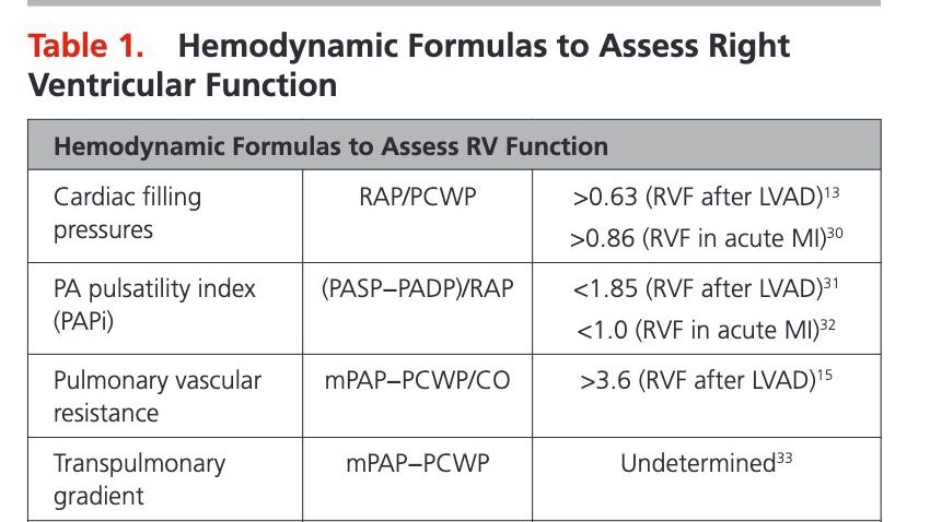
🫀What is PAPI?
PA Pulsatility Index = (PASP - PADP) / RAP
A quick invasive marker of RV function!
It reflects the RV’s ability to generate pulsatile flow vs its filling pressure.
So PAPI assesses the RV’s ability to generate pulsatile flow relative to its filling pressure.
📉 Low PAPI suggests RV failure:
<1.85 → RVF after LVAD
<1.0 → RVF in acute MI
🔍 Want to interpret invasive hemodynamics? Check out https://t.co/RDDlpd7VNb — an incredible resource by @BrototoD@ruey_hu@ElliottMillerMD@Gastanadui_MG @kiaragoldwag
#CardioNuggets #Hemodynamics #CardioTwitter #SwanGanz #MedEd
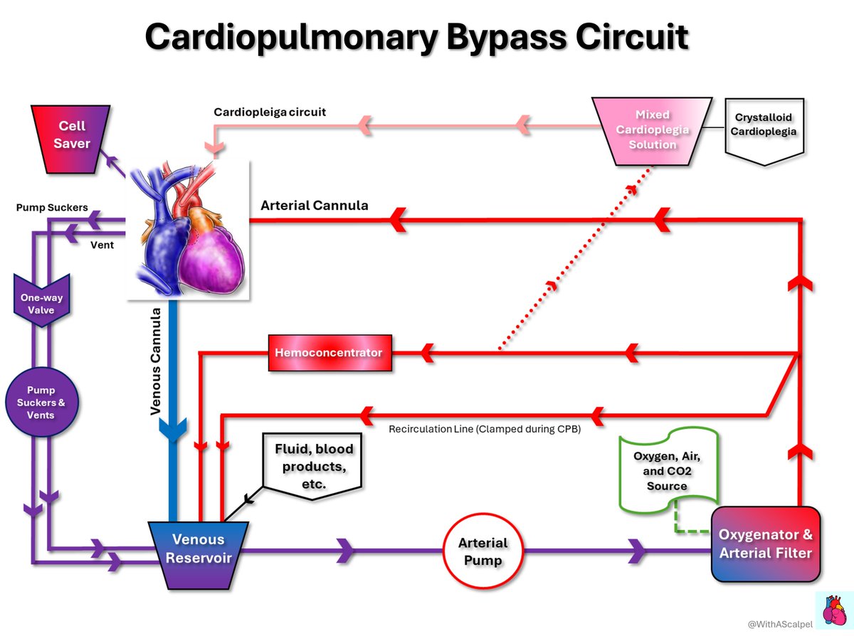
Cardiopulmonary Bypass Circuit🤖🫀🫁
👉Venous blood drains into the reservoir, then passes through the pump, oxygenator, and arterial filter before returning to the aorta.
👉The oxygenator provides gas exchange while the arterial filter helps remove microbubbles, making this segment central to gaseous microemboli control during CPB.
A practical review of pneumatosis intestinalis integrating imaging, labs and clinical evidence to distinguish benign from pathologic cases and guide bedside decision-making.
https://t.co/SNfO13b00F
What is VTI (Velocity Time Integral) in echocardiography?
VTI measures the distance a column of blood travels during systole through the LVOT. It’s a vital parameter to estimate stroke volume and cardiac output noninvasively.
How is VTI measured?
1️⃣ Obtain an apical 5-chamber view (5C).
2️⃣ Place PW Doppler just below the aortic valve (at LVOT).
3️⃣ Measure the area under the velocity curve during systole➡️ that’s the VTI (in cm).
Calculation of Stroke Volume (SV):
SV = VTI × LVOT area
LVOT area = π × (LVOT diameter/2)²
Requires accurate LVOT diameter from parasternal long-axis view.
Then,
Cardiac Output (CO) = SV × HR
♦️Normal LVOT VTI: ~18–22 cm (adults)
▶️A low VTI often means reduced stroke volume, requiring further hemodynamic assessment.
I do👂to most🧠neurology podcasts.
But this ⓵ is way beyond.
It takes us into the family rooms of the patients & then picks up the event from there.
Last Seen Normal.
The⏰ticks.
Every second.
#FAST: Awareness is the🔑
This is for public & neurologists in general🙏
Exemplary!


![MarlonVFZR's tweet photo. 🫀🚨 𝗜𝗻𝘀𝘂𝗳𝗶𝗰𝗶𝗲𝗻𝗰𝗶𝗮 𝗰𝗮𝗿𝗱𝗶𝗮𝗰𝗮 𝗮𝗴𝘂𝗱𝗮 ‼️
@JACCJournals @ACCinTouch
👇🏼👇🏼👇🏼👇🏼
📑🔗🔑🔓
https://t.co/89ht2mBRSA
⬇️⬇️⬇️⬇️
🧵👇
La revisión State-of-the-Art de 𝙅𝘼𝘾𝘾 𝟮𝟬𝟮𝟲 propone un cambio de paradigma:
💧 aliviar congestión rápidamente
🔍 identificar y tratar el precipitante
🧬 definir la cardiopatía subyacente
💊 iniciar precozmente terapia modificadora de enfermedad
📈 planear la trayectoria después del alta
El acrónimo 𝙎𝙋𝙇𝘼𝙎𝙃 resume la evaluación inicial:
𝙎ymptoms
𝙋ast history
𝙇ife signs
𝘼ssess congestion
𝙎TEMI/equivalente
𝙃ypoperfusion
🔥 La hospitalización es una 𝙫𝙚𝙣𝙩𝙖𝙣𝙖 𝙩𝙚𝙧𝙖𝙥𝙚́𝙪𝙩𝙞𝙘𝙖 𝙥𝙖𝙧𝙖 𝙢𝙤𝙙𝙞𝙛𝙞𝙘𝙖𝙧 𝙚𝙡 𝙥𝙧𝙤𝙣𝙤́𝙨𝙩𝙞𝙘𝙤, no simplemente para hacer desaparecer la disnea.
💉💧 𝙁𝙪𝙧𝙤𝙨𝙚𝙢𝙞𝙙𝙖: 𝙣𝙤 𝙗𝙖𝙨𝙩𝙖 𝙘𝙤𝙣 𝙖𝙙𝙢𝙞𝙣𝙞𝙨𝙩𝙧𝙖𝙧𝙡𝙖. 𝙃𝙖𝙮 𝙦𝙪𝙚 𝙨𝙖𝙗𝙚𝙧 𝙚𝙣 𝟮 𝙝𝙤𝙧𝙖𝙨 𝙨𝙞 𝙛𝙪𝙣𝙘𝙞𝙤𝙣𝙤́
Si existe alta sospecha de IC aguda con congestión, el artículo recomienda iniciar el diurético IV 𝙨𝙞𝙣 𝙚𝙨𝙥𝙚𝙧𝙖𝙧 𝙖 𝙘𝙤𝙢𝙥𝙡𝙚𝙩𝙖𝙧 𝙩𝙤𝙙𝙖𝙨 𝙡𝙖𝙨 𝙥𝙧𝙪𝙚𝙗𝙖𝙨 𝙙𝙞𝙖𝙜𝙣𝙤́𝙨𝙩𝙞𝙘𝙖𝙨.
💊 Sin tratamiento previo o dosis baja: 𝙛𝙪𝙧𝙤𝙨𝙚𝙢𝙞𝙙𝙖 𝟰𝟬 𝙢𝙜 𝙄𝙑
💊 Dosis oral crónica alta: ≥𝟴𝟬 𝙢𝙜 𝙄𝙑 𝙤 ≈𝟮× 𝙡𝙖 𝙙𝙤𝙨𝙞𝙨 𝙤𝙧𝙖𝙡
A las 𝟮 𝙝𝙤𝙧𝙖𝙨, evaluar:
✅ 𝘿𝙞𝙪𝙧𝙚𝙨𝙞𝙨 ≥𝟯𝟬𝟬 𝙢𝙇
o
✅ 𝙉𝙖 𝙪𝙧𝙞𝙣𝙖𝙧𝙞𝙤 ≥𝟳𝟬–𝟴𝟬 𝙢𝙢𝙤𝙡/𝙇
Si no se alcanza:
⬆️ duplicar la dosis de asa
➕ considerar 𝙖𝙘𝙚𝙩𝙖𝙯𝙤𝙡𝙖𝙢𝙞𝙙𝙖 𝙤 𝙩𝙞𝙖𝙯𝙞𝙙𝙖
🔍 excluir causas intrarrenales/postrenales
🔥 Esperar 24 horas para descubrir que el diurético no funcionó es perder tiempo de descongestión.
🫘📈 𝘾𝙧𝙚𝙖𝙩𝙞𝙣𝙞𝙣𝙖 𝙦𝙪𝙚 𝙖𝙪𝙢𝙚𝙣𝙩𝙖 𝙙𝙪𝙧𝙖𝙣𝙩𝙚 𝙪𝙣𝙖 𝙙𝙚𝙨𝙘𝙤𝙣𝙜𝙚𝙨𝙩𝙞𝙤́𝙣 𝙚𝙛𝙞𝙘𝙖𝙯 ≠ 𝙛𝙧𝙖𝙘𝙖𝙨𝙤 𝙩𝙚𝙧𝙖𝙥𝙚́𝙪𝙩𝙞𝙘𝙤
Uno de los errores más frecuentes es reducir o suspender diuréticos automáticamente ante una elevación moderada de creatinina.
La revisión enfatiza:
🫘 en ERC pueden necesitarse 𝙙𝙤𝙨𝙞𝙨 𝙢𝙖𝙮𝙤𝙧𝙚𝙨 de diuréticos
📈 la creatinina puede aumentar transitoriamente durante una descongestión efectiva
🚫 esto no obliga por sí solo a detener el tratamiento
🎯 el contexto de 𝙘𝙤𝙣𝙜𝙚𝙨𝙩𝙞𝙤́𝙣 𝙧𝙚𝙨𝙞𝙙𝙪𝙖𝙡 𝙮 𝙧𝙚𝙨𝙥𝙪𝙚𝙨𝙩𝙖 𝙙𝙞𝙪𝙧𝙚́𝙩𝙞𝙘𝙖 importa más que una cifra aislada
Además, la congestión persistente al alta se asocia con peor pronóstico.
📌 Una reducción de aproximadamente 𝟯𝟬% 𝙙𝙚𝙡 𝙉𝙏-𝙥𝙧𝙤𝘽𝙉𝙋 desde el ingreso al alta se considera compatible con descongestión adecuada dentro de una valoración multimodal.
🔥 En IC aguda, proteger la creatinina a costa de dejar al paciente congestionado puede ser una falsa victoria.
💊⚡ 𝙀𝙡 𝙜𝙧𝙖𝙣 𝙘𝙖𝙢𝙗𝙞𝙤: 𝙡𝙖 𝙂𝘿𝙈𝙏 𝙙𝙚𝙗𝙚 𝙘𝙤𝙢𝙚𝙣𝙯𝙖𝙧 𝙙𝙪𝙧𝙖𝙣𝙩𝙚 𝙡𝙖 𝙝𝙤𝙨𝙥𝙞𝙩𝙖𝙡𝙞𝙯𝙖𝙘𝙞𝙤́𝙣, 𝙣𝙤 “𝙖𝙡𝙜𝙪́𝙣 𝙙𝙞́𝙖” 𝙚𝙣 𝙘𝙤𝙣𝙨𝙪𝙡𝙩𝙖 𝙚𝙭𝙩𝙚𝙧𝙣𝙖
Secuencia rápida después de la estabilización:
🔹 𝙞𝙎𝙂𝙇𝙏𝟮 → desde los primeros días si eGFR ≥20 mL/min/1,73 m²
🔹 𝘼𝙍𝙈 → tempranamente si presión, función renal y K⁺ lo permiten
🔹 𝘼𝙍𝙉𝙄/𝙍𝘼𝙎𝙄 → tras estabilización, preferentemente con congestión leve o euvolemia
🔹 β-𝙗𝙡𝙤𝙦𝙪𝙚𝙖𝙙𝙤𝙧 → mantener si estable; iniciar en HFrEF cuando mejore la congestión
📊 La evidencia citada es potente:
⚡ 𝙋𝙄𝙊𝙉𝙀𝙀𝙍-𝙃𝙁: sacubitrilo/valsartán → ↓66% del endpoint clínico compuesto exploratorio
⚡ 𝙀𝙈𝙋𝙐𝙇𝙎𝙀: empagliflozina → ↓31% de muerte CV/eventos de IC
⚡ 𝙎𝙊𝙇𝙊𝙄𝙎𝙏-𝙒𝙃𝙁: sotagliflozina → ↓33%
⚡ 𝙎𝙏𝙍𝙊𝙉𝙂-𝙃𝙁: estrategia intensiva → ↓34% de muerte o rehospitalización por IC
🔥 El predictor más fuerte de que un paciente NO reciba GDMT después del alta es que 𝙣𝙤 𝙨𝙚 𝙡𝙖 𝙝𝙖𝙮𝙖𝙣 𝙞𝙣𝙞𝙘𝙞𝙖𝙙𝙤 𝙙𝙪𝙧𝙖𝙣𝙩𝙚 𝙡𝙖 𝙝𝙤𝙨𝙥𝙞𝙩𝙖𝙡𝙞𝙯𝙖𝙘𝙞𝙤́𝙣.
🎯 𝙀𝙡 𝙖𝙡𝙩𝙖 𝙣𝙤 𝙩𝙚𝙧𝙢𝙞𝙣𝙖 𝙚𝙡 𝙩𝙧𝙖𝙩𝙖𝙢𝙞𝙚𝙣𝙩𝙤: 𝙘𝙤𝙢𝙞𝙚𝙣𝙯𝙖 𝙡𝙖 “𝙛𝙖𝙨𝙚 𝙫𝙪𝙡𝙣𝙚𝙧𝙖𝙗𝙡𝙚”
La estrategia contemporánea exige salir del hospital con:
✅ 𝙚𝙪𝙫𝙤𝙡𝙚𝙢𝙞𝙖 𝙤 𝙙𝙚𝙨𝙘𝙤𝙣𝙜𝙚𝙨𝙩𝙞𝙤́𝙣 𝙤́𝙥𝙩𝙞𝙢𝙖
✅ GDMT iniciada y plan de titulación
✅ educación del paciente y cuidadores
✅ evaluación de hierro, función renal y comorbilidades
✅ plan para dispositivos cuando corresponda
✅ seguimiento 𝙨𝙚𝙢𝙖𝙣𝙖𝙡 𝙤 𝙘𝙖𝙙𝙖 𝟮 𝙨𝙚𝙢𝙖𝙣𝙖𝙨 𝙙𝙪𝙧𝙖𝙣𝙩𝙚 𝙚𝙡 𝙥𝙧𝙞𝙢𝙚𝙧 𝙢𝙚𝙨
Objetivo:
🚀 alcanzar la implementación completa y dosis toleradas de las terapias indicadas en 𝙨𝙚𝙢𝙖𝙣𝙖𝙨, idealmente antes de 𝟯 𝙢𝙚𝙨𝙚𝙨.
❌ paradigma antiguo:
𝙘𝙤𝙣𝙜𝙚𝙨𝙩𝙞𝙤́𝙣 → 𝙙𝙞𝙪𝙧𝙚́𝙩𝙞𝙘𝙤 → 𝙖𝙡𝙩𝙖 → 𝙣𝙪𝙚𝙫𝙖 𝙘𝙤𝙣𝙜𝙚𝙨𝙩𝙞𝙤́𝙣
✅ paradigma moderno:
𝙙𝙚𝙨𝙘𝙤𝙣𝙜𝙚𝙨𝙩𝙞𝙤́𝙣 → 𝙂𝘿𝙈𝙏 𝙧𝙖́𝙥𝙞𝙙𝙖 → 𝙧𝙚𝙢𝙤𝙙𝙚𝙡𝙖𝙙𝙤 𝙧𝙚𝙫𝙚𝙧𝙨𝙤 → 𝙢𝙚𝙣𝙤𝙧 𝙧𝙞𝙚𝙨𝙜𝙤 𝙙𝙚 𝙣𝙪𝙚𝙫𝙖 𝙄𝘾 → 𝙥𝙤𝙨𝙞𝙗𝙡𝙚 𝙧𝙚𝙢𝙞𝙨𝙞𝙤́𝙣
🔥 𝙈𝙚𝙣𝙨𝙖𝙟𝙚 𝙛𝙞𝙣𝙖𝙡: el tratamiento contemporáneo de la insuficiencia cardiaca aguda ya no termina cuando el paciente deja de tener edema. El verdadero objetivo es utilizar la hospitalización para 𝙩𝙧𝙖𝙣𝙨𝙛𝙤𝙧𝙢𝙖𝙧 𝙪𝙣𝙖 𝙙𝙚𝙨𝙘𝙤𝙢𝙥𝙚𝙣𝙨𝙖𝙘𝙞𝙤́𝙣 𝙖𝙜𝙪𝙙𝙖 𝙚𝙣 𝙚𝙡 𝙞𝙣𝙞𝙘𝙞𝙤 𝙙𝙚 𝙪𝙣𝙖 𝙚𝙨𝙩𝙧𝙖𝙩𝙚𝙜𝙞𝙖 𝙙𝙚 𝙧𝙚𝙢𝙞𝙨𝙞𝙤́𝙣 𝙙𝙚 𝙡𝙖 𝙚𝙣𝙛𝙚𝙧𝙢𝙚𝙙𝙖𝙙
📚📖 Más en 𝕏 @MarlonVFZR y en el blog 👉 [https://t.co/wbCJFKHCXL]
‼️Si te sirve: ❤️ Me gusta | 🔁 Repost | ➕ Follow para más👇🏼👇🏼👇🏼👇🏼
📚📖#ClubCrit👨🏻⚕️👨🏻🏫🧠🫶
#InsuficienciaCardiaca #AcuteHeartFailure #Cardiología
#FOAMed #FOAMcc #CriticalCare #CriticalCare #CuidadoCritico #MedTwitter #MedX #IntensiveCare #MedXCommunity #MedED](https://pbs.twimg.com/media/HPrz5WEXIAAJNs-.jpg)
![MarlonVFZR's tweet photo. 🫀🚨 𝗜𝗻𝘀𝘂𝗳𝗶𝗰𝗶𝗲𝗻𝗰𝗶𝗮 𝗰𝗮𝗿𝗱𝗶𝗮𝗰𝗮 𝗮𝗴𝘂𝗱𝗮 ‼️
@JACCJournals @ACCinTouch
👇🏼👇🏼👇🏼👇🏼
📑🔗🔑🔓
https://t.co/89ht2mBRSA
⬇️⬇️⬇️⬇️
🧵👇
La revisión State-of-the-Art de 𝙅𝘼𝘾𝘾 𝟮𝟬𝟮𝟲 propone un cambio de paradigma:
💧 aliviar congestión rápidamente
🔍 identificar y tratar el precipitante
🧬 definir la cardiopatía subyacente
💊 iniciar precozmente terapia modificadora de enfermedad
📈 planear la trayectoria después del alta
El acrónimo 𝙎𝙋𝙇𝘼𝙎𝙃 resume la evaluación inicial:
𝙎ymptoms
𝙋ast history
𝙇ife signs
𝘼ssess congestion
𝙎TEMI/equivalente
𝙃ypoperfusion
🔥 La hospitalización es una 𝙫𝙚𝙣𝙩𝙖𝙣𝙖 𝙩𝙚𝙧𝙖𝙥𝙚́𝙪𝙩𝙞𝙘𝙖 𝙥𝙖𝙧𝙖 𝙢𝙤𝙙𝙞𝙛𝙞𝙘𝙖𝙧 𝙚𝙡 𝙥𝙧𝙤𝙣𝙤́𝙨𝙩𝙞𝙘𝙤, no simplemente para hacer desaparecer la disnea.
💉💧 𝙁𝙪𝙧𝙤𝙨𝙚𝙢𝙞𝙙𝙖: 𝙣𝙤 𝙗𝙖𝙨𝙩𝙖 𝙘𝙤𝙣 𝙖𝙙𝙢𝙞𝙣𝙞𝙨𝙩𝙧𝙖𝙧𝙡𝙖. 𝙃𝙖𝙮 𝙦𝙪𝙚 𝙨𝙖𝙗𝙚𝙧 𝙚𝙣 𝟮 𝙝𝙤𝙧𝙖𝙨 𝙨𝙞 𝙛𝙪𝙣𝙘𝙞𝙤𝙣𝙤́
Si existe alta sospecha de IC aguda con congestión, el artículo recomienda iniciar el diurético IV 𝙨𝙞𝙣 𝙚𝙨𝙥𝙚𝙧𝙖𝙧 𝙖 𝙘𝙤𝙢𝙥𝙡𝙚𝙩𝙖𝙧 𝙩𝙤𝙙𝙖𝙨 𝙡𝙖𝙨 𝙥𝙧𝙪𝙚𝙗𝙖𝙨 𝙙𝙞𝙖𝙜𝙣𝙤́𝙨𝙩𝙞𝙘𝙖𝙨.
💊 Sin tratamiento previo o dosis baja: 𝙛𝙪𝙧𝙤𝙨𝙚𝙢𝙞𝙙𝙖 𝟰𝟬 𝙢𝙜 𝙄𝙑
💊 Dosis oral crónica alta: ≥𝟴𝟬 𝙢𝙜 𝙄𝙑 𝙤 ≈𝟮× 𝙡𝙖 𝙙𝙤𝙨𝙞𝙨 𝙤𝙧𝙖𝙡
A las 𝟮 𝙝𝙤𝙧𝙖𝙨, evaluar:
✅ 𝘿𝙞𝙪𝙧𝙚𝙨𝙞𝙨 ≥𝟯𝟬𝟬 𝙢𝙇
o
✅ 𝙉𝙖 𝙪𝙧𝙞𝙣𝙖𝙧𝙞𝙤 ≥𝟳𝟬–𝟴𝟬 𝙢𝙢𝙤𝙡/𝙇
Si no se alcanza:
⬆️ duplicar la dosis de asa
➕ considerar 𝙖𝙘𝙚𝙩𝙖𝙯𝙤𝙡𝙖𝙢𝙞𝙙𝙖 𝙤 𝙩𝙞𝙖𝙯𝙞𝙙𝙖
🔍 excluir causas intrarrenales/postrenales
🔥 Esperar 24 horas para descubrir que el diurético no funcionó es perder tiempo de descongestión.
🫘📈 𝘾𝙧𝙚𝙖𝙩𝙞𝙣𝙞𝙣𝙖 𝙦𝙪𝙚 𝙖𝙪𝙢𝙚𝙣𝙩𝙖 𝙙𝙪𝙧𝙖𝙣𝙩𝙚 𝙪𝙣𝙖 𝙙𝙚𝙨𝙘𝙤𝙣𝙜𝙚𝙨𝙩𝙞𝙤́𝙣 𝙚𝙛𝙞𝙘𝙖𝙯 ≠ 𝙛𝙧𝙖𝙘𝙖𝙨𝙤 𝙩𝙚𝙧𝙖𝙥𝙚́𝙪𝙩𝙞𝙘𝙤
Uno de los errores más frecuentes es reducir o suspender diuréticos automáticamente ante una elevación moderada de creatinina.
La revisión enfatiza:
🫘 en ERC pueden necesitarse 𝙙𝙤𝙨𝙞𝙨 𝙢𝙖𝙮𝙤𝙧𝙚𝙨 de diuréticos
📈 la creatinina puede aumentar transitoriamente durante una descongestión efectiva
🚫 esto no obliga por sí solo a detener el tratamiento
🎯 el contexto de 𝙘𝙤𝙣𝙜𝙚𝙨𝙩𝙞𝙤́𝙣 𝙧𝙚𝙨𝙞𝙙𝙪𝙖𝙡 𝙮 𝙧𝙚𝙨𝙥𝙪𝙚𝙨𝙩𝙖 𝙙𝙞𝙪𝙧𝙚́𝙩𝙞𝙘𝙖 importa más que una cifra aislada
Además, la congestión persistente al alta se asocia con peor pronóstico.
📌 Una reducción de aproximadamente 𝟯𝟬% 𝙙𝙚𝙡 𝙉𝙏-𝙥𝙧𝙤𝘽𝙉𝙋 desde el ingreso al alta se considera compatible con descongestión adecuada dentro de una valoración multimodal.
🔥 En IC aguda, proteger la creatinina a costa de dejar al paciente congestionado puede ser una falsa victoria.
💊⚡ 𝙀𝙡 𝙜𝙧𝙖𝙣 𝙘𝙖𝙢𝙗𝙞𝙤: 𝙡𝙖 𝙂𝘿𝙈𝙏 𝙙𝙚𝙗𝙚 𝙘𝙤𝙢𝙚𝙣𝙯𝙖𝙧 𝙙𝙪𝙧𝙖𝙣𝙩𝙚 𝙡𝙖 𝙝𝙤𝙨𝙥𝙞𝙩𝙖𝙡𝙞𝙯𝙖𝙘𝙞𝙤́𝙣, 𝙣𝙤 “𝙖𝙡𝙜𝙪́𝙣 𝙙𝙞́𝙖” 𝙚𝙣 𝙘𝙤𝙣𝙨𝙪𝙡𝙩𝙖 𝙚𝙭𝙩𝙚𝙧𝙣𝙖
Secuencia rápida después de la estabilización:
🔹 𝙞𝙎𝙂𝙇𝙏𝟮 → desde los primeros días si eGFR ≥20 mL/min/1,73 m²
🔹 𝘼𝙍𝙈 → tempranamente si presión, función renal y K⁺ lo permiten
🔹 𝘼𝙍𝙉𝙄/𝙍𝘼𝙎𝙄 → tras estabilización, preferentemente con congestión leve o euvolemia
🔹 β-𝙗𝙡𝙤𝙦𝙪𝙚𝙖𝙙𝙤𝙧 → mantener si estable; iniciar en HFrEF cuando mejore la congestión
📊 La evidencia citada es potente:
⚡ 𝙋𝙄𝙊𝙉𝙀𝙀𝙍-𝙃𝙁: sacubitrilo/valsartán → ↓66% del endpoint clínico compuesto exploratorio
⚡ 𝙀𝙈𝙋𝙐𝙇𝙎𝙀: empagliflozina → ↓31% de muerte CV/eventos de IC
⚡ 𝙎𝙊𝙇𝙊𝙄𝙎𝙏-𝙒𝙃𝙁: sotagliflozina → ↓33%
⚡ 𝙎𝙏𝙍𝙊𝙉𝙂-𝙃𝙁: estrategia intensiva → ↓34% de muerte o rehospitalización por IC
🔥 El predictor más fuerte de que un paciente NO reciba GDMT después del alta es que 𝙣𝙤 𝙨𝙚 𝙡𝙖 𝙝𝙖𝙮𝙖𝙣 𝙞𝙣𝙞𝙘𝙞𝙖𝙙𝙤 𝙙𝙪𝙧𝙖𝙣𝙩𝙚 𝙡𝙖 𝙝𝙤𝙨𝙥𝙞𝙩𝙖𝙡𝙞𝙯𝙖𝙘𝙞𝙤́𝙣.
🎯 𝙀𝙡 𝙖𝙡𝙩𝙖 𝙣𝙤 𝙩𝙚𝙧𝙢𝙞𝙣𝙖 𝙚𝙡 𝙩𝙧𝙖𝙩𝙖𝙢𝙞𝙚𝙣𝙩𝙤: 𝙘𝙤𝙢𝙞𝙚𝙣𝙯𝙖 𝙡𝙖 “𝙛𝙖𝙨𝙚 𝙫𝙪𝙡𝙣𝙚𝙧𝙖𝙗𝙡𝙚”
La estrategia contemporánea exige salir del hospital con:
✅ 𝙚𝙪𝙫𝙤𝙡𝙚𝙢𝙞𝙖 𝙤 𝙙𝙚𝙨𝙘𝙤𝙣𝙜𝙚𝙨𝙩𝙞𝙤́𝙣 𝙤́𝙥𝙩𝙞𝙢𝙖
✅ GDMT iniciada y plan de titulación
✅ educación del paciente y cuidadores
✅ evaluación de hierro, función renal y comorbilidades
✅ plan para dispositivos cuando corresponda
✅ seguimiento 𝙨𝙚𝙢𝙖𝙣𝙖𝙡 𝙤 𝙘𝙖𝙙𝙖 𝟮 𝙨𝙚𝙢𝙖𝙣𝙖𝙨 𝙙𝙪𝙧𝙖𝙣𝙩𝙚 𝙚𝙡 𝙥𝙧𝙞𝙢𝙚𝙧 𝙢𝙚𝙨
Objetivo:
🚀 alcanzar la implementación completa y dosis toleradas de las terapias indicadas en 𝙨𝙚𝙢𝙖𝙣𝙖𝙨, idealmente antes de 𝟯 𝙢𝙚𝙨𝙚𝙨.
❌ paradigma antiguo:
𝙘𝙤𝙣𝙜𝙚𝙨𝙩𝙞𝙤́𝙣 → 𝙙𝙞𝙪𝙧𝙚́𝙩𝙞𝙘𝙤 → 𝙖𝙡𝙩𝙖 → 𝙣𝙪𝙚𝙫𝙖 𝙘𝙤𝙣𝙜𝙚𝙨𝙩𝙞𝙤́𝙣
✅ paradigma moderno:
𝙙𝙚𝙨𝙘𝙤𝙣𝙜𝙚𝙨𝙩𝙞𝙤́𝙣 → 𝙂𝘿𝙈𝙏 𝙧𝙖́𝙥𝙞𝙙𝙖 → 𝙧𝙚𝙢𝙤𝙙𝙚𝙡𝙖𝙙𝙤 𝙧𝙚𝙫𝙚𝙧𝙨𝙤 → 𝙢𝙚𝙣𝙤𝙧 𝙧𝙞𝙚𝙨𝙜𝙤 𝙙𝙚 𝙣𝙪𝙚𝙫𝙖 𝙄𝘾 → 𝙥𝙤𝙨𝙞𝙗𝙡𝙚 𝙧𝙚𝙢𝙞𝙨𝙞𝙤́𝙣
🔥 𝙈𝙚𝙣𝙨𝙖𝙟𝙚 𝙛𝙞𝙣𝙖𝙡: el tratamiento contemporáneo de la insuficiencia cardiaca aguda ya no termina cuando el paciente deja de tener edema. El verdadero objetivo es utilizar la hospitalización para 𝙩𝙧𝙖𝙣𝙨𝙛𝙤𝙧𝙢𝙖𝙧 𝙪𝙣𝙖 𝙙𝙚𝙨𝙘𝙤𝙢𝙥𝙚𝙣𝙨𝙖𝙘𝙞𝙤́𝙣 𝙖𝙜𝙪𝙙𝙖 𝙚𝙣 𝙚𝙡 𝙞𝙣𝙞𝙘𝙞𝙤 𝙙𝙚 𝙪𝙣𝙖 𝙚𝙨𝙩𝙧𝙖𝙩𝙚𝙜𝙞𝙖 𝙙𝙚 𝙧𝙚𝙢𝙞𝙨𝙞𝙤́𝙣 𝙙𝙚 𝙡𝙖 𝙚𝙣𝙛𝙚𝙧𝙢𝙚𝙙𝙖𝙙
📚📖 Más en 𝕏 @MarlonVFZR y en el blog 👉 [https://t.co/wbCJFKHCXL]
‼️Si te sirve: ❤️ Me gusta | 🔁 Repost | ➕ Follow para más👇🏼👇🏼👇🏼👇🏼
📚📖#ClubCrit👨🏻⚕️👨🏻🏫🧠🫶
#InsuficienciaCardiaca #AcuteHeartFailure #Cardiología
#FOAMed #FOAMcc #CriticalCare #CriticalCare #CuidadoCritico #MedTwitter #MedX #IntensiveCare #MedXCommunity #MedED](https://pbs.twimg.com/media/HPrz5TIWUAEAHUa.jpg)
![MarlonVFZR's tweet photo. 🫀🚨 𝗜𝗻𝘀𝘂𝗳𝗶𝗰𝗶𝗲𝗻𝗰𝗶𝗮 𝗰𝗮𝗿𝗱𝗶𝗮𝗰𝗮 𝗮𝗴𝘂𝗱𝗮 ‼️
@JACCJournals @ACCinTouch
👇🏼👇🏼👇🏼👇🏼
📑🔗🔑🔓
https://t.co/89ht2mBRSA
⬇️⬇️⬇️⬇️
🧵👇
La revisión State-of-the-Art de 𝙅𝘼𝘾𝘾 𝟮𝟬𝟮𝟲 propone un cambio de paradigma:
💧 aliviar congestión rápidamente
🔍 identificar y tratar el precipitante
🧬 definir la cardiopatía subyacente
💊 iniciar precozmente terapia modificadora de enfermedad
📈 planear la trayectoria después del alta
El acrónimo 𝙎𝙋𝙇𝘼𝙎𝙃 resume la evaluación inicial:
𝙎ymptoms
𝙋ast history
𝙇ife signs
𝘼ssess congestion
𝙎TEMI/equivalente
𝙃ypoperfusion
🔥 La hospitalización es una 𝙫𝙚𝙣𝙩𝙖𝙣𝙖 𝙩𝙚𝙧𝙖𝙥𝙚́𝙪𝙩𝙞𝙘𝙖 𝙥𝙖𝙧𝙖 𝙢𝙤𝙙𝙞𝙛𝙞𝙘𝙖𝙧 𝙚𝙡 𝙥𝙧𝙤𝙣𝙤́𝙨𝙩𝙞𝙘𝙤, no simplemente para hacer desaparecer la disnea.
💉💧 𝙁𝙪𝙧𝙤𝙨𝙚𝙢𝙞𝙙𝙖: 𝙣𝙤 𝙗𝙖𝙨𝙩𝙖 𝙘𝙤𝙣 𝙖𝙙𝙢𝙞𝙣𝙞𝙨𝙩𝙧𝙖𝙧𝙡𝙖. 𝙃𝙖𝙮 𝙦𝙪𝙚 𝙨𝙖𝙗𝙚𝙧 𝙚𝙣 𝟮 𝙝𝙤𝙧𝙖𝙨 𝙨𝙞 𝙛𝙪𝙣𝙘𝙞𝙤𝙣𝙤́
Si existe alta sospecha de IC aguda con congestión, el artículo recomienda iniciar el diurético IV 𝙨𝙞𝙣 𝙚𝙨𝙥𝙚𝙧𝙖𝙧 𝙖 𝙘𝙤𝙢𝙥𝙡𝙚𝙩𝙖𝙧 𝙩𝙤𝙙𝙖𝙨 𝙡𝙖𝙨 𝙥𝙧𝙪𝙚𝙗𝙖𝙨 𝙙𝙞𝙖𝙜𝙣𝙤́𝙨𝙩𝙞𝙘𝙖𝙨.
💊 Sin tratamiento previo o dosis baja: 𝙛𝙪𝙧𝙤𝙨𝙚𝙢𝙞𝙙𝙖 𝟰𝟬 𝙢𝙜 𝙄𝙑
💊 Dosis oral crónica alta: ≥𝟴𝟬 𝙢𝙜 𝙄𝙑 𝙤 ≈𝟮× 𝙡𝙖 𝙙𝙤𝙨𝙞𝙨 𝙤𝙧𝙖𝙡
A las 𝟮 𝙝𝙤𝙧𝙖𝙨, evaluar:
✅ 𝘿𝙞𝙪𝙧𝙚𝙨𝙞𝙨 ≥𝟯𝟬𝟬 𝙢𝙇
o
✅ 𝙉𝙖 𝙪𝙧𝙞𝙣𝙖𝙧𝙞𝙤 ≥𝟳𝟬–𝟴𝟬 𝙢𝙢𝙤𝙡/𝙇
Si no se alcanza:
⬆️ duplicar la dosis de asa
➕ considerar 𝙖𝙘𝙚𝙩𝙖𝙯𝙤𝙡𝙖𝙢𝙞𝙙𝙖 𝙤 𝙩𝙞𝙖𝙯𝙞𝙙𝙖
🔍 excluir causas intrarrenales/postrenales
🔥 Esperar 24 horas para descubrir que el diurético no funcionó es perder tiempo de descongestión.
🫘📈 𝘾𝙧𝙚𝙖𝙩𝙞𝙣𝙞𝙣𝙖 𝙦𝙪𝙚 𝙖𝙪𝙢𝙚𝙣𝙩𝙖 𝙙𝙪𝙧𝙖𝙣𝙩𝙚 𝙪𝙣𝙖 𝙙𝙚𝙨𝙘𝙤𝙣𝙜𝙚𝙨𝙩𝙞𝙤́𝙣 𝙚𝙛𝙞𝙘𝙖𝙯 ≠ 𝙛𝙧𝙖𝙘𝙖𝙨𝙤 𝙩𝙚𝙧𝙖𝙥𝙚́𝙪𝙩𝙞𝙘𝙤
Uno de los errores más frecuentes es reducir o suspender diuréticos automáticamente ante una elevación moderada de creatinina.
La revisión enfatiza:
🫘 en ERC pueden necesitarse 𝙙𝙤𝙨𝙞𝙨 𝙢𝙖𝙮𝙤𝙧𝙚𝙨 de diuréticos
📈 la creatinina puede aumentar transitoriamente durante una descongestión efectiva
🚫 esto no obliga por sí solo a detener el tratamiento
🎯 el contexto de 𝙘𝙤𝙣𝙜𝙚𝙨𝙩𝙞𝙤́𝙣 𝙧𝙚𝙨𝙞𝙙𝙪𝙖𝙡 𝙮 𝙧𝙚𝙨𝙥𝙪𝙚𝙨𝙩𝙖 𝙙𝙞𝙪𝙧𝙚́𝙩𝙞𝙘𝙖 importa más que una cifra aislada
Además, la congestión persistente al alta se asocia con peor pronóstico.
📌 Una reducción de aproximadamente 𝟯𝟬% 𝙙𝙚𝙡 𝙉𝙏-𝙥𝙧𝙤𝘽𝙉𝙋 desde el ingreso al alta se considera compatible con descongestión adecuada dentro de una valoración multimodal.
🔥 En IC aguda, proteger la creatinina a costa de dejar al paciente congestionado puede ser una falsa victoria.
💊⚡ 𝙀𝙡 𝙜𝙧𝙖𝙣 𝙘𝙖𝙢𝙗𝙞𝙤: 𝙡𝙖 𝙂𝘿𝙈𝙏 𝙙𝙚𝙗𝙚 𝙘𝙤𝙢𝙚𝙣𝙯𝙖𝙧 𝙙𝙪𝙧𝙖𝙣𝙩𝙚 𝙡𝙖 𝙝𝙤𝙨𝙥𝙞𝙩𝙖𝙡𝙞𝙯𝙖𝙘𝙞𝙤́𝙣, 𝙣𝙤 “𝙖𝙡𝙜𝙪́𝙣 𝙙𝙞́𝙖” 𝙚𝙣 𝙘𝙤𝙣𝙨𝙪𝙡𝙩𝙖 𝙚𝙭𝙩𝙚𝙧𝙣𝙖
Secuencia rápida después de la estabilización:
🔹 𝙞𝙎𝙂𝙇𝙏𝟮 → desde los primeros días si eGFR ≥20 mL/min/1,73 m²
🔹 𝘼𝙍𝙈 → tempranamente si presión, función renal y K⁺ lo permiten
🔹 𝘼𝙍𝙉𝙄/𝙍𝘼𝙎𝙄 → tras estabilización, preferentemente con congestión leve o euvolemia
🔹 β-𝙗𝙡𝙤𝙦𝙪𝙚𝙖𝙙𝙤𝙧 → mantener si estable; iniciar en HFrEF cuando mejore la congestión
📊 La evidencia citada es potente:
⚡ 𝙋𝙄𝙊𝙉𝙀𝙀𝙍-𝙃𝙁: sacubitrilo/valsartán → ↓66% del endpoint clínico compuesto exploratorio
⚡ 𝙀𝙈𝙋𝙐𝙇𝙎𝙀: empagliflozina → ↓31% de muerte CV/eventos de IC
⚡ 𝙎𝙊𝙇𝙊𝙄𝙎𝙏-𝙒𝙃𝙁: sotagliflozina → ↓33%
⚡ 𝙎𝙏𝙍𝙊𝙉𝙂-𝙃𝙁: estrategia intensiva → ↓34% de muerte o rehospitalización por IC
🔥 El predictor más fuerte de que un paciente NO reciba GDMT después del alta es que 𝙣𝙤 𝙨𝙚 𝙡𝙖 𝙝𝙖𝙮𝙖𝙣 𝙞𝙣𝙞𝙘𝙞𝙖𝙙𝙤 𝙙𝙪𝙧𝙖𝙣𝙩𝙚 𝙡𝙖 𝙝𝙤𝙨𝙥𝙞𝙩𝙖𝙡𝙞𝙯𝙖𝙘𝙞𝙤́𝙣.
🎯 𝙀𝙡 𝙖𝙡𝙩𝙖 𝙣𝙤 𝙩𝙚𝙧𝙢𝙞𝙣𝙖 𝙚𝙡 𝙩𝙧𝙖𝙩𝙖𝙢𝙞𝙚𝙣𝙩𝙤: 𝙘𝙤𝙢𝙞𝙚𝙣𝙯𝙖 𝙡𝙖 “𝙛𝙖𝙨𝙚 𝙫𝙪𝙡𝙣𝙚𝙧𝙖𝙗𝙡𝙚”
La estrategia contemporánea exige salir del hospital con:
✅ 𝙚𝙪𝙫𝙤𝙡𝙚𝙢𝙞𝙖 𝙤 𝙙𝙚𝙨𝙘𝙤𝙣𝙜𝙚𝙨𝙩𝙞𝙤́𝙣 𝙤́𝙥𝙩𝙞𝙢𝙖
✅ GDMT iniciada y plan de titulación
✅ educación del paciente y cuidadores
✅ evaluación de hierro, función renal y comorbilidades
✅ plan para dispositivos cuando corresponda
✅ seguimiento 𝙨𝙚𝙢𝙖𝙣𝙖𝙡 𝙤 𝙘𝙖𝙙𝙖 𝟮 𝙨𝙚𝙢𝙖𝙣𝙖𝙨 𝙙𝙪𝙧𝙖𝙣𝙩𝙚 𝙚𝙡 𝙥𝙧𝙞𝙢𝙚𝙧 𝙢𝙚𝙨
Objetivo:
🚀 alcanzar la implementación completa y dosis toleradas de las terapias indicadas en 𝙨𝙚𝙢𝙖𝙣𝙖𝙨, idealmente antes de 𝟯 𝙢𝙚𝙨𝙚𝙨.
❌ paradigma antiguo:
𝙘𝙤𝙣𝙜𝙚𝙨𝙩𝙞𝙤́𝙣 → 𝙙𝙞𝙪𝙧𝙚́𝙩𝙞𝙘𝙤 → 𝙖𝙡𝙩𝙖 → 𝙣𝙪𝙚𝙫𝙖 𝙘𝙤𝙣𝙜𝙚𝙨𝙩𝙞𝙤́𝙣
✅ paradigma moderno:
𝙙𝙚𝙨𝙘𝙤𝙣𝙜𝙚𝙨𝙩𝙞𝙤́𝙣 → 𝙂𝘿𝙈𝙏 𝙧𝙖́𝙥𝙞𝙙𝙖 → 𝙧𝙚𝙢𝙤𝙙𝙚𝙡𝙖𝙙𝙤 𝙧𝙚𝙫𝙚𝙧𝙨𝙤 → 𝙢𝙚𝙣𝙤𝙧 𝙧𝙞𝙚𝙨𝙜𝙤 𝙙𝙚 𝙣𝙪𝙚𝙫𝙖 𝙄𝘾 → 𝙥𝙤𝙨𝙞𝙗𝙡𝙚 𝙧𝙚𝙢𝙞𝙨𝙞𝙤́𝙣
🔥 𝙈𝙚𝙣𝙨𝙖𝙟𝙚 𝙛𝙞𝙣𝙖𝙡: el tratamiento contemporáneo de la insuficiencia cardiaca aguda ya no termina cuando el paciente deja de tener edema. El verdadero objetivo es utilizar la hospitalización para 𝙩𝙧𝙖𝙣𝙨𝙛𝙤𝙧𝙢𝙖𝙧 𝙪𝙣𝙖 𝙙𝙚𝙨𝙘𝙤𝙢𝙥𝙚𝙣𝙨𝙖𝙘𝙞𝙤́𝙣 𝙖𝙜𝙪𝙙𝙖 𝙚𝙣 𝙚𝙡 𝙞𝙣𝙞𝙘𝙞𝙤 𝙙𝙚 𝙪𝙣𝙖 𝙚𝙨𝙩𝙧𝙖𝙩𝙚𝙜𝙞𝙖 𝙙𝙚 𝙧𝙚𝙢𝙞𝙨𝙞𝙤́𝙣 𝙙𝙚 𝙡𝙖 𝙚𝙣𝙛𝙚𝙧𝙢𝙚𝙙𝙖𝙙
📚📖 Más en 𝕏 @MarlonVFZR y en el blog 👉 [https://t.co/wbCJFKHCXL]
‼️Si te sirve: ❤️ Me gusta | 🔁 Repost | ➕ Follow para más👇🏼👇🏼👇🏼👇🏼
📚📖#ClubCrit👨🏻⚕️👨🏻🏫🧠🫶
#InsuficienciaCardiaca #AcuteHeartFailure #Cardiología
#FOAMed #FOAMcc #CriticalCare #CriticalCare #CuidadoCritico #MedTwitter #MedX #IntensiveCare #MedXCommunity #MedED](https://pbs.twimg.com/media/HPrz5R0W8AAWBvQ.jpg)












![MarlonVFZR's tweet photo. 🫀🚨 𝗜𝗻𝘀𝘂𝗳𝗶𝗰𝗶𝗲𝗻𝗰𝗶𝗮 𝗰𝗮𝗿𝗱𝗶𝗮𝗰𝗮 𝗮𝗴𝘂𝗱𝗮 ‼️
@JACCJournals @ACCinTouch
👇🏼👇🏼👇🏼👇🏼
📑🔗🔑🔓
https://t.co/89ht2mBRSA
⬇️⬇️⬇️⬇️
🧵👇
La revisión State-of-the-Art de 𝙅𝘼𝘾𝘾 𝟮𝟬𝟮𝟲 propone un cambio de paradigma:
💧 aliviar congestión rápidamente
🔍 identificar y tratar el precipitante
🧬 definir la cardiopatía subyacente
💊 iniciar precozmente terapia modificadora de enfermedad
📈 planear la trayectoria después del alta
El acrónimo 𝙎𝙋𝙇𝘼𝙎𝙃 resume la evaluación inicial:
𝙎ymptoms
𝙋ast history
𝙇ife signs
𝘼ssess congestion
𝙎TEMI/equivalente
𝙃ypoperfusion
🔥 La hospitalización es una 𝙫𝙚𝙣𝙩𝙖𝙣𝙖 𝙩𝙚𝙧𝙖𝙥𝙚́𝙪𝙩𝙞𝙘𝙖 𝙥𝙖𝙧𝙖 𝙢𝙤𝙙𝙞𝙛𝙞𝙘𝙖𝙧 𝙚𝙡 𝙥𝙧𝙤𝙣𝙤́𝙨𝙩𝙞𝙘𝙤, no simplemente para hacer desaparecer la disnea.
💉💧 𝙁𝙪𝙧𝙤𝙨𝙚𝙢𝙞𝙙𝙖: 𝙣𝙤 𝙗𝙖𝙨𝙩𝙖 𝙘𝙤𝙣 𝙖𝙙𝙢𝙞𝙣𝙞𝙨𝙩𝙧𝙖𝙧𝙡𝙖. 𝙃𝙖𝙮 𝙦𝙪𝙚 𝙨𝙖𝙗𝙚𝙧 𝙚𝙣 𝟮 𝙝𝙤𝙧𝙖𝙨 𝙨𝙞 𝙛𝙪𝙣𝙘𝙞𝙤𝙣𝙤́
Si existe alta sospecha de IC aguda con congestión, el artículo recomienda iniciar el diurético IV 𝙨𝙞𝙣 𝙚𝙨𝙥𝙚𝙧𝙖𝙧 𝙖 𝙘𝙤𝙢𝙥𝙡𝙚𝙩𝙖𝙧 𝙩𝙤𝙙𝙖𝙨 𝙡𝙖𝙨 𝙥𝙧𝙪𝙚𝙗𝙖𝙨 𝙙𝙞𝙖𝙜𝙣𝙤́𝙨𝙩𝙞𝙘𝙖𝙨.
💊 Sin tratamiento previo o dosis baja: 𝙛𝙪𝙧𝙤𝙨𝙚𝙢𝙞𝙙𝙖 𝟰𝟬 𝙢𝙜 𝙄𝙑
💊 Dosis oral crónica alta: ≥𝟴𝟬 𝙢𝙜 𝙄𝙑 𝙤 ≈𝟮× 𝙡𝙖 𝙙𝙤𝙨𝙞𝙨 𝙤𝙧𝙖𝙡
A las 𝟮 𝙝𝙤𝙧𝙖𝙨, evaluar:
✅ 𝘿𝙞𝙪𝙧𝙚𝙨𝙞𝙨 ≥𝟯𝟬𝟬 𝙢𝙇
o
✅ 𝙉𝙖 𝙪𝙧𝙞𝙣𝙖𝙧𝙞𝙤 ≥𝟳𝟬–𝟴𝟬 𝙢𝙢𝙤𝙡/𝙇
Si no se alcanza:
⬆️ duplicar la dosis de asa
➕ considerar 𝙖𝙘𝙚𝙩𝙖𝙯𝙤𝙡𝙖𝙢𝙞𝙙𝙖 𝙤 𝙩𝙞𝙖𝙯𝙞𝙙𝙖
🔍 excluir causas intrarrenales/postrenales
🔥 Esperar 24 horas para descubrir que el diurético no funcionó es perder tiempo de descongestión.
🫘📈 𝘾𝙧𝙚𝙖𝙩𝙞𝙣𝙞𝙣𝙖 𝙦𝙪𝙚 𝙖𝙪𝙢𝙚𝙣𝙩𝙖 𝙙𝙪𝙧𝙖𝙣𝙩𝙚 𝙪𝙣𝙖 𝙙𝙚𝙨𝙘𝙤𝙣𝙜𝙚𝙨𝙩𝙞𝙤́𝙣 𝙚𝙛𝙞𝙘𝙖𝙯 ≠ 𝙛𝙧𝙖𝙘𝙖𝙨𝙤 𝙩𝙚𝙧𝙖𝙥𝙚́𝙪𝙩𝙞𝙘𝙤
Uno de los errores más frecuentes es reducir o suspender diuréticos automáticamente ante una elevación moderada de creatinina.
La revisión enfatiza:
🫘 en ERC pueden necesitarse 𝙙𝙤𝙨𝙞𝙨 𝙢𝙖𝙮𝙤𝙧𝙚𝙨 de diuréticos
📈 la creatinina puede aumentar transitoriamente durante una descongestión efectiva
🚫 esto no obliga por sí solo a detener el tratamiento
🎯 el contexto de 𝙘𝙤𝙣𝙜𝙚𝙨𝙩𝙞𝙤́𝙣 𝙧𝙚𝙨𝙞𝙙𝙪𝙖𝙡 𝙮 𝙧𝙚𝙨𝙥𝙪𝙚𝙨𝙩𝙖 𝙙𝙞𝙪𝙧𝙚́𝙩𝙞𝙘𝙖 importa más que una cifra aislada
Además, la congestión persistente al alta se asocia con peor pronóstico.
📌 Una reducción de aproximadamente 𝟯𝟬% 𝙙𝙚𝙡 𝙉𝙏-𝙥𝙧𝙤𝘽𝙉𝙋 desde el ingreso al alta se considera compatible con descongestión adecuada dentro de una valoración multimodal.
🔥 En IC aguda, proteger la creatinina a costa de dejar al paciente congestionado puede ser una falsa victoria.
💊⚡ 𝙀𝙡 𝙜𝙧𝙖𝙣 𝙘𝙖𝙢𝙗𝙞𝙤: 𝙡𝙖 𝙂𝘿𝙈𝙏 𝙙𝙚𝙗𝙚 𝙘𝙤𝙢𝙚𝙣𝙯𝙖𝙧 𝙙𝙪𝙧𝙖𝙣𝙩𝙚 𝙡𝙖 𝙝𝙤𝙨𝙥𝙞𝙩𝙖𝙡𝙞𝙯𝙖𝙘𝙞𝙤́𝙣, 𝙣𝙤 “𝙖𝙡𝙜𝙪́𝙣 𝙙𝙞́𝙖” 𝙚𝙣 𝙘𝙤𝙣𝙨𝙪𝙡𝙩𝙖 𝙚𝙭𝙩𝙚𝙧𝙣𝙖
Secuencia rápida después de la estabilización:
🔹 𝙞𝙎𝙂𝙇𝙏𝟮 → desde los primeros días si eGFR ≥20 mL/min/1,73 m²
🔹 𝘼𝙍𝙈 → tempranamente si presión, función renal y K⁺ lo permiten
🔹 𝘼𝙍𝙉𝙄/𝙍𝘼𝙎𝙄 → tras estabilización, preferentemente con congestión leve o euvolemia
🔹 β-𝙗𝙡𝙤𝙦𝙪𝙚𝙖𝙙𝙤𝙧 → mantener si estable; iniciar en HFrEF cuando mejore la congestión
📊 La evidencia citada es potente:
⚡ 𝙋𝙄𝙊𝙉𝙀𝙀𝙍-𝙃𝙁: sacubitrilo/valsartán → ↓66% del endpoint clínico compuesto exploratorio
⚡ 𝙀𝙈𝙋𝙐𝙇𝙎𝙀: empagliflozina → ↓31% de muerte CV/eventos de IC
⚡ 𝙎𝙊𝙇𝙊𝙄𝙎𝙏-𝙒𝙃𝙁: sotagliflozina → ↓33%
⚡ 𝙎𝙏𝙍𝙊𝙉𝙂-𝙃𝙁: estrategia intensiva → ↓34% de muerte o rehospitalización por IC
🔥 El predictor más fuerte de que un paciente NO reciba GDMT después del alta es que 𝙣𝙤 𝙨𝙚 𝙡𝙖 𝙝𝙖𝙮𝙖𝙣 𝙞𝙣𝙞𝙘𝙞𝙖𝙙𝙤 𝙙𝙪𝙧𝙖𝙣𝙩𝙚 𝙡𝙖 𝙝𝙤𝙨𝙥𝙞𝙩𝙖𝙡𝙞𝙯𝙖𝙘𝙞𝙤́𝙣.
🎯 𝙀𝙡 𝙖𝙡𝙩𝙖 𝙣𝙤 𝙩𝙚𝙧𝙢𝙞𝙣𝙖 𝙚𝙡 𝙩𝙧𝙖𝙩𝙖𝙢𝙞𝙚𝙣𝙩𝙤: 𝙘𝙤𝙢𝙞𝙚𝙣𝙯𝙖 𝙡𝙖 “𝙛𝙖𝙨𝙚 𝙫𝙪𝙡𝙣𝙚𝙧𝙖𝙗𝙡𝙚”
La estrategia contemporánea exige salir del hospital con:
✅ 𝙚𝙪𝙫𝙤𝙡𝙚𝙢𝙞𝙖 𝙤 𝙙𝙚𝙨𝙘𝙤𝙣𝙜𝙚𝙨𝙩𝙞𝙤́𝙣 𝙤́𝙥𝙩𝙞𝙢𝙖
✅ GDMT iniciada y plan de titulación
✅ educación del paciente y cuidadores
✅ evaluación de hierro, función renal y comorbilidades
✅ plan para dispositivos cuando corresponda
✅ seguimiento 𝙨𝙚𝙢𝙖𝙣𝙖𝙡 𝙤 𝙘𝙖𝙙𝙖 𝟮 𝙨𝙚𝙢𝙖𝙣𝙖𝙨 𝙙𝙪𝙧𝙖𝙣𝙩𝙚 𝙚𝙡 𝙥𝙧𝙞𝙢𝙚𝙧 𝙢𝙚𝙨
Objetivo:
🚀 alcanzar la implementación completa y dosis toleradas de las terapias indicadas en 𝙨𝙚𝙢𝙖𝙣𝙖𝙨, idealmente antes de 𝟯 𝙢𝙚𝙨𝙚𝙨.
❌ paradigma antiguo:
𝙘𝙤𝙣𝙜𝙚𝙨𝙩𝙞𝙤́𝙣 → 𝙙𝙞𝙪𝙧𝙚́𝙩𝙞𝙘𝙤 → 𝙖𝙡𝙩𝙖 → 𝙣𝙪𝙚𝙫𝙖 𝙘𝙤𝙣𝙜𝙚𝙨𝙩𝙞𝙤́𝙣
✅ paradigma moderno:
𝙙𝙚𝙨𝙘𝙤𝙣𝙜𝙚𝙨𝙩𝙞𝙤́𝙣 → 𝙂𝘿𝙈𝙏 𝙧𝙖́𝙥𝙞𝙙𝙖 → 𝙧𝙚𝙢𝙤𝙙𝙚𝙡𝙖𝙙𝙤 𝙧𝙚𝙫𝙚𝙧𝙨𝙤 → 𝙢𝙚𝙣𝙤𝙧 𝙧𝙞𝙚𝙨𝙜𝙤 𝙙𝙚 𝙣𝙪𝙚𝙫𝙖 𝙄𝘾 → 𝙥𝙤𝙨𝙞𝙗𝙡𝙚 𝙧𝙚𝙢𝙞𝙨𝙞𝙤́𝙣
🔥 𝙈𝙚𝙣𝙨𝙖𝙟𝙚 𝙛𝙞𝙣𝙖𝙡: el tratamiento contemporáneo de la insuficiencia cardiaca aguda ya no termina cuando el paciente deja de tener edema. El verdadero objetivo es utilizar la hospitalización para 𝙩𝙧𝙖𝙣𝙨𝙛𝙤𝙧𝙢𝙖𝙧 𝙪𝙣𝙖 𝙙𝙚𝙨𝙘𝙤𝙢𝙥𝙚𝙣𝙨𝙖𝙘𝙞𝙤́𝙣 𝙖𝙜𝙪𝙙𝙖 𝙚𝙣 𝙚𝙡 𝙞𝙣𝙞𝙘𝙞𝙤 𝙙𝙚 𝙪𝙣𝙖 𝙚𝙨𝙩𝙧𝙖𝙩𝙚𝙜𝙞𝙖 𝙙𝙚 𝙧𝙚𝙢𝙞𝙨𝙞𝙤́𝙣 𝙙𝙚 𝙡𝙖 𝙚𝙣𝙛𝙚𝙧𝙢𝙚𝙙𝙖𝙙
📚📖 Más en 𝕏 @MarlonVFZR y en el blog 👉 [https://t.co/wbCJFKHCXL]
‼️Si te sirve: ❤️ Me gusta | 🔁 Repost | ➕ Follow para más👇🏼👇🏼👇🏼👇🏼
📚📖#ClubCrit👨🏻⚕️👨🏻🏫🧠🫶
#InsuficienciaCardiaca #AcuteHeartFailure #Cardiología
#FOAMed #FOAMcc #CriticalCare #CriticalCare #CuidadoCritico #MedTwitter #MedX #IntensiveCare #MedXCommunity #MedED](https://pbs.twimg.com/media/HPrz5WgW8AAXauO.jpg)