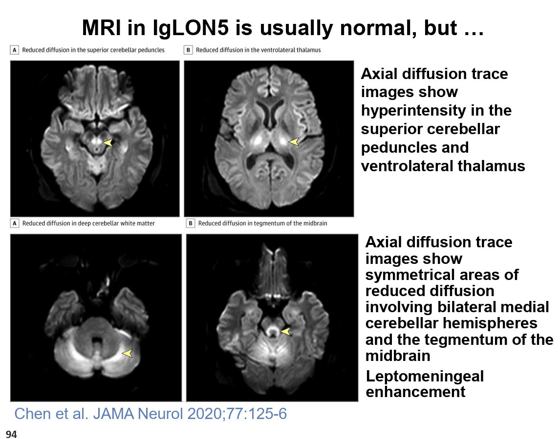
One of the messages from Joe Jankovic's outstanding Aspen Movement Course lecture today on autoimmune-associated movement disorders that stuck with me was the importance of resisting the temptation to over-diagnose rare conditions. With the growing awareness of anti-IgLON5 disease, many of us have probably been ordering the antibody test more often than we should. Joe reminded us that testing is most appropriate when the clinical picture includes the characteristic sleep disorder, which remains one of the strongest clues to the diagnosis. He also shared an important case where the MRI showed abnormalities, underscoring that imaging can occasionally be helpful, although MRI is normal in most patients with IgLON5 disease. Perhaps he and Alberto Espay's most practical pearl was one we should all remember in clinic: if a patient looks like they have classic progressive supranuclear palsy (PSP), they probably have classic PSP, not IgLON5 disease. As new biomarkers emerge, thoughtful clinical judgment remains our most valuable diagnostic tool.
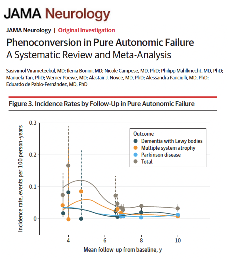
From autonomic failure to Parkinson’s and beyond: tracking the early warning signs of synuclein related disease. Phenoconversion means the transition from one clinical condition to another over time, in this case from pure autonomic failure to disorders like Parkinson’s disease, dementia with Lewy bodies or multiple system atrophy. Virameteekul and colleagues describe in a new paper in JAMA Neurology how frequently and under what conditions folks w/ pure autonomic failure develop central α-synucleinopathies.
Key points:
- Approximately 30% of individuals w/ pure autonomic failure developed a central α-synucleinopathy over follow-up, w/ an annual conversion rate of about 5%.
- Multiple system atrophy tended to emerge earlier, while Parkinson’s disease and dementia with Lewy bodies showed a more gradual and sustained risk over time.
- Clinical features such as REM sleep behavior disorder, subtle motor signs and hyposmia (smell loss) were among the most consistent predictors of conversion.
My take: This study reinforces something many of us have suspected for years: the autonomic nervous system may be one of the earliest windows into neurodegeneration. The ability to identify who is at highest risk and when conversion may occur could reshape how we think about early diagnosis and intervention.
Here are 5 points that resonated w/ me:
1- Autonomic symptoms may represent an early stage of Parkinson’s biology rather than a separate condition.
2- A 5% annual conversion rate is meaningful and highlights the need for close longitudinal follow-up.
3- Timing matters as different diseases emerge on different trajectories.
4- Simple clinical features like smell loss and dream enactment may help stratify risk today.
5- The future will likely combine clinical signs w/ biomarkers to identify the right folks for early intervention and disease modifying trials.
https://t.co/TcDtHeU1wG @JAMANetwork@jamaneuro@ParkinsonDotOrg #parkinson
In Alzheimer’s imaging, there are some things you just can’t forget!
New Alzheimer’s treatments are changing the way we look at these scans!
Removal of amyloid beta proteins from vessel walls by anti-amyloid antibodies leads increased vascular permeability.
This can cause edema or hemorrhage, called ARIA (Amyloid Related Imaging Abnormalities)
Grading of the degree of ARIA is important bc it can change management. Asymptomatic ARIA is usually treated by pausing treatment if it’s moderate to severe
For ARIA-E (edema) or ARIA-H (microhemorrhage), if the edema measure > 5 cm or number of microhemorrhages are >5, it is moderate
For superficial siderosis, 2 regions is considered moderate.
Remember it by this little rhyme:
--If ARIA findings are great than 5, then the medication deprive
--Or if siderosis is 2, then medication break for you!
Hopefully, now the grading of ARIA will stay in your memory!!
Distinguishing #MS from #NMOSD and #MOGAD is critical—treatments and outcomes differ. Drs Robertson & Khanna with A. Demers review diagnostic criteria, antibody testing, & key imaging patterns that guide accurate classification.
https://t.co/POc0fpfsgP
@USFHealthMed@mssociety@mscare@ACTRIMS@AANmember
#STROKE Advances ⭐️ in neurocritical care, by @sheth_kevin & Jha: from endovascular therapy for chronic SDH to BP management in ICH, fever prevention, hemoglobin targets in aSAH/ABI, and insights into cognitive motor dissociation. https://t.co/Ob2w96kMu8
Many people with Parkinson’s disease continue to drive safely long after their diagnosis, but the ability to drive with PD depends on a person’s specific symptoms as well as the presence of other age-related changes. 🚙
These tips can help you determine if you (or your loved one) can continue driving safely. For more information on driving with Parkinson's, visit: https://t.co/TnX4g19oFg
The distinctive psychopathology of NMDAR-antibody encephalitis compared with primary psychoses: an international, multicentre, retrospective phenotypic analysis - The Lancet Psychiatry https://t.co/lKI3MuLmlw
New in Cephalalgia!
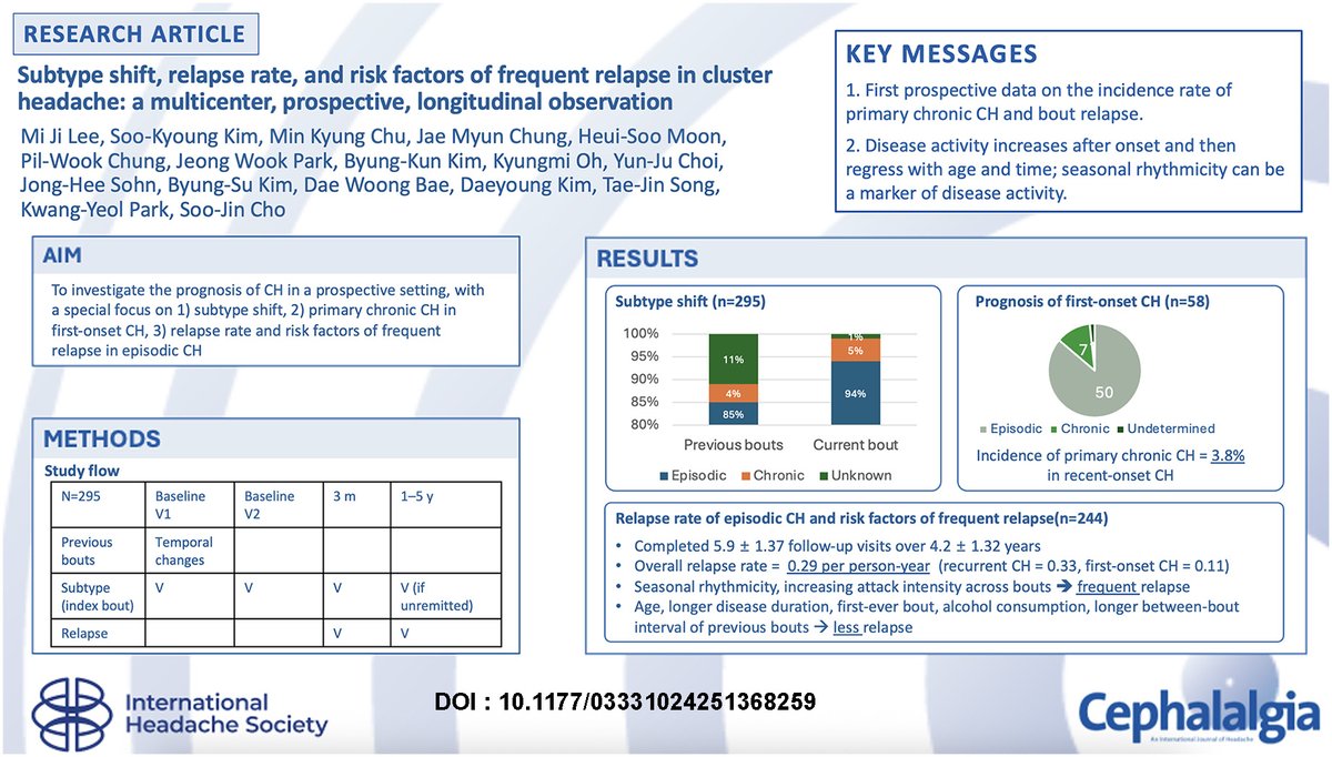
New data on the prognosis of cluster headache (CH) have been released, including subtype shift, incidence rate of primary chronic CH, and relapse rate of bouts, based on a prospective observation of 295 patients with CH.
https://t.co/wtNcH6mOw8
#Clusterheadache #headache #brain #pain #neurology #neuroscience
If you are on a dopamine agonist for Parkinson’s pay attention to this long term follow-up. The occurrence of impulse control disorders cumulatively increases over time and is a shocking 46% in those followed for at least 5 years. Regina Katzenschlager said this was the most important slide to remember from her MDS plenary talk this morning in Hawaii. @fixelinstitute@movedisorder@ParkinsonDotOrg
How to treat orthostatic hypotension in Parkinson and Parkinsonism? Dizziness on standing? Melissa Armstrong nails it in one slide during her plenary lecture this morning at MDS Hawaii. @movedisorder @FixelInstitute
From a Thai-inspired chilled capellini with seafood to a ‘naughty’ eggplant ‘parmigiana’, San Lou Pasta Bar is a star in PJ for Italian-Asian fusion cuisine https://t.co/lepArbZGXe
Is cranial nerve anatomy making you nervous??!!
High resolution SSFP imaging can visualize cranial nerves, typically for pts w/microvascular compression
But knowing their anatomy & location can help you on any imaging—bc if you know where they are, you know if they may be involved
Here’s what you can see on MRI:
Olfactory: along anterior cranial fossa above the cribriform plate
Optic: from globe, posteriorly through the orbit until intracranially optic nerves join to form chiasm
Oculomotor: from between the cerebral peduncles to cavernous sinus
Trochlear: From back of the midbrain at the aqueduct to cavernous sinus
Trigeminal: From pons at the middle cerebellar peduncles into meckels cave
Abducens: From undersurface of inferior pons anteriorly to dorello canal along clivus
7th/8th complex: From undersurface of inferior pons laterally to the IAC
Glossopharyngeal & vagus: from medulla where it looks like a clover to the jugular foramen
Hypoglossal: from medulla where it looks round like the spinal cord to the Hypoglossal foramen
Hopefully all this info of cranial nerves didn’t get on your last nerve!!
Differentials of L-dopa responsive juvenile onset Dystonia- Parkinsonism
# To keep Juvenile form of NIID in this list with L-dopa induced dyskinesia mimicking PRKN
#Neurotwitter@AlbertoEspay@AMahajanMD@LucaMarsili1
https://t.co/0Mzs1Tsal9