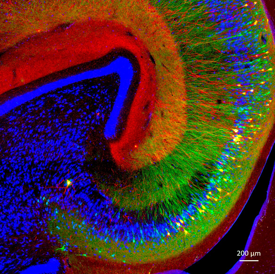
Happy #MicroscopyMonday! The photo below is from our Tau monkey model of Alzheimer's disease! Tau is a protein that gets misfolded and accumulates inside neurons in #Alzheimer's and other neurodegenerative diseases. The photo below shows two tau phosphorylations observed in the disease in red (AT8) and green (pS422). Healthy neurons in blue only. Everything in red and green are dying neurons and its projections. A colorful death.
Confocal image taken with @zeiss_micro
Thesis work rewarded by publication in mBio 3 days before defense ! 🥳 🍾
Featuring HTLV-1 biofilms and host cell tetraspanins working together for an efficient viral transmission 🦠🔬
Many thanks to all collaborators, and to my two guides @MuriauxD & @HeleneDutartre 👇🏼
If you are interested in Respirator syncitia virus and it’s Specific lipids target..This work of mine for you.This is our 1st collaborative work with @monika form INRAE, Many more to come. Thanks to all Coauthors. Always thank to @MuriauxD@VirAssemblyLab https://t.co/3dLc4Wagbg
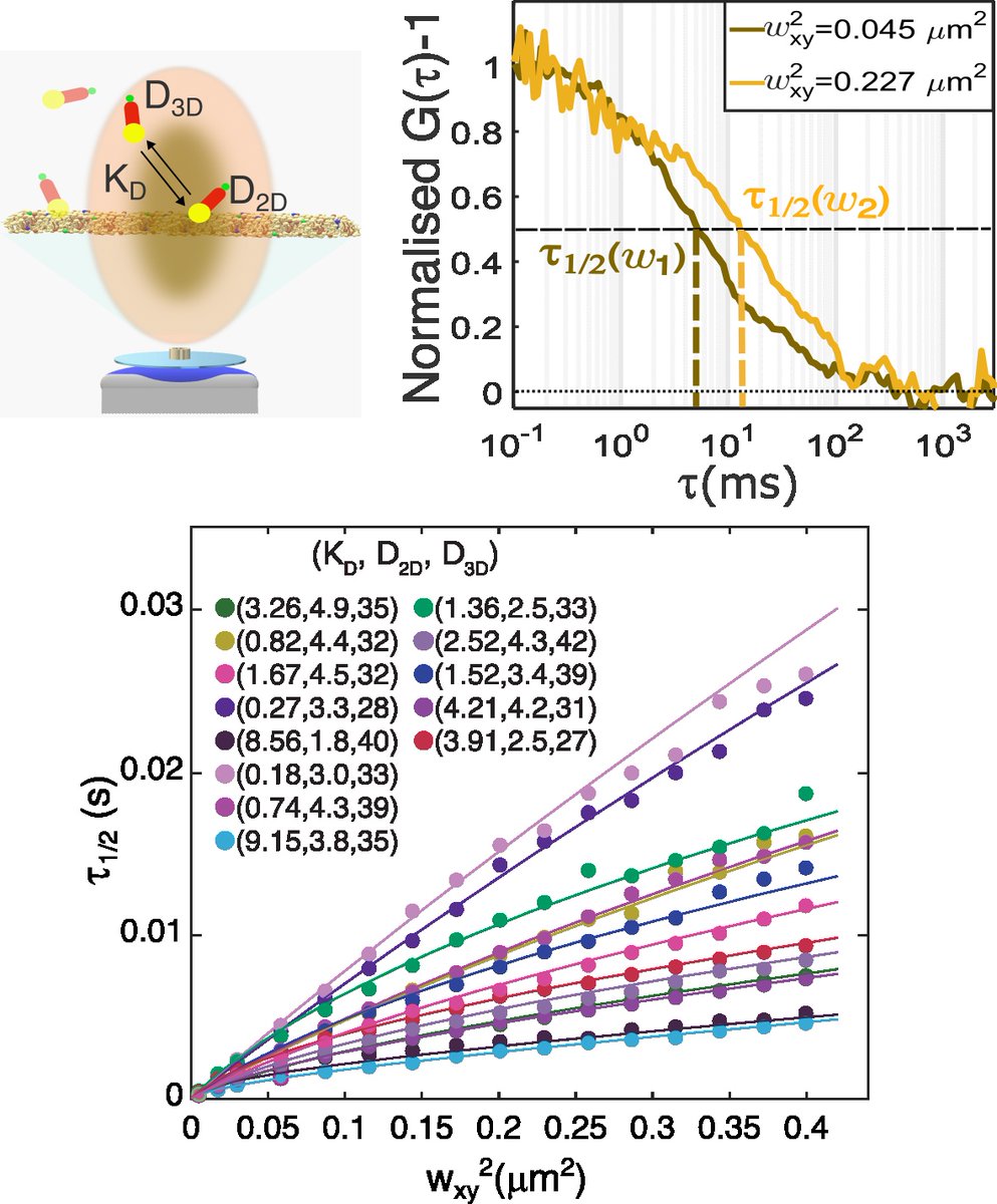
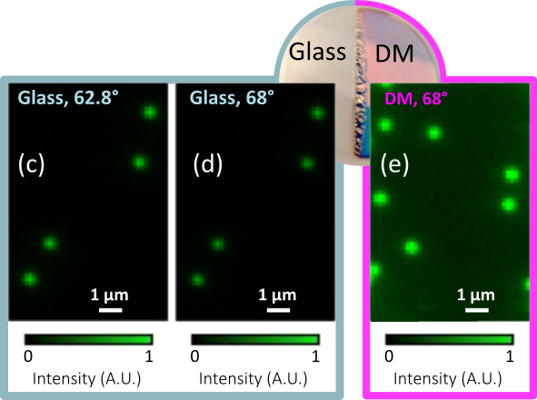
Interested in binding/diffusion on biological membranes? We found how to quantify both using spot variation FCS. Read it in @BiophysJ :
https://t.co/ANWPwsUaan
# See details in the thread
Big congrats to #Manon & @MuriauxD your BioRXiv paper on #SARSCoV2 VLPs optimization is already highlighted!! A whole team effort! @IRIM_life
https://t.co/NsuNcSaJaw