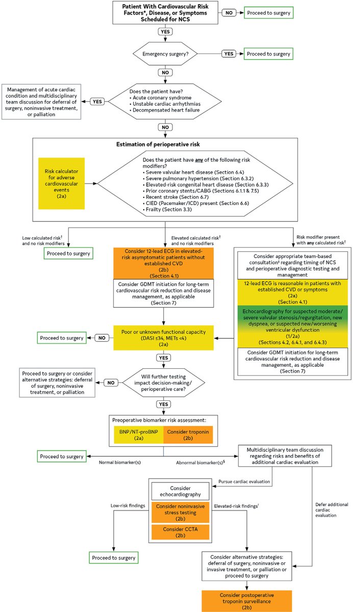
The 2026 AHA/ACC Multisociety Guideline for #Perioperative CV Management for Noncardiac Surgery reaffirms the 2024 guideline. The title reflects the reaffirmation, while recommendations & supporting content remain unchanged. https://t.co/DsgvuOLEqp #JACC
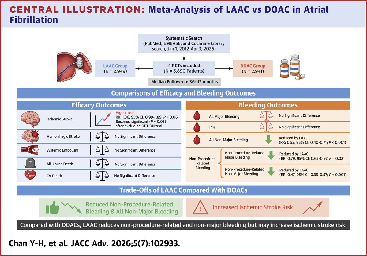
Can LAAC replace DOACs for #stroke prevention in #AFib?
New in #JACCAdvances: This meta-analysis of randomized trials found LAAC reduced nonprocedure-related bleeding but may come at the cost of a higher risk of ischemic stroke. https://t.co/T9cebrziis
#NOAC#cvLAAOccluder
🫀💊 Smarter risk stratification after anthracyclines! The RE-ACT score adds post-treatment prognostic value to HFA-ICOS, supporting a practical 2-step approach to personalize #CardioOnc surveillance 🎯📊 https://t.co/cnRa4k8bBN
#JACCCardioOnc#Anthracycline#Cardiotoxicity
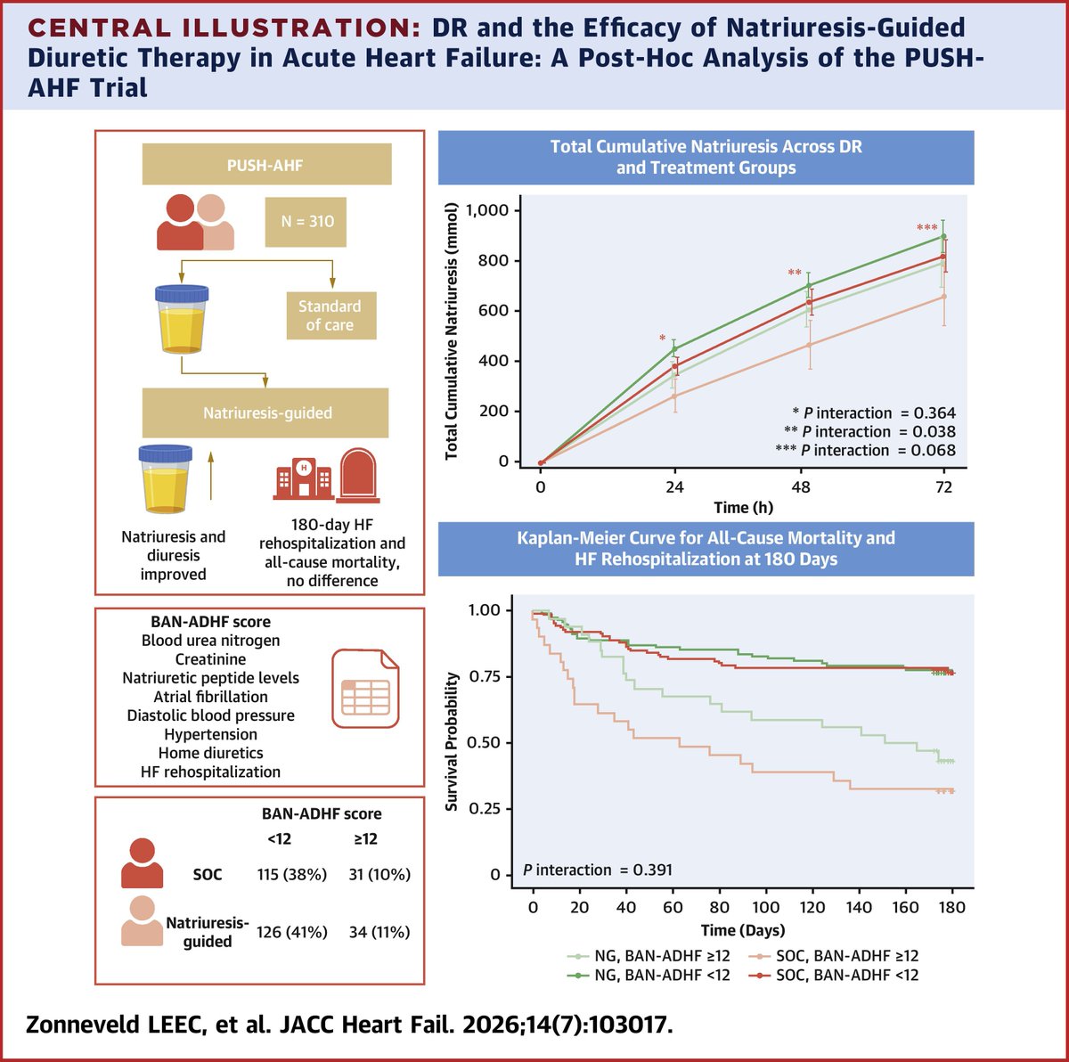
High BAN-ADHF scores identified patients with impaired natriuresis and worse outcomes. Natriuresis-guided therapy improved 24-hour natriuresis, especially in those at highest diuretic resistance risk. https://t.co/SX4L5T2980 #JACCHF#HeartFailure
🩸🎗️ Bleeding risk remains a major challenge in cancer-assoc #VTE.
The new ONCO-DOAC BLEED score helps identify pts at higher risk of major bleeding while on #DOAC therapy, supporting more personalized anticoagulation decisions https://t.co/REAm5NBBqo #JACCCardioOnc#CardioOnc
We evaluated and compared the diagnostic performance of 5 echocardiographic formulas for estimating mPAP in a large cohort of consecutive patients undergoing both RHC & TTE, applying the updated hemodynamic PH definition.
Read our @JournalASEcho article: https://t.co/NJE98bn09b
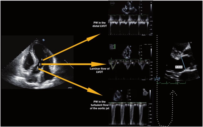
🫀 Did you know where the recommendation to place the pulsed wave Doppler sample volume 0.5–1 cm from the aortic valve to measure LVOT VTI comes from?
The answer is more interesting than it seems. It doesn’t come from a single study or an experiment designed for that purpose. It comes from a historical chain spanning nearly 40 years:
🔬 1982–1984 — The physical foundation
Pasipoularides and Murgo demonstrated using invasive catheters and mathematical models that in aortic stenosis there is a real zone of flow acceleration in the LVOT, just proximal to the valve, without any second anatomic obstruction. Pure hemodynamics — no Doppler yet.
👉 Bird et al. Circulation 1982 → https://t.co/oCXGNgWsPF
👉 Pasipoularides et al. Am J Physiol 1984 → https://t.co/cfvNMpEgzG
📐 1984 — The apical 5-chamber view
Lewis, Kuo and Quinones were the first to validate cardiac output measurement using pulsed wave Doppler from the cardiac apex. They described placing the sample volume “immediately proximal to the aortic valve leaflets” — but without specifying any distance in centimeters.
👉 Lewis et al. Circulation 1984 → https://t.co/lRVjLuYMEs
📏 1985 — The first numerical distance
Skjaerpe, Hegrenaes and Hatle (the Norwegian group) were the first to quantify this in Doppler: they empirically observed that flow acceleration began 0.5 to 1.5 cm proximal to the valve, and placed the sample volume just proximal to that zone. They directly cited Pasipoularides as supporting evidence. This was the first time a numerical distance appeared in the technique.
👉 Skjaerpe et al. Circulation 1985 → https://t.co/rf7l2FQUs4
📊 1986–1988 — Practical consolidation
Otto et al. used ~1.0 cm. Oh, Tajik and the Mayo Clinic group explicitly established the range of 0.5 to 1.0 cm in 100 patients, justifying it as necessary to avoid the subvalvular acceleration zone. This is the figure we all recognize today.
👉 Otto et al. JACC 1986 → https://t.co/avke1qOy4x
👉 Zoghbi et al. Circulation 1986 → https://t.co/KFiChfBNuc
👉 Oh et al. JACC 1988 → https://t.co/1C8sHRSMPq
📋 2002 — It becomes “official”
Quinones, Otto, Zoghbi and colleagues codified it in the ASE guidelines as “~5 mm proximal to the aortic valve”… but without citing any specific study to support it. It had already become expert consensus.
👉 Quiñones et al. JASE 2002 → https://t.co/U3hXFns4jx
⚔️ 2017 — The debate reopens
Baumgartner et al. (EACVI/ASE) maintained the 0.5–1 cm recommendation. However, Hahn and Pibarot responded with a critical letter pointing out that the original articles from the 1980s measured at the aortic annulus, not 0.5–1 cm below it, and that moving away from the annulus introduces errors due to the elliptical and irregular shape of the subannular LVOT.
👉 Baumgartner et al. Eur Heart J Cardiovasc Imaging 2017 → https://t.co/iu07xBTwQS
👉 Hahn & Pibarot. JASE 2017 → https://t.co/uqgcv8kzO7
💡 Bottom line:
The 0.5–1 cm figure was never experimentally validated as the optimal distance. It emerged from empirical observations in the 1980s aimed at avoiding a flow acceleration zone that had been demonstrated with invasive catheters. It was adopted through accumulated clinical practice and later elevated to a formal recommendation by consensus. The debate over whether to measure at the annulus or 0.5–1 cm below it remains open to this day.
One of those recommendations we all follow but few know where it actually came from 🙂
Dr Benigno Valderrábano Salas
@MDBeni@JaeKOh2@ottoecho@WilliamZoghbi@ASE360@EACVIPresident@NephroP@iamritu@PPibarot@hahn_rt@MAecocardio@SISIACOficial@SONECOM_AC@VazyurVasquez@Cardiotweets83@HEARTof_echo@echobasics
48M with prior PFO closure, rheumatoid arthritis, and tobacco use presented with months of atypical chest pain and fatigue. CCTA with FFR showed multivessel #atherosclerosis with functionally significant stenoses.
Get the details: https://t.co/j9K6WhX23I
#JACCCaseReports
Cardiac imaging in oncology: the detection of cardiotoxicity
In this episode of the Heart podcast, Digital Media Editor, Professor James Rudd, is joined by Professor Kazuaki Negishi from Sydney, Australia. They discuss the optimal use of imaging to detect cardiac effects of cancer therapies. If you enjoy the show, please leave us a positive review wherever you get your podcasts. It helps us to reach more people - thanks!
Podcast: https://t.co/4FRyqcIy2w
Paper: https://t.co/S8sGXaJo4c
Cardiac involvement in parasitic infections. This State-of-the-Art review just published in #EHJ provides current evidence on epidemiology, clinical manifestations, diagnosis, and management of parasitic cardiac disease.
https://t.co/JGFumBGzBx
@escardio@ESC_Journals
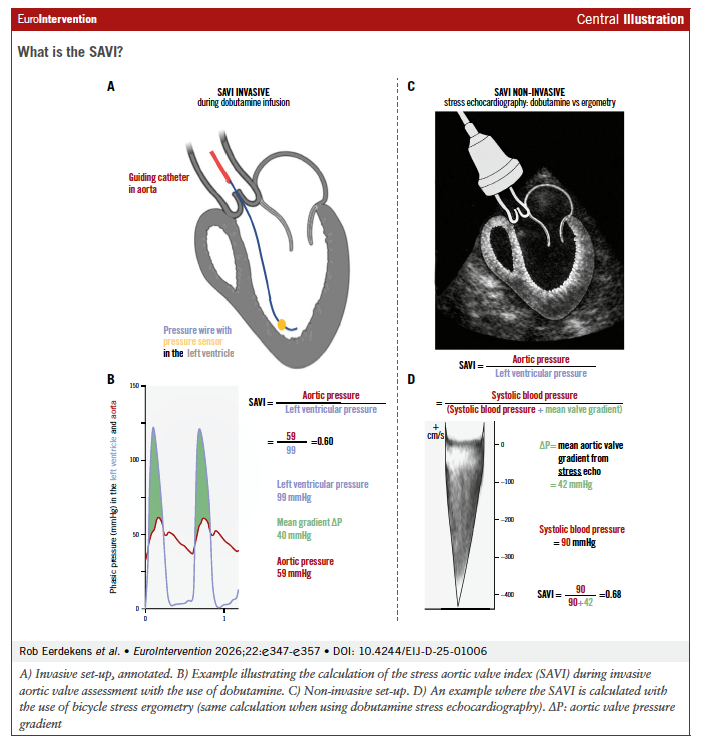
In symptomatic moderate AS, the stress aortic valve index (SAVI) identifies a high-risk subgroup not captured by conventional metrics and independently predicts the need for valve intervention (HR 5.7).
https://t.co/90M4bBCnBJ
@RobEerdekens@RikAdrMD@drnvanmieghem@AshkanEftekhar1
#AorticStenosis #Cardiology #StructuralHeart
#EHJCVI 🫀 Can LA parameters help risk-stratify embolic stroke of undetermined source? Patients show larger LA volumes, reduced reservoir and contractile strain, and increased LA circularity, features suggesting atrial cardiomyopathy may play a role 🔍 👉 https://t.co/WdFLOxDTgZ