After about 25 minutes of sitting, the muscles in your thighs are almost completely quiet. A new study in npj Exercise Medicine and Health tracked this in real life, and it may help explain why long sitting is so hard on metabolic health.
Prolonged sitting raises the risk of diabetes, heart disease, and some cancers. Most studies measure how long people sit, usually with accelerometers. That tells you nothing about what the muscles are doing. Not all sitting is the same, and muscle activity may be the missing piece.
Researchers from Australia, Finland, and the UK used shorts with electrodes woven into the fabric. This is electromyography, or EMG, which detects the electrical signals muscles produce when they switch on. They tracked 84 adults across three cohorts, including people with type 2 diabetes, during 7,684 sitting bouts in normal daily life.
Why muscle activity matters: muscles are a major site where your body clears sugar from the blood. When they contract, they pull in glucose with less need for insulin. When they go quiet for long stretches, that disposal system idles.
What they found:
• Muscle activity drops fast. Detectable activity fell to about 10% at 9 minutes, 5% at 12 minutes, and 1% at roughly 26 minutes into a sitting bout. The decline follows a power-law curve, steep early and then flattening out near zero.
• Long sitters were quieter overall. People who usually sat for 25 minutes or more at a time had about 6.5 minutes of muscle activity per hour of sitting. People who sat in shorter bouts had about 10.4 minutes.
• What you did before sitting mattered. Sitting bouts that followed longer (over 10 minutes) or more intense activity had more muscle activity. Bouts that followed only standing had the least.
• Breaking up sitting added up. More frequent and more intense interruptions meant about 5 extra minutes of muscle activity per hour of sitting.
The practical takeaway is that the first 10 to 25 minutes of a sitting bout is when your muscles switch off. Interrupting sitting before that window closes, and doing it with more than just standing, keeps them engaged.
There are limits. This was observational, so it shows how muscle activity behaves, not that boosting it directly improves blood sugar or disease outcomes. The sample was also small, and very long sitting bouts were rare, so estimates there are noisier.
But it shifts the question. Instead of asking how many hours you sit, it may be more useful to ask how long your muscles stay silent. That is a more specific target for the metabolic damage sitting seems to cause.
For longevity, the implications are practical but untested, since the study didn't test any intervention. Skeletal muscle is the largest site of glucose disposal, and it is also a tissue that declines with age. Hours of silent muscle each day may add up to a chronic reduction in glucose clearance and in the signals that keep muscle healthy. If so, a short walk, a set of squats, or a few calf raises every 20 minutes may matter more than one hard workout followed by eight hours in a chair. That second point is my inference, not something the study tested.
It also means exercise and sitting are not the same variable. You can train regularly and still spend most of your day with idle muscles. Daily habits in your 30s, 40s, and 50s shape insulin sensitivity and muscle quality in your 70s and 80s.
Which "zone 2" are you talking about?
It drives me a little crazy when folks ask "what do you think about zone 2?"
Because, they are often, talking about different things.
A quick 🧵
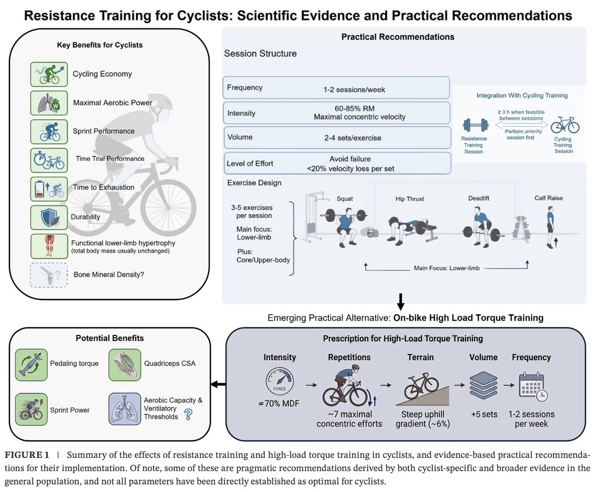
🚨New publication: Resistance Training for Cyclists: Scientific Evidence and Practical Recommendations
.
A review of the potential benefits of strength training for cyclists (strength training in the gym and on the bike)
.
OA: https://t.co/xHLsBuukmT
LOW VS HIGH INTENSITY TRAINING STUDIES
1/2 — WHAT DO THEY SAY
If a study compares 30 min of low-intensity vs 30 min of high-intensity training and the latter produces greater adaptations, it shows that high intensity produces greater adaptations per unit of time.
However👇
"I'm just a non-responder."
I can't stand that phrase. In all my years working with people, I've never met anyone who didn't respond to exercise.
"Non-responder" is a research term, and it gets used far too loosely. If we're going to use it at all, it needs to be tied to the dose. Not "they don't respond to exercise," but "they didn't respond to that program, at that dose, on that test."
The research backs this up.
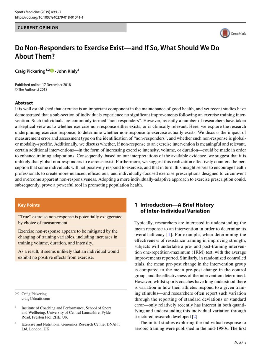
A 2019 paper in Sports Medicine by Pickering and Kiely asked whether non-responders actually exist. Their answer: probably not.
Much of what looks like "not responding" is something else:
��� Normal testing error and day-to-day ups and downs • A bad test day or low effort on a max test • Measuring one or two things, when exercise improves dozens • A dose that simply wasn't enough
When researchers tracked several outcomes, almost everyone improved on something. When people trained more often, harder or longer, the "non-responders" largely disappeared. In one study, adding two sessions per week turned every non-responder into a responder.
The authors do support individualized training, but read why. They aren't arguing that people need different principles. They're arguing for adjusting the dose: the amount, intensity, frequency, length or type of training, based on things like training history, sleep, stress, nutrition, recovery and whether someone can complete the work.
That's not "you're unique, so you need a special plan."
That's "same principles, different dose."
You're not a non-responder.
You just haven't found the right dose yet.
Source: Pickering C, Kiely J. Sports Medicine, 2019;49:1–7.
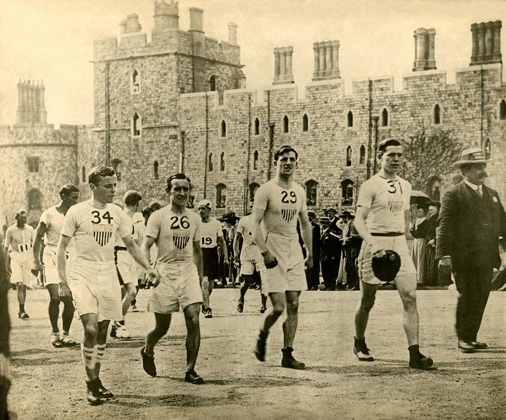
Frank Shorter is shown here with the late Kenny Moore. Please read Kenny's stories on the 1971 Fukuoka Marathon and the 1972 Olympic marathon in #BestEfforts, a book every real distance runner should have, along with John Parker's #OnceARunner and Ron Clarke's
#TheLonelyBreed. Thanks @geric99018902 for your wonderful photos--they bring up so many wonderful memories.
At 82 years old, USA Triathlon Hall of Famer Bob Plant has survived crashed bikes, broken bones, and frigid waters. Now, as the oldest competitor at the 2026 Ironman World Championship in Kona, he isn't just looking to finish- he's racing to win. https://t.co/9h4j8PtboE
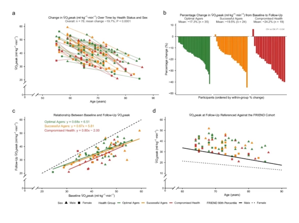
A 9-year longitudinal investigation into physiological aging in highly active older adults
“longitudinal data support the concept that even with emerging disease, exercise confers good physiological function in a body that is declining with age.”
https://t.co/trCGAqFvnD
@GeroScienceAGE
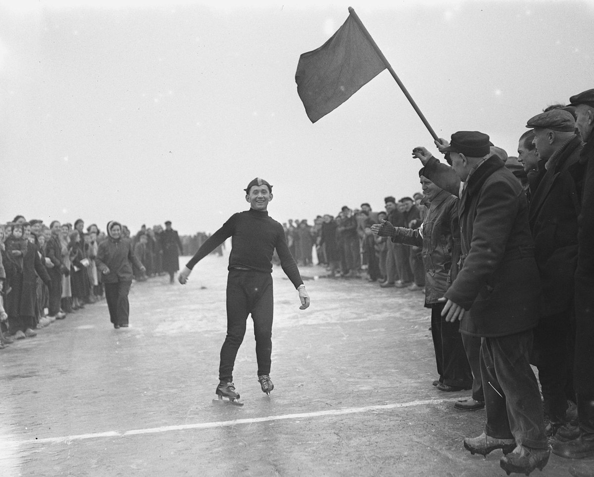
Op 5 oktober 2001 overleed schaatser Egbert van ’t Oever, Nederlands kampioen van 1954. Hij was coach van Yvonne van Gennip, Marianne Timmer en Hilbert van der Duim. Op zijn 25e sterfdag staan nog steeds 17 baanrecords op zijn naam.
Mijn verhaal staat op https://t.co/IbqnzD0W4z
Scientists have known since the 1970s that muscle fibers shrink with age and disuse. They just didn't know why. A new study finally answers that question by looking inside individual muscle fibers.
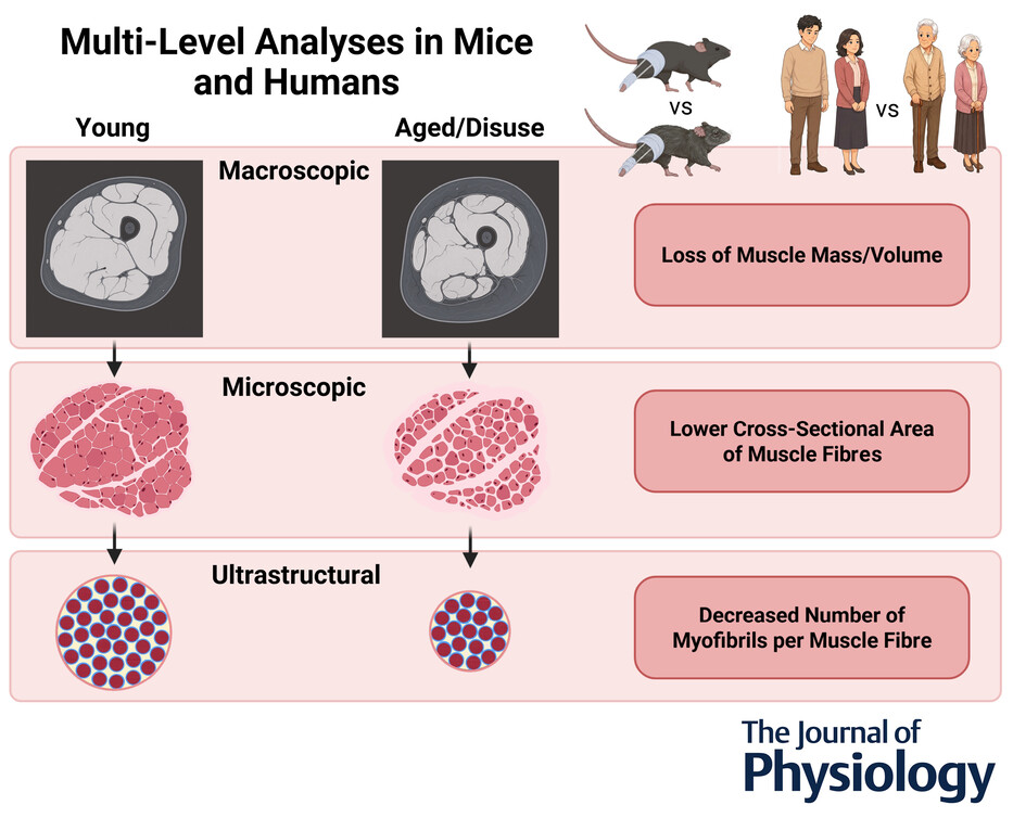
Researchers at University of Wisconsin-Madison published findings in The Journal of Physiology examining muscle structure at three distinct scales: whole muscle, individual fibers, and the contractile units inside those fibers called myofibrils.
The central question: when muscle fibers get smaller with aging or disuse, is that because the myofibrils themselves shrink, or because you're losing myofibrils?
Think of it this way. A muscle fiber is like a cable containing thousands of individual threads. When the cable gets thinner, either the threads are getting smaller, or you're losing threads. The mechanism matters because it tells you what's actually breaking down.
The study examined muscle samples from young adults in their 20s and older adults in their 70s, along with parallel experiments in young and aged mice. Some mice also underwent 10 days of hindlimb immobilization to model disuse.
Three levels of analysis
Macroscopic level: MRI imaging measured whole muscle volume and cross-sectional area in humans. Individual muscles were weighed in mice.
Microscopic level: Standard immunohistochemistry identified individual muscle fibers and measured their cross-sectional area.
Ultrastructural level: A newly developed imaging technique called FIM-ID allowed automated counting and measurement of thousands of myofibrils per sample.
That last part is critical. Previous studies relied on electron microscopy and manual tracing, which meant even modest analyses required hundreds of hours of labor. FIM-ID uses fluorescence imaging and computational clearing to generate high-contrast images that can be automatically quantified.
The aging findings in humans
Older adults showed 34% lower quadriceps muscle volume compared to young adults. The largest cross-sectional area along the muscle was 32% smaller.
At the fiber level, aging caused selective atrophy of Type II fibers, the fast-twitch fibers that contain SERCA1 pumps. These fibers were 23% smaller in older adults. Type I slow-twitch fibers were preserved.
Here's where it gets interesting. When researchers measured the myofibrils inside those Type II fibers, they found no significant change in the size of individual myofibrils. Instead, older adults had significantly fewer myofibrils per fiber.
The atrophy was driven almost entirely by myofibril loss, not myofibril shrinkage.
This was consistent across both male and female participants. Sex didn't significantly influence the aging-induced changes in muscle structure.
The findings in mice largely replicated the human results. Aged mice showed lower muscle mass in flexor muscles, smaller whole muscle cross-sectional area, and radial atrophy of Type II fibers driven primarily by fewer myofibrils per fiber.
One difference: aged mice also showed a small decrease in myofibril size, particularly in highly glycolytic Type IIb fibers. That fiber type doesn't exist in humans, which might explain the discrepancy.
The disuse findings:
Ten days of hindlimb immobilization in mice caused significant muscle atrophy in both young and aged animals, though the effect was blunted in the aged group.
Disuse-induced atrophy followed the same basic pattern as aging: smaller whole muscle, smaller individual fibers, and critically, fewer myofibrils per fiber.
Disuse also caused a small decrease in myofibril size, unlike aging in humans. This suggests disuse operates through slightly different mechanisms than aging, affecting both myofibril number and size.
Why this matters:
The fact that atrophy is driven primarily by myofibril loss rather than shrinkage points to a defect in myofibril turnover. Your body isn't just degrading existing contractile machinery uniformly. It's failing to maintain the number of contractile units.
The study also revealed that tubular aggregates, abnormal accumulations of sarcoplasmic reticulum, were prevalent in aged mouse muscle but rare in humans. These were found almost exclusively in highly glycolytic Type IIb fibers, reinforcing that this fiber type shows distinct vulnerability to aging.
Key takeaways:
• Aging reduces quadriceps volume by 34% in humans, driven by selective Type II fiber atrophy
• Fiber shrinkage results primarily from losing myofibrils, not from myofibrils getting smaller
• This mechanism is conserved across humans and mice for aging-induced atrophy
• Disuse causes similar myofibril loss but also produces small decreases in myofibril size
• Type I slow-twitch fibers are preserved during aging in humans
What this means for training:
The study didn't test any intervention, so what follows is my inference.
The fibers that shrink are the fast-twitch Type II fibers. Those respond best to heavy resistance work and power training, so steady-state cardio alone is unlikely to protect them.
Because the problem is losing contractile units rather than shrinking them, the goal is to keep building and replacing myofibrils. Mechanical load and adequate protein are the best-supported stimuli for myofibril synthesis.
Disuse produced the same myofibril loss in just 10 days. Injury, illness, travel, and hospital stays are the windows when this likely accelerates, so keeping some loading in those periods may matter more than most people assume.
Fiber size can also hide what's happening inside. Strength and power measures, like leg press, jump height, and sit-to-stand speed, may track the quality of your contractile machinery better than muscle size alone.
What the study can't tell us
The human analysis was restricted to the vastus lateralis, so muscle-specific differences across the body wouldn't be captured. The study was cross-sectional, preventing conclusions about causality or the temporal sequence of changes.
The immobilization model provided mechanistic insight into disuse, but differences in fiber-type composition between species, particularly the presence of Type IIb fibers in mice, limit extrapolation of certain findings.
The imaging technique provided robust ultrastructural measurements but didn't reveal the molecular mechanisms driving the observed changes. That requires follow-up work on signaling pathways and protein turnover rates.
The study answers a fundamental question that's been unresolved since the 1970s: muscle fiber atrophy during aging and disuse is mediated primarily by losing contractile units, not by those units getting uniformly smaller.
For longevity, that makes the decades before 70 the important ones. The myofibril reserve you build and protect in your 30s, 40s, and 50s shapes how much muscle machinery you have left in your 70s.