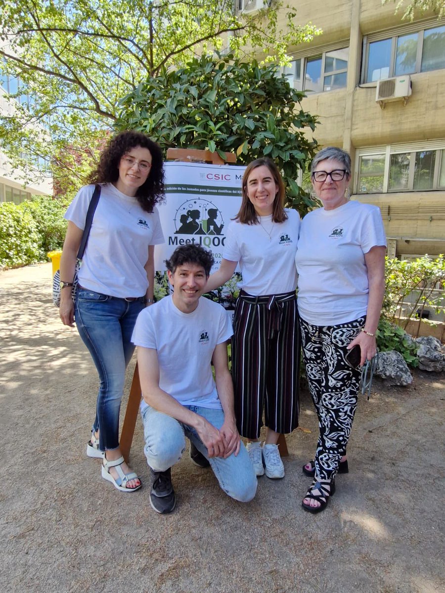
El 3 de junio tuvo lugar la II edición del MeetIQOG, donde nuestros jóvenes investigadores compartieron sus resultados y avances científicos. La jornada fue un éxito gracias a la participación, el entusiasmo y la calidad de las presentaciones. ¡Gracias a todos!👏🔬
Physical activity is the most effective intervention available for promoting brain health and longevity. Recent research demonstrates that regular exercise, even in modest amounts, is effective for enhancing brain volume, improving memory, and reducing the risk of neurodegenerative diseases. Beyond its cognitive benefits, moderate physical activity also contributes to extended lifespan and better overall health outcomes.
Even regular walking has shown to have powerful impacts on brain structure and function:
*️⃣Activity levels equivalent to 4,000 steps per day have been linked to measurable increases in gray matter and white matter volumes (PMID: 38073389)
*️⃣A 2011 study showed that 40 minutes of walking, three times per week lead to a 2% increase in hippocampal volume over one year, effectively reversing 1–2 years of age-related atrophy and enhancing spatial memory performance (PMID: 21282661).
*️⃣A more recent study suggests that activity levels equivalent to walking just under 3 hours daily for the least active populations could extend lifespan by 11 years on average (doi: 10.1136/bjsports-2024-108125).
Even walking is an accessible, non-pharmacological intervention for promoting brain health, reducing disease risk, and enhancing longevity.
Cellular senescence and its role in chronic disease
This figure shows how cellular senescence, the permanent arrest of cell division that occurs after stress or damage, contributes to chronic low-grade inflammation and age-related disease across multiple organs. Senescence acts as a protective mechanism against cancer early in life but becomes harmful when senescent cells accumulate and disrupt normal tissue function.
1️⃣ Initiation of senescence
Cells enter senescence in response to DNA damage, oxidative stress, mitochondrial dysfunction, or telomere shortening. Tumor suppressor pathways such as p53 and p16ⁱⁿᵏ⁴ᵃ halt the cell cycle and prevent further division.
🟢 Example: Mitochondrial dysfunction increases reactive oxygen species (ROS), which damage DNA and reinforce the senescent state.
2️⃣ Senescence-associated secretory phenotype (SASP)
Senescent cells remain metabolically active and release a combination of cytokines, chemokines, growth factors, and proteases known as the SASP. These molecules sustain inflammation, remodel the extracellular matrix, and alter the behavior of nearby cells.
🟢 Example: Cytokines such as TNF-α and IL-6 maintain chronic inflammation, while matrix metalloproteinases (MMPs) degrade tissue structure and promote fibrosis.
3️⃣ Interactions with immune and tissue systems
Senescent cells attract immune cells through chemokines like CCLs and CXCLs but often avoid destruction by producing immune-evasive proteins. Their secretions can also induce nearby healthy cells to become senescent, amplifying tissue inflammation.
🟢 Example: In adipose tissue, senescent fat cells release IL-6 and MCP-1, which recruit macrophages and worsen insulin resistance.
4️⃣ Organ-specific effects
Chronic accumulation of senescent cells drives pathology in multiple organs by altering local signaling and energy metabolism.
🟢 Example: In the brain, SASP factors are linked to neurodegenerative disorders and diabetic macular degeneration.
🟢 Example: In skeletal muscle and bone, senescence contributes to sarcopenia, osteoarthritis, and osteoporosis.
🟢 Example: In blood vessels, senescent endothelial cells promote atherosclerosis and thrombosis through ROS and matrix remodeling.
Cellular senescence is a hallmark of aging that bridges molecular damage with systemic inflammation. Its persistence fuels metabolic, cardiovascular, and degenerative diseases, making targeted removal or suppression of senescent cells an emerging focus of longevity research.
How do the neurons in our brain connect to underlie thought? New free AI-powered software from @columbia and @ohiostate can map these links automatically, rapidly and accurately to help shed light on how the brain works. #neuroscience
https://t.co/CEyLDoEfzW
Through the development of metal–organic frameworks, 2025 chemistry laureates Susumu Kitagawa, Richard Robson and Omar Yaghi have provided chemists with new opportunities for solving some of the challenges we face.
Following the laureates’ groundbreaking discoveries, researchers have created numerous different and functional metal–organic frameworks (MOF). So far, in most cases, the materials have only been used on a small scale. To harness the benefits of MOF materials for humanity, many companies are now investing in their mass production and commercialisation. Some have succeeded. For example, the electronics industry can now use MOF materials to contain some of the toxic gases required to produce semiconductors. Another MOF can instead break down harmful gases, including some that can be used as chemical weapons. Numerous companies are also testing materials that can capture carbon dioxide from factories and power stations, to reduce greenhouse gas emissions.
Some researchers believe that metal–organic frameworks have such huge potential that they will be the material of the twenty-first century.
#NobelPrize
BREAKING NEWS
The Royal Swedish Academy of Sciences has decided to award the 2025 #NobelPrize in Chemistry to Susumu Kitagawa, Richard Robson and Omar M. Yaghi “for the development of metal–organic frameworks.”
A "simple" guide to how your brain makes energy
Your brain is an energy-hungry organ. It uses ~20% of your body’s fuel even though it’s only 2% of your weight. Here’s how it actually powers itself:
1️⃣ Glucose as the primary fuel
Most brain energy comes from glucose, transported across the blood–brain barrier into astrocytes and neurons.
🟢 Example: A drop in blood sugar can quickly cause brain fog or dizziness.
2️⃣ Astrocytes as the middlemen
Astrocytes take up glucose and break it into lactate, which they shuttle to neurons. Neurons then burn lactate for ATP.
🟢 Example: This “lactate shuttle” explains why lactate isn’t just a waste product. Your brain uses it as fuel.
3️⃣ Neurons as the power users
Neurons convert lactate (or glucose directly) into acetyl-CoA, feeding the Krebs cycle and electron transport chain in mitochondria to produce ATP.
🟢 Example: Each neuron needs thousands of ATP molecules per second to fire signals.
4️⃣ Ketones as backup fuel
During fasting, ketogenic diets, or low glucose, the brain can switch to ketones (like β-hydroxybutyrate) for energy.
🟢 Example: This is why fasting or ketosis often feels mentally “clear." ketones are a "clean" brain fuel that you can use rapidly.
5️⃣ Insulin sensitivity matters
Neurons need insulin signaling to use glucose effectively. Resistance here has been linked to Alzheimer’s (“type 3 diabetes”).
🟢 Example: Poor insulin sensitivity may impair memory and cognitive performance.
6️⃣ Aging shifts the balance
With age, glucose use in the brain often declines. The ability to use lactate and ketones becomes more important.
🟢 Example: Imaging studies show reduced glucose uptake in Alzheimer’s brains — but ketone uptake is preserved.
7️⃣ The big picture
Brain metabolism is flexible: glucose dominates, astrocytes supply lactate, ketones step in during fasting, and mitochondria tie it all together.
🟢 Example: Whether you eat carbs, fast, or use fat, your brain has backup plans to keep neurons firing.
Your brain doesn’t just “run on sugar.” It draws on glucose, lactate, and ketones. With astrocytes and neurons working as a team to keep energy flowing.
A simple guide to understanding how neurons make neurotransmitters
Your brain has different classes of neurons, each defined by the chemical messenger (neurotransmitter) they release. These transmitters are built from specific precursors using unique enzymes and cofactors.
1️⃣ Cholinergic Neurons (green)
Main transmitter: Acetylcholine (ACh).
Source: Built from acetyl-CoA and choline.
Special enzyme: ChAT (choline acetyltransferase).
🟢 Example: These neurons drive muscle contraction and attention.
2️⃣ GABAergic Neurons (blue)
Main transmitter: GABA (gamma-aminobutyric acid).
Source: Derived from glutamate.
Special enzyme: GAD (glutamate decarboxylase).
🟢 Example: GABA is the brain’s main “calm-down” signal.
3️⃣ Glutamatergic Neurons (orange)
Main transmitter: Glutamate.
Source: Recycled from other cells and stored in vesicles.
🟢 Example: Glutamate is the brain’s main “go” signal for learning and memory.
4️⃣ Serotonergic Neurons (green-teal)
Main transmitter: Serotonin (5-HT).
Source: Made from tryptophan → 5-HTP → serotonin.
Special enzymes: TPH (tryptophan hydroxylase), DDC (decarboxylase).
Cofactor: BH₄ (tetrahydrobiopterin)
🟢 Example: Serotonin regulates mood, sleep, and appetite.
5️⃣ Dopaminergic Neurons (light blue)
Main transmitter: Dopamine.
Source: Made from tyrosine → L-DOPA → dopamine.
Special enzyme: TH (tyrosine hydroxylase), DDC.
Cofactor: BH₄.
🟢 Example: Dopamine drives motivation, reward, and movement.
6️⃣ Noradrenergic / Octopaminergic Neurons (red)
Main transmitter: Norepinephrine (mammals) or Octopamine (in invertebrates).
Source: Derived from tyrosine/tyramine.
Special enzyme: TBH (tyramine β-hydroxylase).
Cofactor: BH₄.
🟢 Example: Linked to arousal and “fight or flight.”
7️⃣ Tyraminergic Neurons (orange-brown)
Main transmitter: Tyramine.
Source: Made from tyrosine → tyramine.
Special enzyme: TDC (tyrosine decarboxylase).
🟢 Example: Acts as a trace amine, modulating dopamine and serotonin systems.
Supporting Pathways (Panels B & C):
Acetyl-CoA production (B): from the citric acid cycle, provides building blocks for acetylcholine.
BH₄ synthesis (C): a critical cofactor for making serotonin, dopamine, and norepinephrine.
Each neurotransmitter system has its own “assembly line”: specific precursors, enzymes, and cofactors. Together, they form the brain’s chemical language — acetylcholine for movement and attention, glutamate and GABA for balance, and monoamines (serotonin, dopamine, norepinephrine) for mood, reward, and arousal.
What if the brain’s massive memory power isn’t just about neurons? MIT researchers reveal that astrocytes, long-overlooked support cells, might be key to our mind’s vast storage capacity. https://t.co/yEiWDe3rSS
"I learned that any difficult problem can be solved by great effort."
Remembering Osamu Shimomura, born #OTD in 1928, who dedicated his life to studying the bioluminescence of the luminous jellyfish Aequorea, and discovered green fluorescent protein, GFP.
🧠La microbiota modula el impacto del ejercicio moderado sobre la memoria
🏃♀️Unos 40 minutos a velocidad media mejoran la diversidad de las bacterias intestinales
🦠Los cambios en la microbiota regulan efectos cognitivos del deporte como la neurogénesis
👉https://t.co/EOVa9CfZ6T
Optimistic people share patterns of brain activity, and make more of a distinction between positive and negative events than pessimists do
https://t.co/7nuQNJjxuv
Optimists’ Brains “Think Alike” When Imagining the Future
A new study shows that optimists’ brains activate in similar ways when imagining future events, while pessimists’ brain patterns are more unique.
Using fMRI, researchers observed that optimists process both positive and negative scenarios with shared neural patterns, suggesting a common mental framework.
This shared way of envisioning the future may help optimists connect socially and feel “on the same wavelength” with others.
In contrast, pessimists showed greater variability, imagining the future in highly individual ways.
Optimists also tended to view negative outcomes more abstractly and less emotionally.
These findings help explain why optimism correlates with stronger social networks.
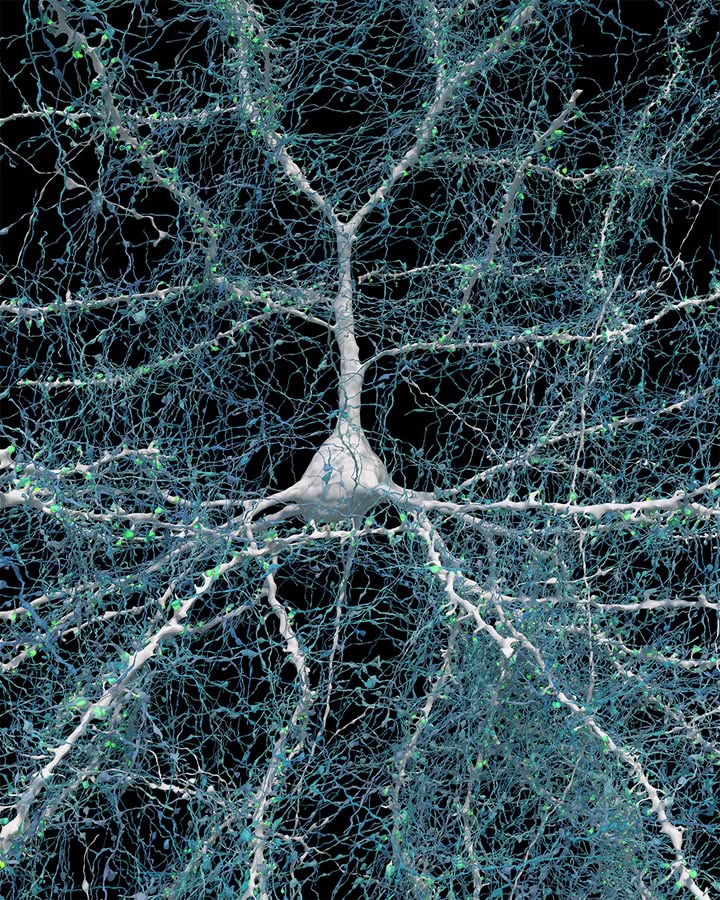
Last year in Science, researchers generated a nanoscale-resolution reconstruction of a millimeter-scale fragment of human cerebral cortex, giving an unprecedented view into the structural organization of brain tissue at the supracellular, cellular, and subcellular levels.
Learn more on #WorldBrainDay: https://t.co/ye8JncNkrJ
🔬 Investigadores del @NeuroAlc (#CSIC@UniversidadMH) identifican un grupo de neuronas implicado en la regulación de la ansiedad y los trastornos sociales
👨🔬 Restaurar el equilibrio en esas neuronas revierte los síntomas en roedores
👉 https://t.co/gA0kb32MFh
Phospholipids contain hydrophilic heads and hydrophobic tails, covalently connecting the hydrophlic/hydrophobic barrier that surrounds each one of our cells.
Learn more at https://t.co/rZjuFXgP1n
#biology#science#3D#animation#education#EdTech#phospholipid
Scientists just used STEM CELLS to restore function in Parkinson's disease - and it worked.
The cells survived, produced dopamine, and improved motor symptoms, reports @Nature.
Here's how they did it 🧵👇