Multiple sclerosis, a common autoimmune disease of the central nervous system, affects the brain, optic nerves, and spinal cord. Its hallmark effects are inflammation, demyelination, and neurodegeneration. 🔗 https://t.co/McH5l7urUG
In traditional descriptions of the disease, the myelin sheaths surrounding nerve-cell axons are the main target; peripheral nerve myelin, although structurally similar, is unaffected.
The disease typically manifests with discrete focal attacks that affect vision, sensation, strength, and balance, and over time the frequency of attacks decreases and progressive worsening with prominent neuronal injury supervenes (seen in figure). In a minority of patients, symptoms are progressive from onset. Diagnosis rests on clinical criteria and magnetic resonance imaging (MRI) findings, supplemented by clonal expansions of immunoglobulin in cerebrospinal fluid and slowing in central nerve conduction velocities as measured by evoked potentials.
Learn more in the Review Article “Advances in Multiple Sclerosis” by Stephen L. Hauser, MD: https://t.co/McH5l7urUG
#STROKE Simultaneous publication at #ESOC2025 🔴🔵
#OPTIMAS trial finds that chronic kidney disease does not modify the effects of early versus delayed DOAC initiation after acute ischemic stroke. #AHAJournals
https://t.co/9oXKCgFtux
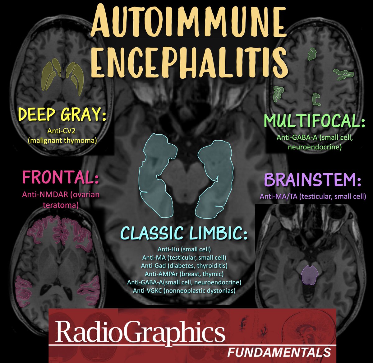
1/Wish that your knowledge of autoimmune encephalitis was automatic?
Do you feel in limbo about limbic encephalitis?
Do you know the patterns?
Read on for what you need to know in this month's @RadioGraphics review!
https://t.co/SQfITzD91w
@cookyscan1@RadG_Editor
Is cranial nerve anatomy making you nervous??!!
High resolution SSFP imaging can visualize cranial nerves, typically for pts w/microvascular compression
But knowing their anatomy & location can help you on any imaging—bc if you know where they are, you know if they may be involved
Here’s what you can see on MRI:
Olfactory: along anterior cranial fossa above the cribriform plate
Optic: from globe, posteriorly through the orbit until intracranially optic nerves join to form chiasm
Oculomotor: from between the cerebral peduncles to cavernous sinus
Trochlear: From back of the midbrain at the aqueduct to cavernous sinus
Trigeminal: From pons at the middle cerebellar peduncles into meckels cave
Abducens: From undersurface of inferior pons anteriorly to dorello canal along clivus
7th/8th complex: From undersurface of inferior pons laterally to the IAC
Glossopharyngeal & vagus: from medulla where it looks like a clover to the jugular foramen
Hypoglossal: from medulla where it looks round like the spinal cord to the Hypoglossal foramen
Hopefully all this info of cranial nerves didn’t get on your last nerve!!
Sink or swim!
Brain is swimming in CSF!
CSF is key for protection, buoyancy, & hormone/waste transport
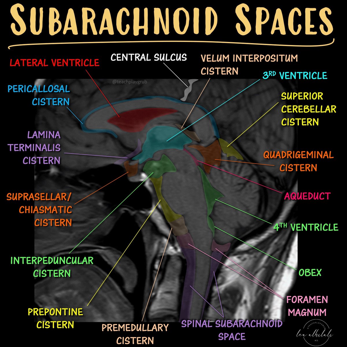
Most know basic ventricular anatomy well, but not subarachnoid anatomy.
How many cisterns do YOU know?
Here’s a quick guide to the key midline cisterns & what to know for each
Cisterna Magna:
Largest of the cisterns
Contains CN 9-11 & vertebral arteries
Remember this bc the LARGEST cistern contains the LARGEST cranial nerve (vagus) and the two cranial nerves around it
Prepontine cistern:
Contains CN 6 & basilar artery
Remember this bc the shape of the number 6 mirrors the curvature of the anterior pons, where this cistern lies
Chiasmatic cistern
Contains CN 2 & pituitary stalk
Easy to remember bc the CHIASM is CN 2
Interpeduncular cistern:
Contains CN 3 & mammillary bodies
Remember this bc if you lay the number 3 on its side it looks both like the two cerebral peduncles (where this cistern lies) & well, two mamillary bodies
Quadrigeminal cistern
Contains CN4 & pineal gland
East to remember bc QUAD is the prefix for FOUR
Lamina terminalis:
Contains the ACOMM
Remember this bc the shape of subarachnoid hemorrhage after ACOMM rupture follows this cistern anteriorly
Now when you look at cisternal anatomy, you will definitely be able to go with the flow!
PhD Students - How to design a research methodology?
You can use the Research Onion model to design your research methodology.
Here is a simple understanding of the different layers of the onion model.
𝟏. 𝐏𝐡𝐢𝐥𝐨𝐬𝐨𝐩𝐡𝐢𝐞𝐬
It refers to the view or stance from which the research has been conducted.
𝟐. 𝐀𝐩𝐩𝐫𝐨𝐚𝐜𝐡𝐞𝐬
Here you will formulate your hypothesis or an observation to test.
𝟑. 𝐒𝐭𝐫𝐚𝐭𝐞𝐠𝐢𝐞𝐬
Here you will choose the concrete method such as action research, survey, interview, or case study, etc.
𝟒. 𝐂𝐡𝐨𝐢𝐜𝐞𝐬
Here you need to specify whether you used one method, two methods, or multiple methods to conduct this research.
𝟓. 𝐓𝐢𝐦𝐞 𝐡𝐨𝐫𝐢𝐳𝐨𝐧𝐬
You need to answer in what time frames the research was conducted e.g., in one go or in various different times.
𝟔. 𝐓𝐞𝐜𝐡𝐧𝐢𝐪𝐮𝐞𝐬 𝐚𝐧𝐝 𝐩𝐫𝐨𝐜𝐞𝐝𝐮𝐫𝐞𝐬
You need to specify what type of data has been collected and how the data has been collected.
Credit: The research onion model by Saunders, Lewis and Thornhill
Anything you'd like to add?
Is Hoover's sign only useful in functional neurological disorders? NOPE. Wonderful experience teaching @UFNeuroRes as the attending this past week. Hoover's can be extremely helpful in the emergency room evaluation of a person with lower extremity weakness. At its core it is a crossed extensor sign. - Flex one hip and the contralateral hip extends. The maneuver can help you to gauge weakness in an emergency or in a consult setting. You don't have to always be looking uncover a functional neurological disorder.
https://t.co/v1Jb4yL1sD #Fixelinstitute
1/Do radiologists sound like they are speaking a different language when they talk about MRI?
T1 shortening what? T2 prolongation who?
Here’s a translation w/an introductory thread to MRI.