To learn more about pulmonary fibrosis and lung ultrasound, get a copy of The POCUS Textbook. It is a complete guide to point-of-care ultrasound of the blood vessels, heart, and lungs.
Get a copy today! https://t.co/jjlccnLKFv
Lung auscultation has been the standard tool for bedside pulmonary diagnosis for 200 years.
What's rarely discussed in clinical training is how poorly it actually performs when tested empirically.
A thread on the evidence.
A couple people who have had an incredible impact on medicine, through agency and deciding the field can change
@ThinkingCC helping dream and innovate on VEXUS
@PulmCrit for creating IBCC, the most widely used critical care resource in the world
@emcrit for "bringing upstairs care downstairs..." and now everywhere, realizing that ED medicine can change
@TheSGEM for innovating around EBM
@COREIMpodcast (Shreya et al.) for bringing innovation and new ideas for IM.
These people are all smart, yes, but most importantly, decided at some point to start something new.
What will you start in 2026?
Nice overview of lung #POCUS in #Nephrology by @AkhilOnX. Wishing him the very best this #residency interview season and for the Match.
🔗https://t.co/98xBO4dOdL
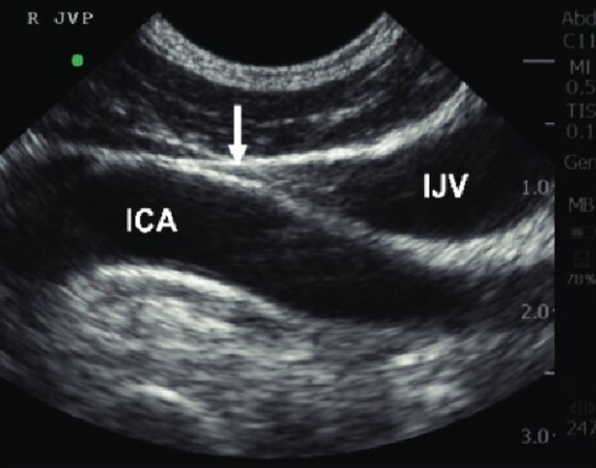
(1/3) What's the worst trend in POCUS for 2025?
Ultrasound to measure the height of the jugular venous pressure.
In fact, this might be the worst trend in POCUS ever👇
Aside from the distinction between static and dynamic air bronchograms, the use of pulsatile color Doppler (lower scale for detecting lower velocities) suggests intrapulmonary shunts and pneumonia versus atelectasis. Nice article in the comments
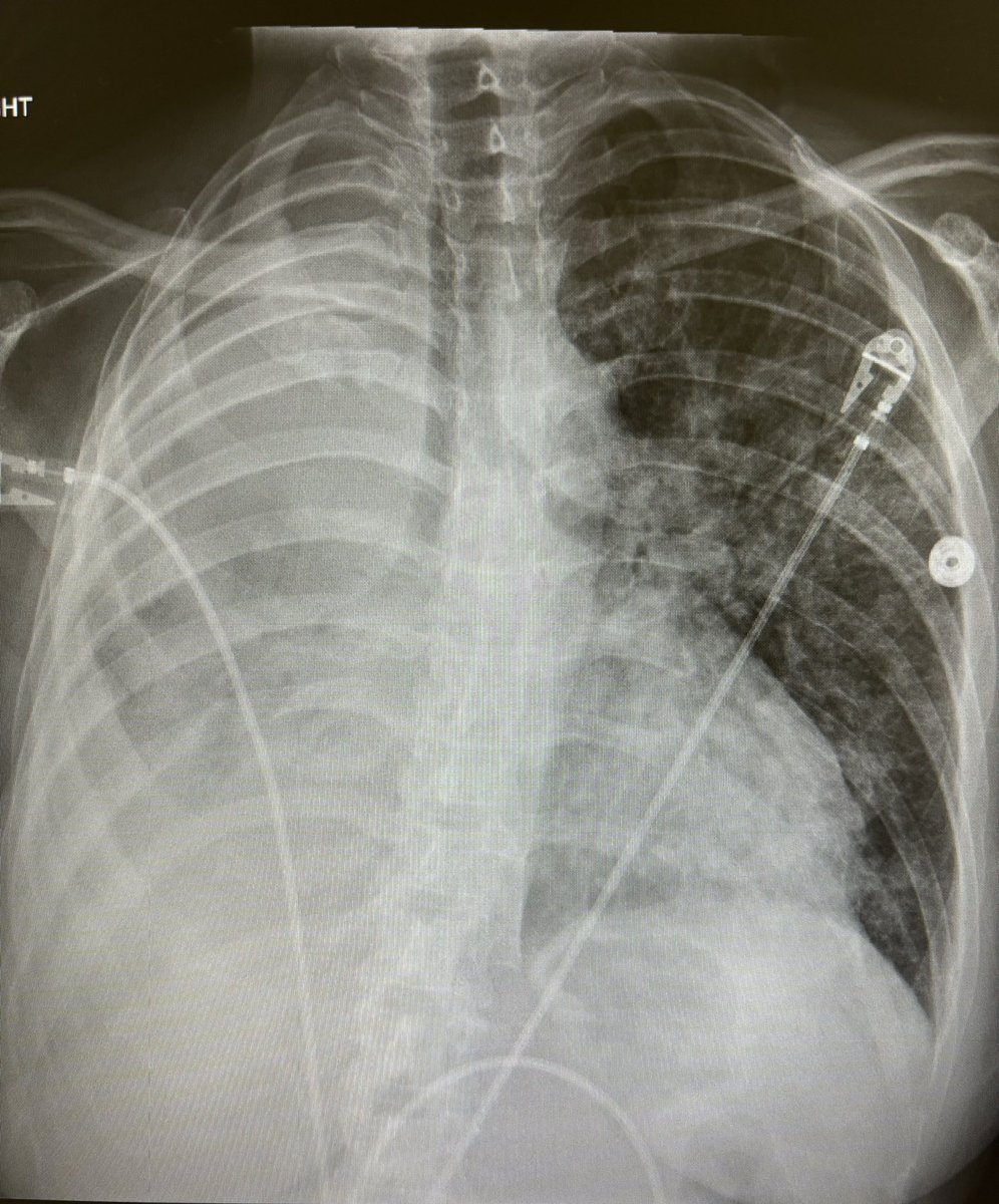
Reason #678376 why #pocus is essential to good medicine: Chest X-rays are not reliable enough.
This is a patient with known lung cancer who presented to a hospital with acutely worsening hypoxic respiratory failure. They have a known large right lung mass. CXR:
Using lung ultrasound to inspect the lung pleura can help differentiate types of pulmonary disease. In cardiogenic pulmonary edema, the lung pleura is smooth and regular with widely spaced B-lines. In contrast, with pulmonary fibrosis (seen here), the pleura is thickened and irregular with many hypoechoic interruptions. Subpleural consolidations may be present, and pleural effusions are usually absent.
In contrast, thickened and irregular pleura with subpleural consolidations or hypoechoic interruptions are seen with pneumonias, fibrosis, or acute respiratory distress syndrome. This was seen in a study of ARDS patients, for example, which found that:
- 100% of patients with ARDS had pleural abnormalities, while only 25% with cardiogenic pulmonary edema did
- Absence or reduction in lung sliding was seen in 100% of ARDS patients and 0% of cardiogenic pulmonary edema
- Subpleural consolidations were seen in 83% of patients with ARDS and 0% with cardiogenic pulmonary edema
Source: https://t.co/ZpSEbvzvcA
@iceman_ex
For rales on auscultation, the odds ratio was 5.1.
For orthopnea, the odds ratio was 6.9
For elevated NT-proBNP, the odds ratio was 14.3
For Multiple B-lines in multiple rib spaces with lung ultrasound, the odds ratio was an astounding 53.7!
The sensitivity and specificity of lung ultrasound to detect cardiogenic pulmonary edema were 100% and 95%, respectively.
Cardiac #POCUS parameters in the assessment of pulmonary hypertension
#FOAMed#Nephpearls
Click 'ALT' for abbreviations
Courtesy: 2022 ESC/ERS pulm HTN Guidelines
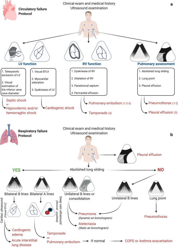
Bedside #POCUS during ward emergencies is associated with improved diagnosis and outcome.
#FOAMed#FOAMcc
🔗 https://t.co/R0S532nbHH
(*lung point should correspond to abolished lung sliding arm of the figure)
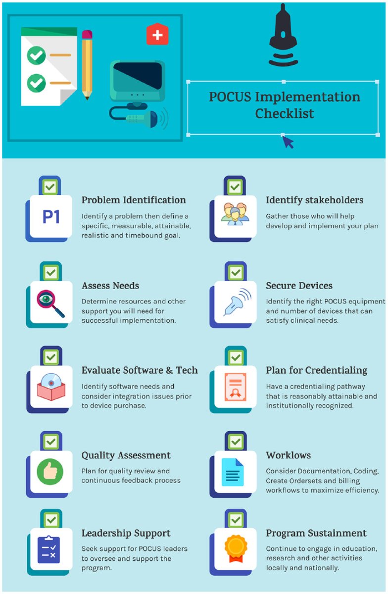
I really like this checklist as a framework to introducing #POCUS to a department or clinical group, as well as tracking how your program is doing
"A Clinician's Guide to Implementation of POCUS in the Outpatient Practice" @OvergaardJosh@BThilagarMD
🔗https://t.co/9bX2YqzX4d