Happy Pride Y’all! I wanted to make sure some incredible LGBTQ+ people that have contributed to science are recognized like Ben Barres, Sara Josephine Baker, Sally Ride, and Alan Turing. Much love and appreciation to all current LGBTQ+ scientists as well!
While we do not study apoptotic bodies in my lab, they are pretty cool! This cell expressing a membrane marker was videoed through a microscope while it was dying by @EmmaKoory. The video captured just the bottom of the cell. The cell rounds up as dying cells tend to do (i.e., it disappears from the region being imaged). Then apoptotic bodies come down and do cool stuff! In case you were wondering...... Yes, I am known internationally for my use of precise scientific language like "cool stuff" :-) #CellBiology
AI 4D Cell Biology!
Watch mitochondria reveal their hidden phenotypes! We built MitoSpace, a self-supervised model trained without labels on terabytes of 4D lattice light-sheet movies of single cells. It learns representations that outperform predefined features, predict membrane potential from 4D form alone (R²=0.91), and generalize zero-shot to unseen drugs and human lung organoids.
🎉 Now published in @CellCellPress!
With @DrvAgwl@ZacharyWang379@ecarkfeld@andremodolo4@Parth_LFC@hhako2@GCMcMahon
📄 Paper: https://t.co/KmoUvFOYAk
🔬 Explorer: https://t.co/asVHY1s028
💻 Code: https://t.co/AUHVSg4aRr
🤗 Model: https://t.co/2vJvQRmDjj
#AI4DCellBiology #cellbiology #microscopy #LLSM #latticelightsheet #mitochondria #deeplearning #phenomics
our 5th publication of the year is now out, and it comes with our shortest title yet: ONE microscopy. https://t.co/7AgYOqBuS3
ONE pushes conventional fluorescence microscopy to the molecular scale. It allows us to directly visualize the shapes of individual proteins, resolve conformational changes of around a nanometer, and achieve nanometer to subnanometer imaging precision, while extending these capabilities from purified molecules to complex cellular and tissue samples.
This Nature Protocols paper provides the complete workflow for doing this, from sample preparation and expansion to imaging and fluctuation analysis. And now there is ONEpy, our new software package that brings the analysis pipeline together and makes it substantially easier to use. Faster to set up, easier to use, and gives us a much better base for what we want to add next.
and this is not all from the team this year. We currently have 3 more papers in revision, and we are preparing a 4th manuscript to submit by the end of the year.
we are only getting started, and I could not ask for a more ambitious, creative and relentless team to build this with.
what started with ONE as something that seemed almost impossible is now becoming a field: optical structural biology.
A large extracellular vesicle, called a "blebbisome," videoed through a microscope. Want to make your blebbisomes extra blebby like this one? Stress them out.
https://t.co/ABCCCZ5fPp
#CellBiology
Membrane blebs form surprisingly fast. This spinning-disk confocal movie of a particularly "blebby" cell was captured over just 35 seconds in real time. Shown here is a single optical (Z) slice. #CellBiology
What if you could watch chromatin states change inside living neurons.....IN REAL TIME? Repost or share if you or someone you know dreams of this!
The Solecki Lab is building an imaging-based biosensor platform to make chromatin biology visible, measurable, and experimentally testable as neurons mature and wire into brain circuits (see enhancer nucleosomes fluttering below).
We’re looking for an Associate Scientist who wants to do more than run experiments. Someone who wants to help build the framework for seeing how the neuronal genome changes as the brain is built.
Our new paper in @PLOSBiology introduces IntAct-U-ExM enabling isoform-specific imaging of actin at nanoscale across yeast, mammalian cells & primary neurons. Fabulous work spearheaded by @anubhav_dhar along with an amazing team! #actin#UExM
https://t.co/6kkrhfb6hz
📣 new preprint multimodal atlas. Imaging + scRNA, 57M cells. 🧬🔬
Cells are complex dynamical systems — but most ways we measure them destroy them. We asked: how does live imaging compare to scRNA-seq, the field’s gold std?
The answer surprised us 🧵
https://t.co/RG0PZ1KTHW
Mesmerizing light sheet imaging of neutrophils swarming at a yeast target!
The neutrophils were labeled with SPY650-DNA (yellow) and a calcium dye (blue).
This movie is part of the recent manuscript by
Evelyn Strickland that we highly recommend reading! https://t.co/U6tn4sfogZ
We worked with @janetiwasa to realize our visualization vision with @jclandoni of how mitochondrial pearling redistributes mitochondrial DNA containing nucleoids. Super fun! Couldn’t have done any of this without support from @HFSP and @ERC_Research
Cells come in many shapes and sizes, with diverse physiological functions. But how do #organelles and their interaction networks remodel during #differentiation of stem cells into different cell types? Here’s what we discovered about neuronal differentiation: 1/13
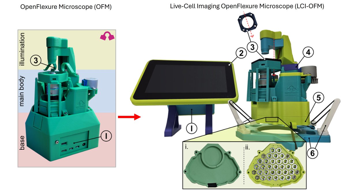
Unleashing live-cell imaging for the masses!
This is MEGA-exciting!!!
Adapting the OpenFlexure Microscope for Affordable Live-Cell Imaging https://t.co/CwPVM2rwKZ
This can fit into existing incubators and capture time-lapse experiments and feeds into existing bioimaging analysis pipelines.
Thanks @wellcometrust for funding
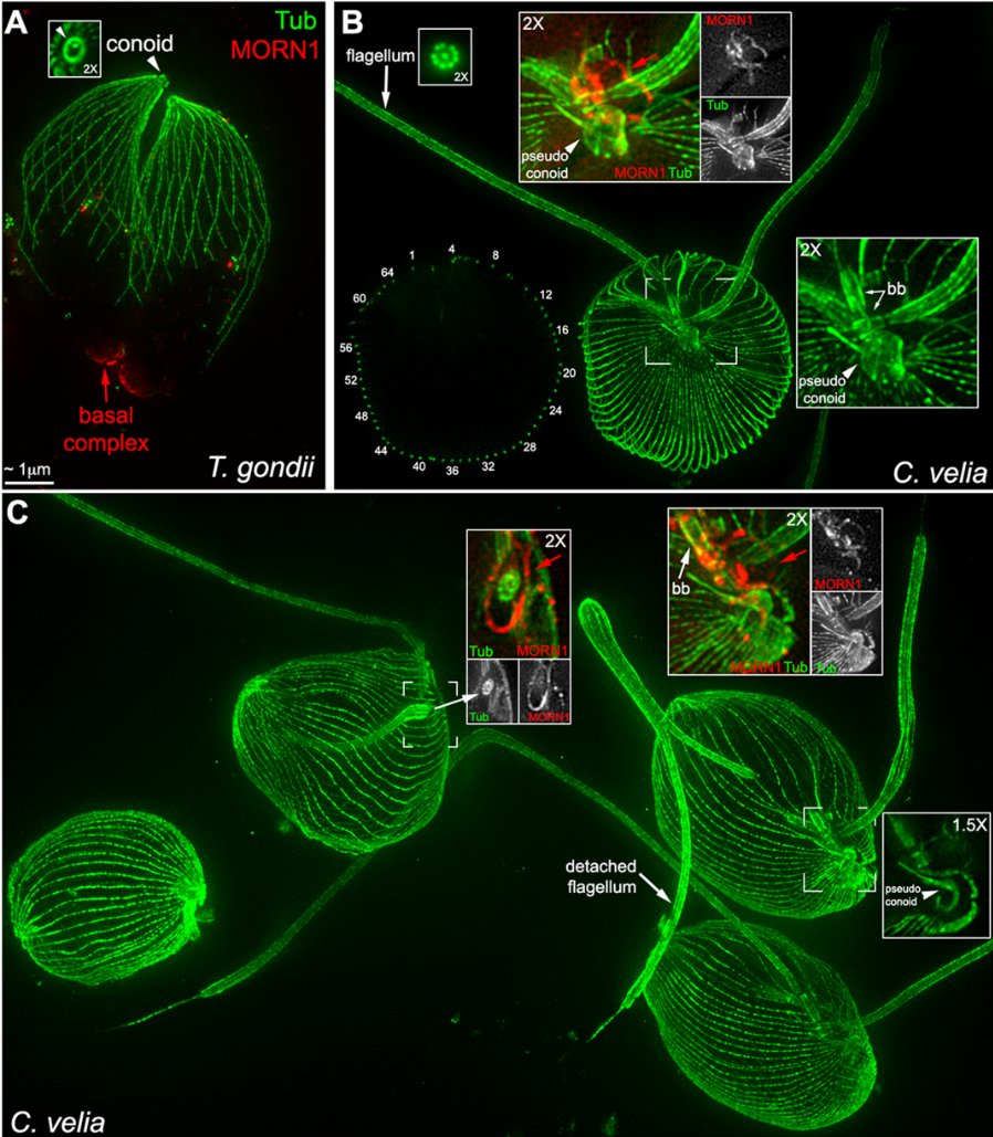
Isadonna Tengganu and Ke Hu develop transfection and ExM protocols for Chromera velia, enabling direct comparisons with its parasitic apicomplexan relative, Toxoplasma gondi.
https://t.co/iEO9bDxrk4
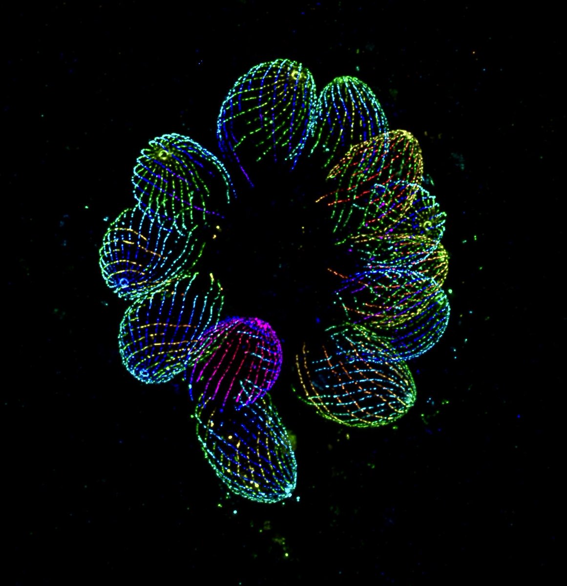
Happy #fluorescencefriday ! Haven’t done one of these in a while, but wanted to share what I’ve been working with in my new post doc! Below is an expansion microscopy image of Toxoplasma gondii stained for microtubules. #microscopy#fluorescence#toxoplasma
Burnette Lab paper: "Pharmacological Inhibition of β Myosin II Disrupts Sarcomere Assembly in Human iPSC-Derived Cardiac Myocytes" is out! This image is of beta myosin II filaments in an iPSC-derived cardiac myocyte. #CellBiology
https://t.co/UvFEH04CU8
"Non-Muscle α-Actinin-4 Couples Sarcomere Function to Cardiac Remodeling" is now online at Circulation Research! Congratulations to Dr. James Hayes on this ambitious work! There is a lot in the paper so let's start with the graphical abstract. @CircRes
https://t.co/OQ4tj9RGnu
Next-generation orange-to-far-red photoconvertible fluorescent protein for single-molecule microscopy and protein dynamic tracking
Oksana M. Subach, et al.
https://t.co/p3ezdkzDjT
A hungry heart muscle cell (cardiac myocyte) videoed through a microscope by Burnette Lab graduate student, @EmmaKoory. What is it eating and why? We may never know....... #CellBiology
A cell with prominent membrane blebs videoed using spinning disk confocal (left) and differential interference contrast (right) microscopy. #CellBiology