We are hiring 📢 Looking to recruit a new lab member (postdoc or PhD student level) to combine brain anatomy & connectivity with in vivo neural recordings & manipulation approaches to study the function of 🧠serotonin circuits.
@scilifelab@Stockholm_Uni
RTs appreciated!
1/2
Esr1+ hypothalamic-habenula neurons shape aversive states. Happy to share our latest results from @meletislab and @CarlenLab! It was a journey with @Daniela_CalvG and @fuzikjanos leading the charge to shed light on subcortical circuits driving aversion.
https://t.co/vmd1rFK10V
Exciting! Our findings in awake mice shed light on the impact of motor activity on striatal sensory processing and how dopamine shapes these interactions. Congratulations to @SilberbergGilad and Maya Ketzef #basalganglia#neuroscience#dopamine
https://t.co/DogmJTZOEb
How are sensory responses in the dorsal striatum shaped by movement? How does dopamine fit in? In vivo whole-cell “optopatcher” recordings and more, in this study with @RobertodelaTor & Maya Ketzef, online today!
https://t.co/IDKSArSqR3
What if you could now build the postsynaptic map of thousands of neurons in your favorite brain region and use it for connectome guided harvesting?!!! 🤯 congrats to the Fuzik Lab
We are thrilled that our latest study is out in @ScienceMagazine: different information processing in neurons with an axon-carrying dendrite (#AcD cells)! 1/13
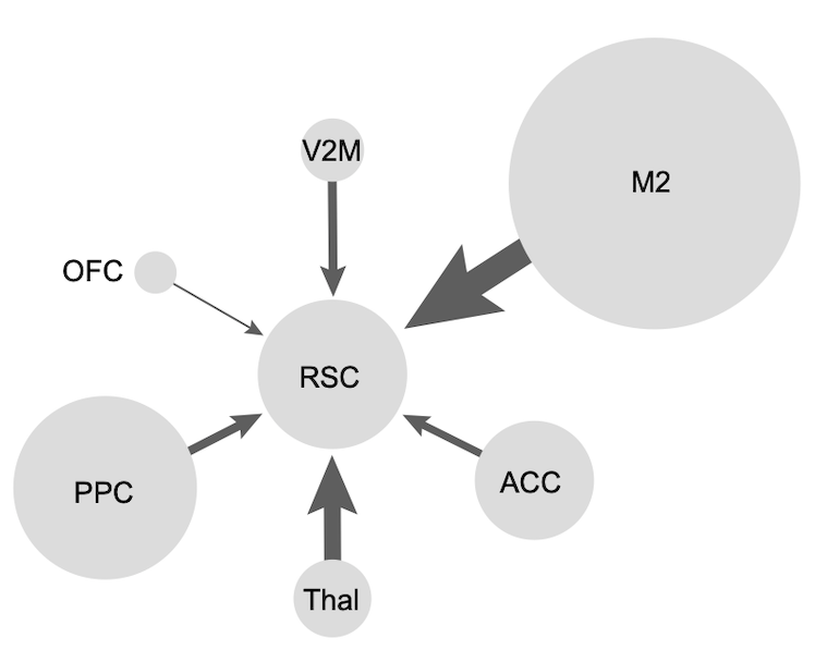
Spatial signals modulate neocortical processing. But where do these come from? Hippocampus? We checked the origin of place signals to the retrosplenial cortex. Surprisingly they originate from nearly all presynaptic circuits, particularly M2 and PPC.
https://t.co/YqMbWrIBDy
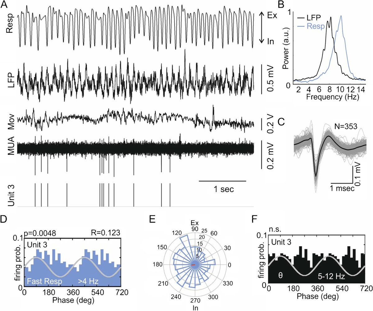
Happy to share our latest work with @DraguhnL et al. on the impact of respiration 🫁 on parietal cortex dynamics and its interaction with theta oscillations 🧠 https://t.co/mJsyHCcbm0
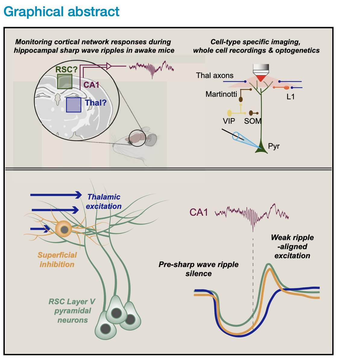
Patching neurons in awake mice, we found that synaptic responses in retrosplenial cortex during sharp wave ripples are widespread but modest. Just before ripples, local inhibition and excitatory input goes quiet, making those small messages stand out more. https://t.co/UCX0oXkWPn
New gradient in #hippocampal-#entorhinal connectivity: both dorsal and ventral hippocampal output signals converge in the dorsal medial EC. The results of a great cooperation with Shinya Ohara, Ken-Ichiro Tsutsui (Japan) and Menno Witter (Norway) @KISNeuro are now online:👇
Very excited to share our new preprint! Using whole-cell patch-clamp in awake mice, we reveal that sensory responses in the dorsolateral striatum are modulated by motor activity in a dopamine-dependent manner. @RobertodelaTor, Maya Ketzef, @SilberbergGilad https://t.co/QZV8VxttKr
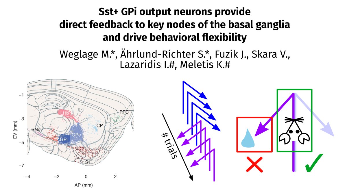
In our new preprint, we show that Sst+ GPi is more anatomically complex than previously described, and that its role in behavioral flexibility is in action execution, not outcome evaluation. w/ @sahrlund @SkaraVasiliki@fuzikjanos@iaklaz @meletislab https://t.co/Phti2ARs7Y 1/8
The Carlenlab is looking to hire! Positions: postdoc x2, PhD, research assistant, all with a focus on the structure and functions of the prefrontal cortex.
Links and information are found in the thread. Come join us @karolinskainst in Stockholm, Sweden!
🙏 for RTs.
#PFC