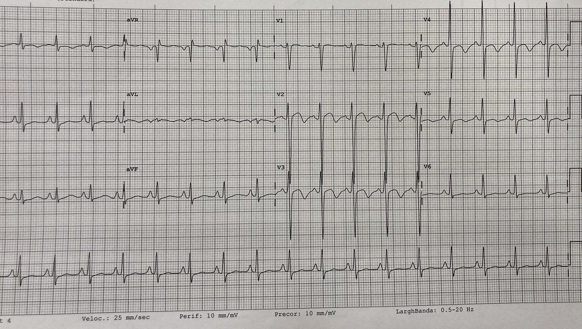
Beautiful typical essudative pericarditis. Don't forget CMR findings: signal hyperintensity of pericardium on T2-W sequences, pericardial LGE , pericardial effusion (in this case with fibrinous shoots).
…A night shift under pressure...
(part 2)
Abdominal ultrasound was performed, evidencing a 7 cm expansive formation, slightly hypoechogenic with regular margins, extending from right kidney to the I and VIII hepatic segment…
Here are the images of the following abdominal MRI:
_…A night shift under pressure...
(part 1)_
👤 30 y.o. man, without CV history
🏥go to the emergency room for epigastralgy for about ten days, increased in the last few hours.
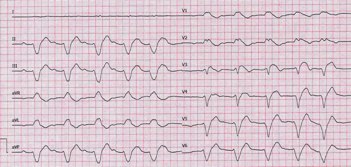
EKG below. PA 250/150 mmHg.❤️🔥
The echo shows a mildly dilated LV, FE 20%.
What's your hypothesis?
1/4
👤61 y/o man with hypertension without any CV history.
🚑 call the emergency because of sudden onset of typical chest pain.
⚡️ EKG below
▶️ cath-lab for primary PCI
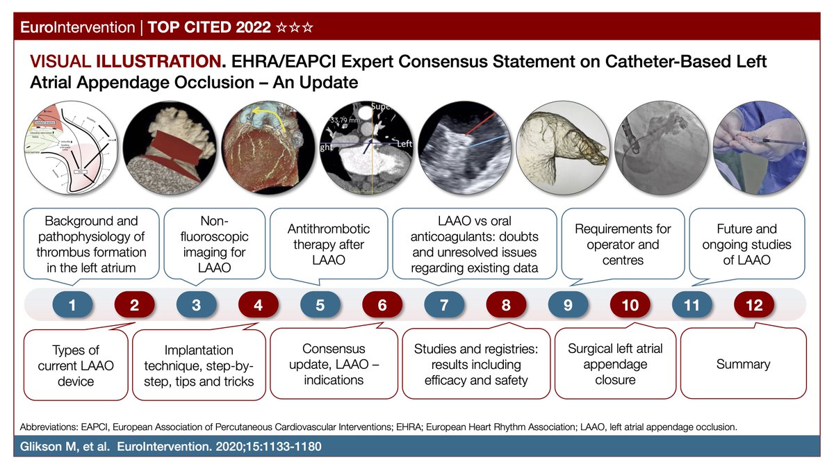
This EHRA/EAPCI consensus document on catheter-based left atrial appendage occlusion is made of 48 pages, 12 chapters, 1 supplement, 24 statements, 20 tables, 44 figures and 24 videos. #EIJTopCited
https://t.co/60cY1OH0gQ
!!Very nice echo/HF case!!
63 yo male, smoker, no past medical history, 1month ago onset of SOB, orthopnea and ankle swelling, no angina; very high NTproBNP, bilateral pleural effusion, sinus tachycardia, Q wave in inferior leads; started furosemide 50mg/die ➡️ NYHA II.
1/2
Superior vena cava syndrome ➡️SVC compressed by right hilar tumor + consequent thrombosis of subclavian dx-anonymous vein
Another great collaboration with vascular surgeon in our cath lab 💪🏻💪🏻 @gladiol_zenunaj
9 Fr right omeral vein + 6 Fr right femoral vein @CarloPenzo1
Immediate hemodynamic collapse following BAV due to severe acute AI is rare, but can be catastrophic. In this high risk patient with refractory HF & cx (-)ve osteo we used an #Impella to bridge to on the fly #TAVR. Pt extubated next AM and doing well mild PVL on f/u TTE @TAVRBot