Excited to see my image on the cover of @NatImmunol!
The scanning electron microscopy image shows lymph node ECM conduits (following removal of cells), which serve as a mechanosensation platform for stromal cells to coordinate organ growth. Read more: https://t.co/VA7V0rA62W
We are hiring a postdoc/Ph.D. student! Join our lab to study cell migration guidance in the context of adaptive immunity.
The details of the position and the instructions for application are found at: https://t.co/L6gXl9jObM
Hello everyone, excited to share our preprint: https://t.co/DoThdFBDLT
----
Everyday, cells like this crawl through your body to protect you. While we know that their front 🔴 and back 🔵 polarity programs work together, how exactly do these two processes communicate? Lets chat!
Guess which of these movies are played forward and which backward? It's not that easy isn't it? We were similarly confused about this, so we started a project with our favourite toy system on entropy in macroscale active matter:
https://t.co/37RlOZzCMh
Let me show you a teaser.
📢The latest issue of the JID is here!
https://t.co/ca0zprQP66
Cover image: depicts a shallow 3D reconstruction of the dermal-epidermal interphase of a human skin section showing the close interaction between #melanocytes and #keratinocytes in the #epidermis. The image was acquired on a dual-laser two-photon microscope, and label-free second-harmonic and third-harmonic imaging (SHG/THG) was used to capture fibrillar collagen (gray) and basal keratinocytes (purple), respectively. Other keratinocyte layers and melanocytes were stained with respective antibodies against Keratin-10 (red) and beta III Tubulin (cyan). Image presented with a transparent background and courtesy of Frank Assen, Ph.D. and Prof. Wolfgang Weninger, M.D., Department of #Dermatology @MedUni_Wien #dermtwitter
6 years of PhD lab work and fantastic collaborators!
I proudly present our recent work: Light-microscopy based dense connectomic reconstruction of mammalian brain.
https://t.co/VSB5BPu2aD
JID November 2023 Cover Video: Visualization of Melanocytes Lining the Hair Follicles and Epidermis.
https://t.co/xo5DrpDMLc
#dermtwitter#dermatology#dermatologist
Fly-Over: Visualization of Melanocytes Lining the Hair Follicles and Epidermis The video showcases a 3D fly-over of immune-stained human skin, highlighting melanocytes (identified by tubulin 3 in white) that line the junction between the hair follicle and the epidermis with the dermis. These melanocytes form intricate protrusions extending into the keratinocyte layers of both the epidermis and hair follicle (indicated by keratin 10 in green). Within the dermis, auto-fluorescent elastin fibers (in red) are visible, accompanied by small nerve fibers (also marked by tubulin 3 in white) that project near the hair follicle and sebaceous gland. The hair and its shaft exhibit additional autofluorescence in white and red.
The November issue of the JID is out now! Take a look at this month's issue at https://t.co/ca0zprQP66 and the striking cover below.
#dermtwitter#dermatology#humanskin
The cover image reveals a highly detailed 3D reconstruction of a 20-micrometer-thick section of immune-stained human skin. It showcases the compact melanocyte lining (indicated by tubulin 3, in white) located at the junction of the epidermal and dermal layers. This lining forms complex protrusions that extend into the keratinocyte strata of the epidermis and hair follicles (marked by keratin 10, in green).
A sebaceous gland is discernible at the base of a hair follicle, and nerve fibers (also marked by tubulin 3, in white) can be found around the base of the hair follicle and within the dermis. Furthermore, auto-fluorescent elastin fibers of the dermis are shown in red.
The image stack was acquired on a confocal fluorescence microscope using a 40x silicon oil objective, stitched together, and rendered using Imaris software.
Image courtesy of Frank Assen, PhD & Prof. Dr. Wolfgang Weninger (Department of Dermatology, @MedUni_Wien Austria)
🧵(1/6)
How do immune cells know where to go? It has been proposed that cells simply respond to externally established gradients of chemokines, but we think that’s not all…
Dendritic cells can “self-shape” the chemotactic gradients that help them transport antigens to lymph nodes, finds a new live imaging study from @Sixt_Lab@EdouardHannezo.
📄: https://t.co/g6ZCFCfCQW
Focus by @hannahdonnelly_@EMandrou@robinsall: https://t.co/1G1xcJdKe4
For this Focus issue, we asked experts in bioimaging from around the world to tell us what excited them about the future of bioimage analysis for the next 5-20 years. (1/n)
All Focus content can also be found here:
https://t.co/ckMtBOofwv
Check out our new paper from first author and recent PhD graduate @LauraBocaM.
See how interkinetic nuclear movements give rise to tissue fluidisation in an epithelium.
https://t.co/2GFjhHxX25
(1/n)
Exciting news! Our latest publication by @karlahuljev et al. shows a hydraulic feedback loop that promotes embryonic axis formation in #zebrafish. Check our tweetorial for a sneak peek. @Dev_Cell: https://t.co/poXkfcen4Y
The directed cell migration GRC is happening next January and there are still some places available! Don’t miss out on an interactive and engaging meeting.
Find out more here: https://t.co/hI4FYtTz7H
NEW PAPER ALERT🚨: As an embryo develops, biochem signaling+mechanical forces drive tissue formation. How these processes are coordinated is unclear. In their collab, @EdouardHannezo group and @HeisenbergCPLab offer imp. insights into #tissuemorphogenesis
https://t.co/BSDuVvB42T
New manuscript out! 🎊 We show that waves of polymerizing actin synchronize in collectively moving endothelial cells. A mechanism that can be mimicked with motorized balls 🤹♀️ An amazing work from @mi_riedl bridging #physics and #biology! check it here 👉 https://t.co/PzBKEO6js8
Excited to share the fruit of my labor of my PhD and my first original paper:
Synchronization in collectively moving inanimate and living active matter: https://t.co/R1CUfQsIQI
Here is the story: (1/12)
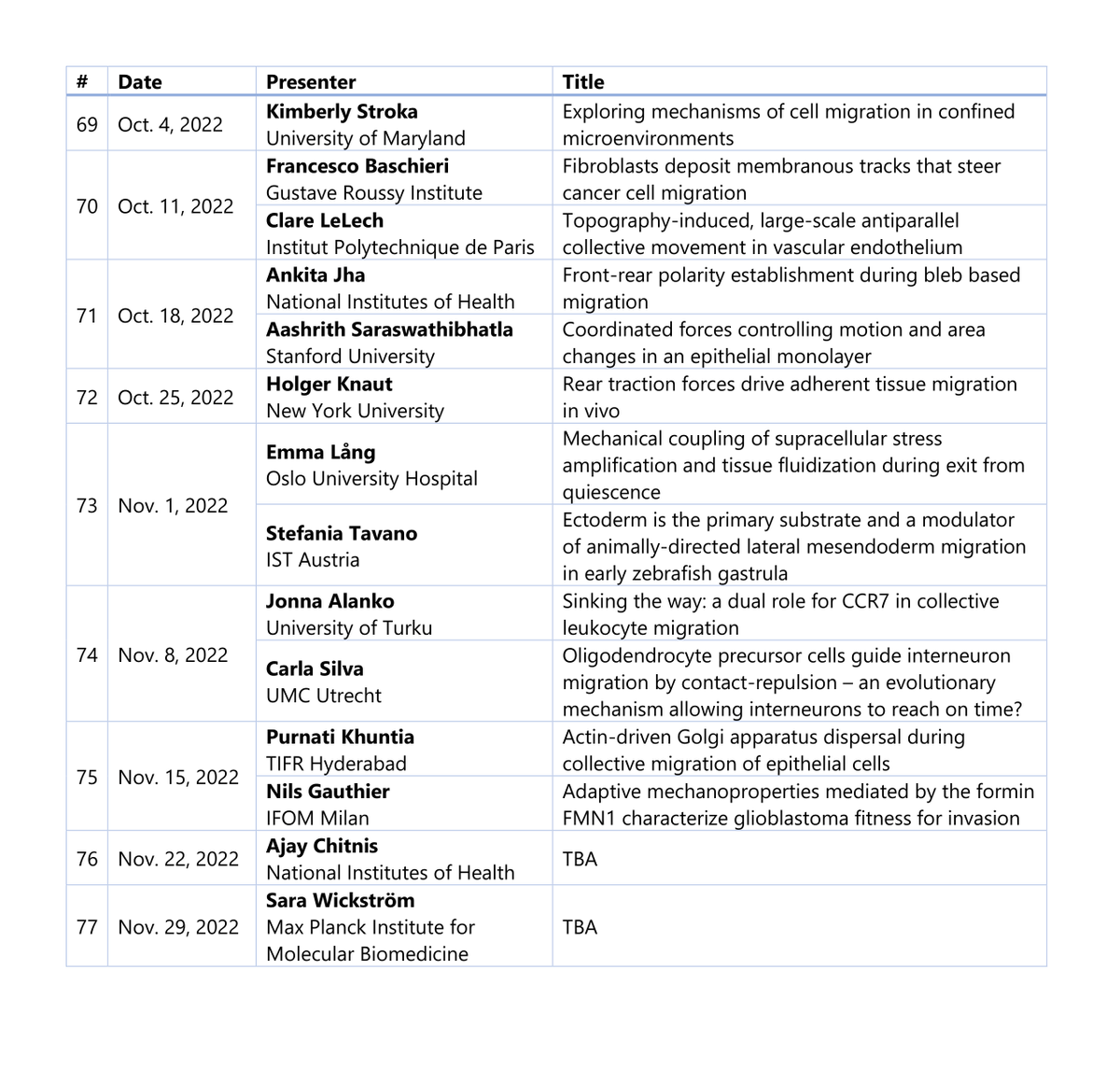
📢Announcing... the Fall '22 Cell Migration Seminars Lineup. We are so excited for all of these talks and can't wait to see you all there!
Tuesdays Oct 4 - Nov 29, 11 AM eastern time, zoom & youtube.
If you need to get on the mailing list, sign up at https://t.co/t4p4XrFGT5