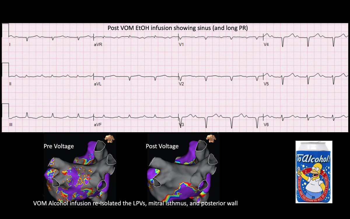
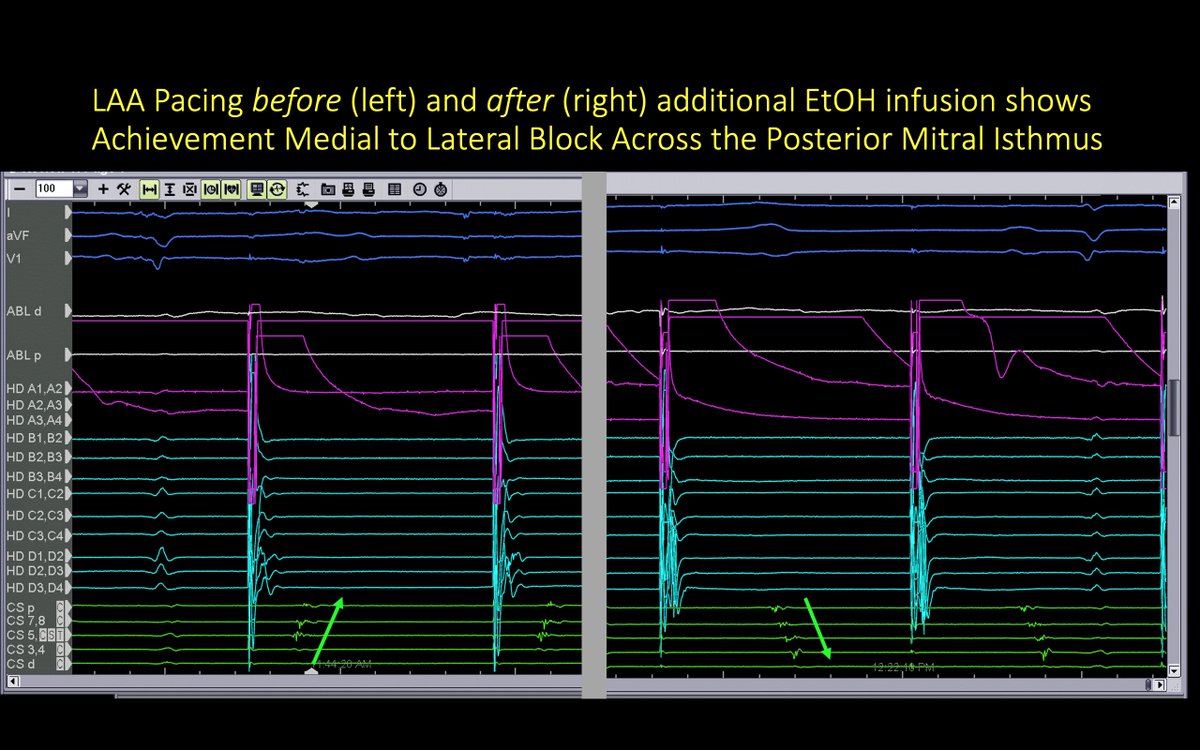
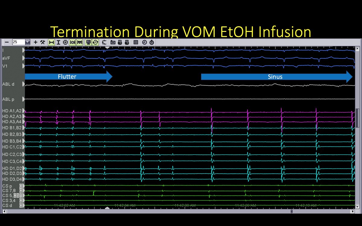
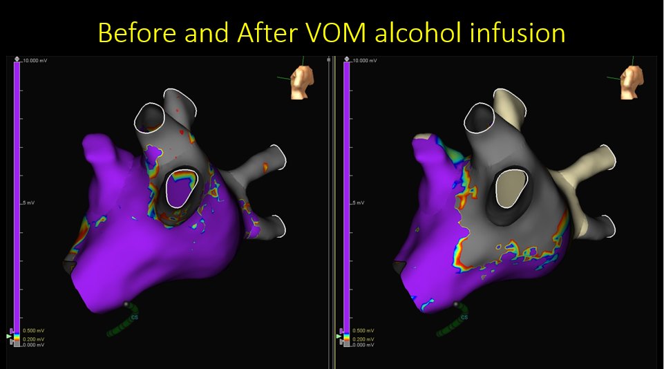
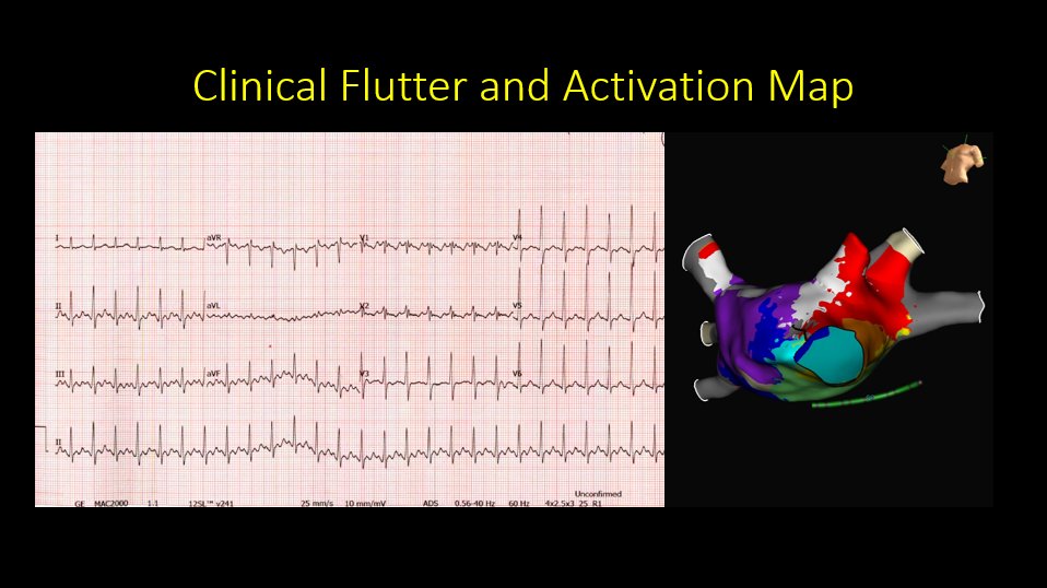
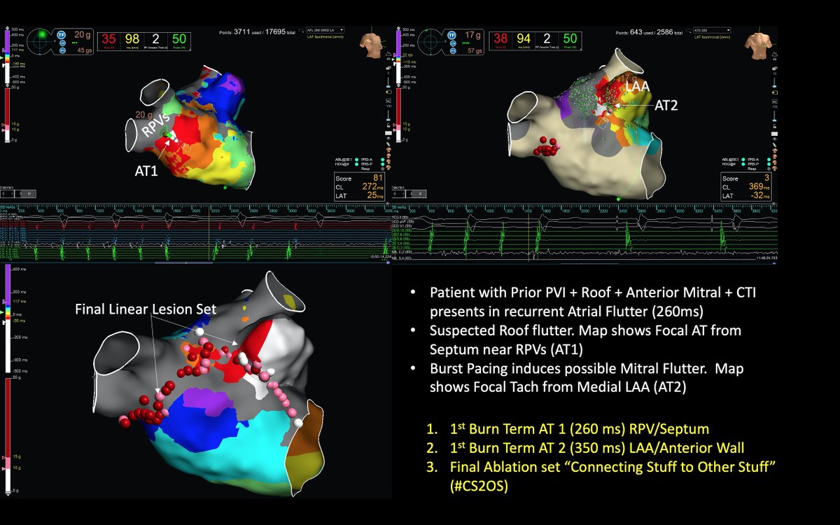
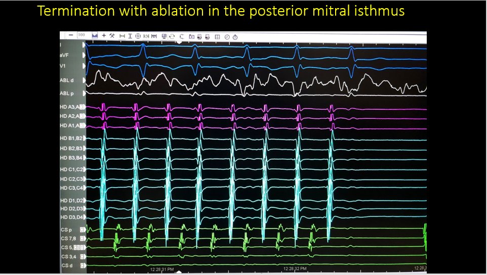
We hoped this would happen but did not expect it...termination of left atrial flutter during VOM ethanol infusion. Hopefully it helps this patient. Nice maps @ryancolemaps@maddyferraro1@arcampado @MiguelVldrbno @TJHeartFellows
Unusual redo because the previous ablation was intact (PVI, roof, anterior mitral line) yet 2 distinct ATs came from areas near the previous ablation🤔. Termination (x2) was quick, but we decided on linear ablation #CS2OS. Thanks @ryancolemaps@TJHeartFellows@AbbottCardio
In our editorial, we argue that stroke risk might diminish after a period of AF-free time. Thus, there is value to investigating the time since the last AF event to the future risk of stroke. @FrischMd @SDikdan @DrHowardWeitz@LiverpoolCCS@TJHeartFellows
https://t.co/vDN5V31cW9
Nice example of propagation from a focal LAA-AT that also shows lateral to septal block across the anterior mitral line (and roof line too). Thanks @ryancolemap and your colorful EnSite X system and @MichaelCoMD (@TJHeartFellows) #EnsiteX
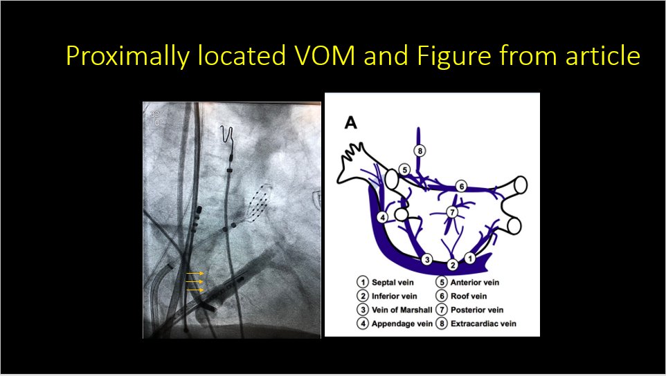
In this interesting case of mitral flutter we were surprised by how proximal the VOM was in the CS with venography. Luckily we had a guide! Thanks @MiguelVldrbno @MichaelCoMD@AbbottCardio@TJHeartFellows https://t.co/XBy3dOiHUn
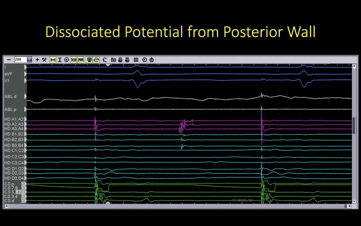
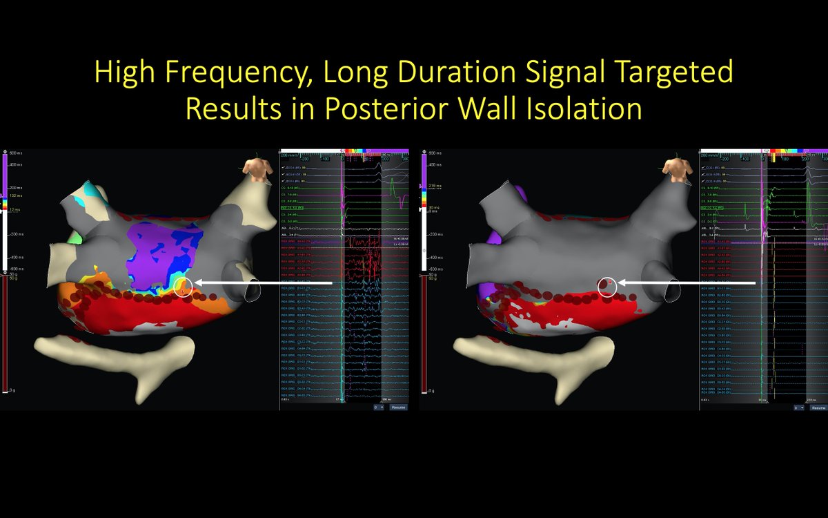
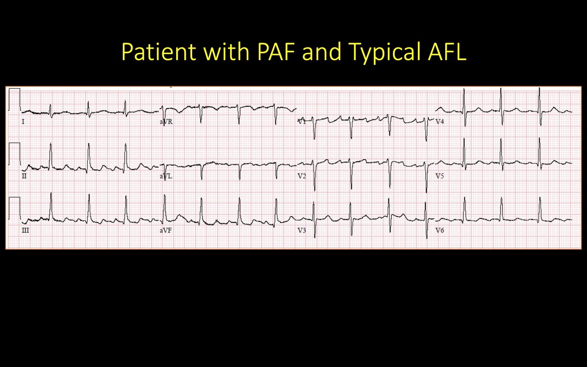
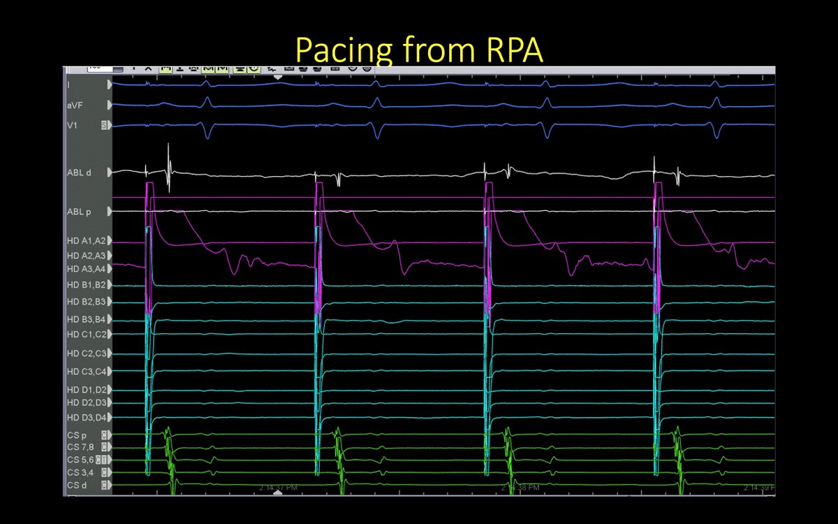
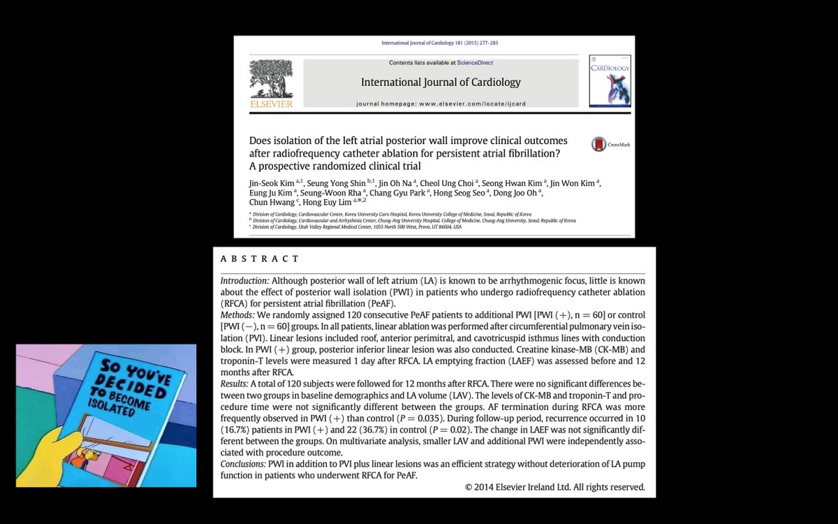
This patient had PAF after prior abl for Persistent AF. PVI was intact. We targeted the posterior wall (PW). An interesting EGM was seen on the grid as the remaining connection and (unusually) we were able to show exit block from the PW @ryancolemaps@AbbottCardio
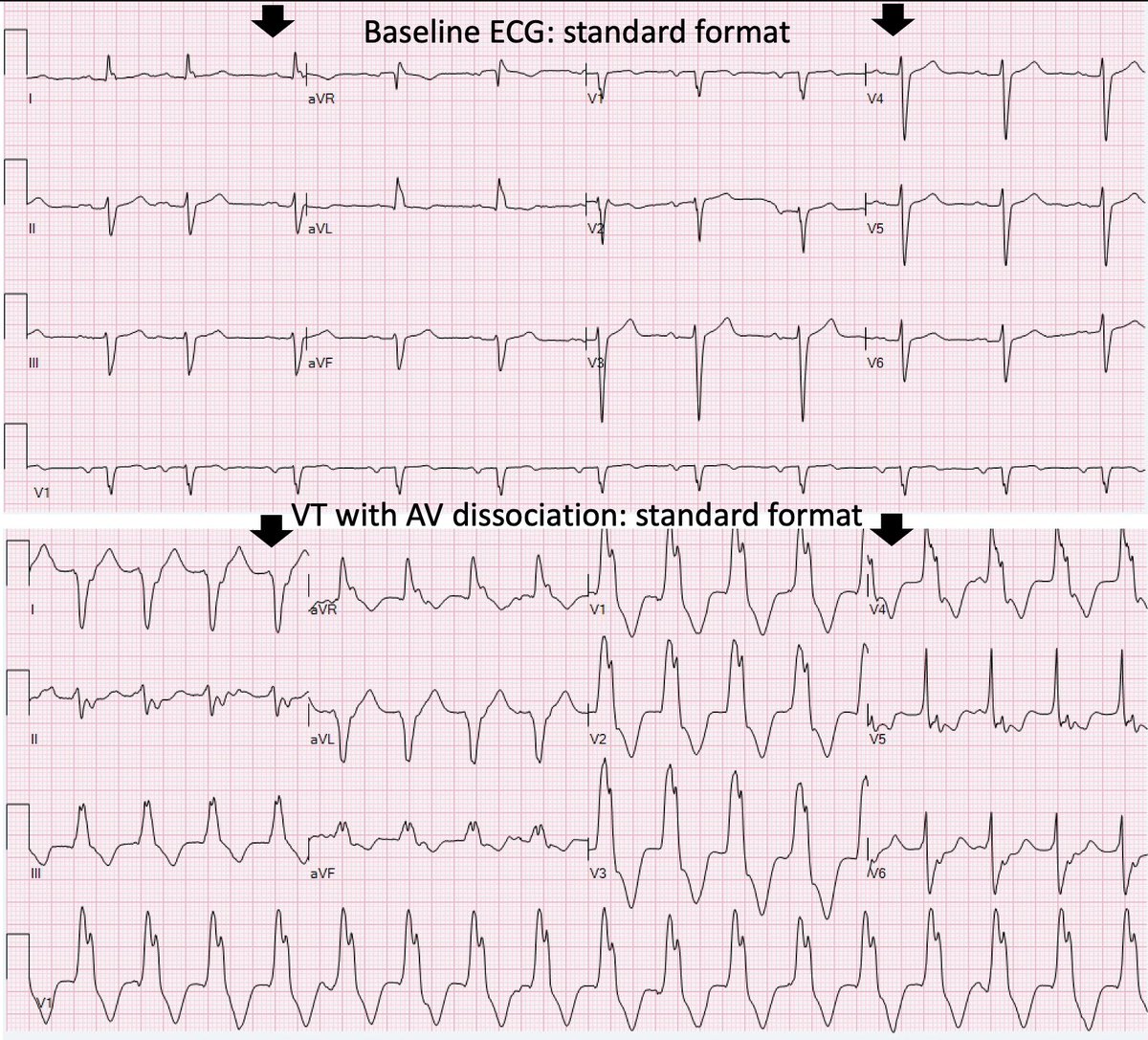
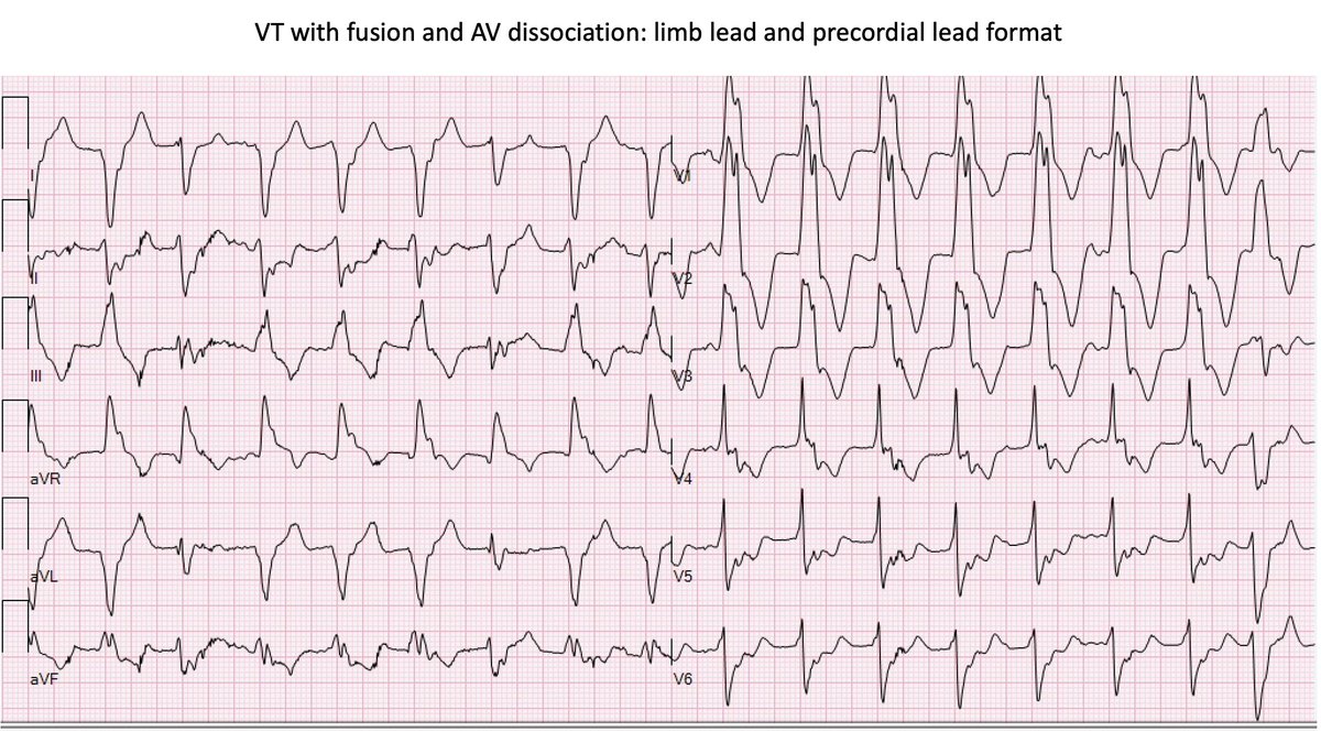
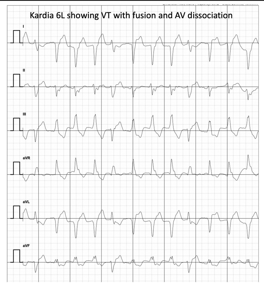
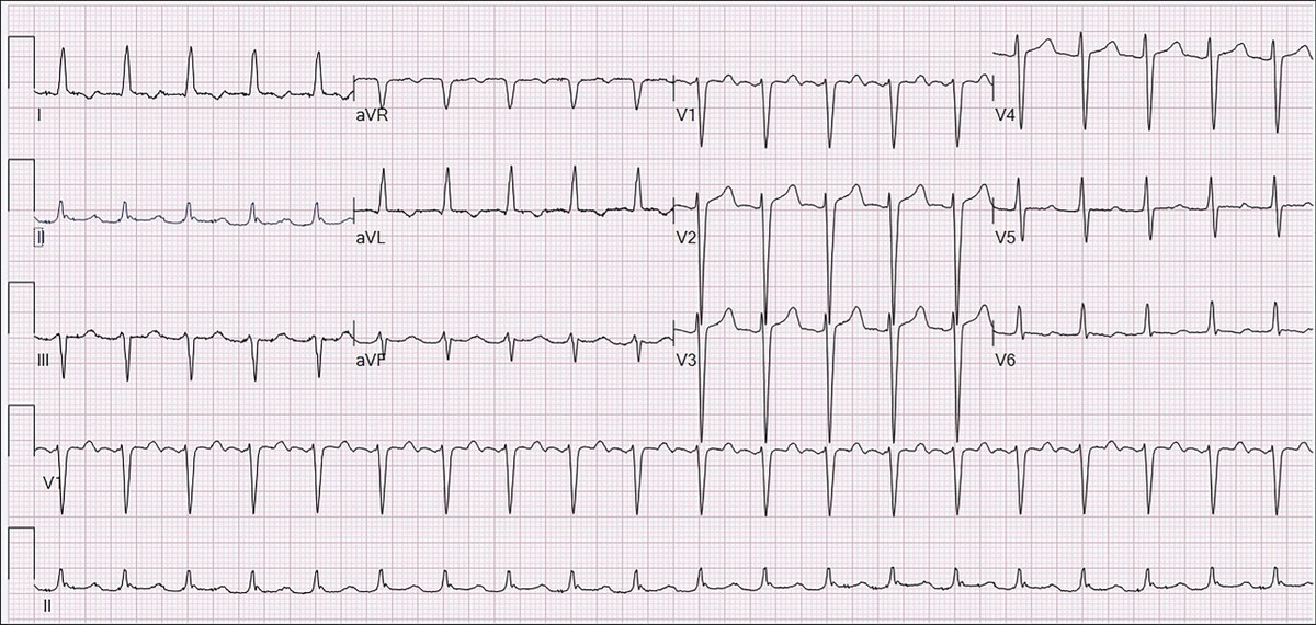
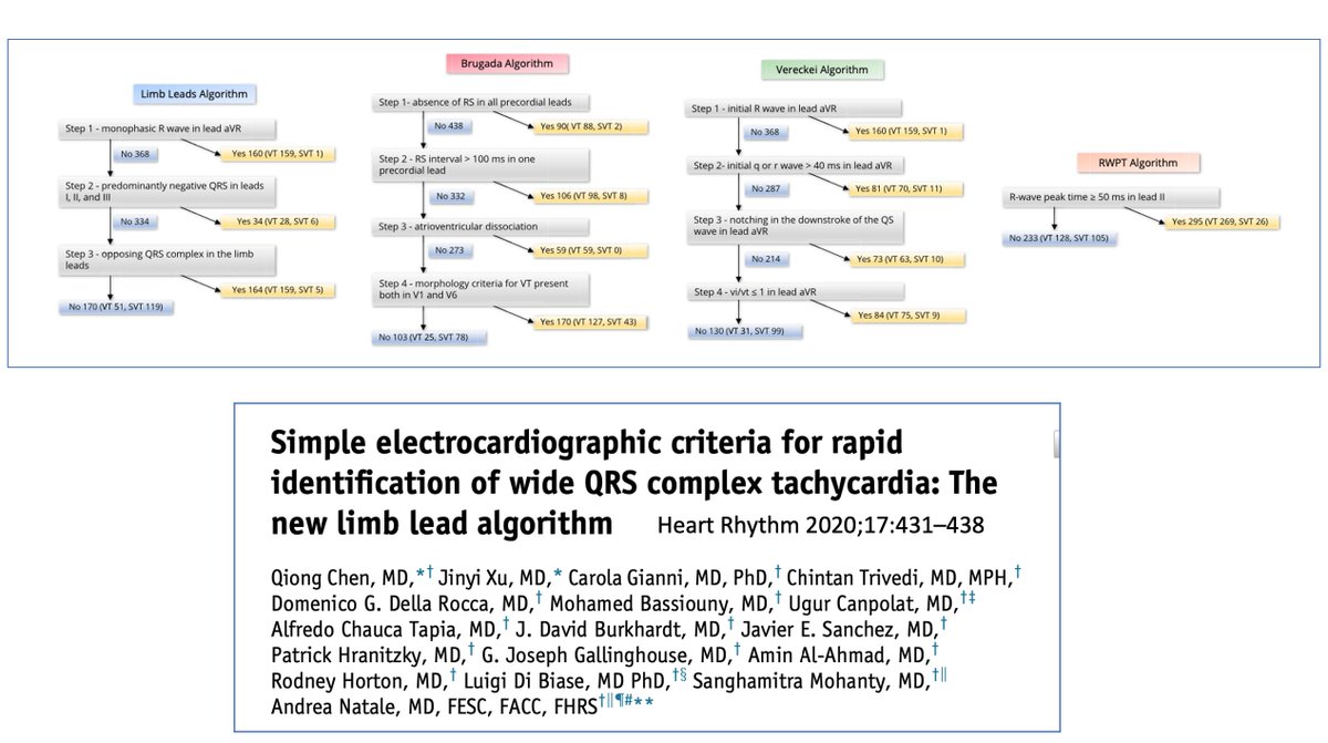
This was an unusual opportunity to record (slow) VT using a Kardia 6L and show the corresponding ECGs. Choose your algorithm… @DrDave01@narrowQRS@TJHeartFellows
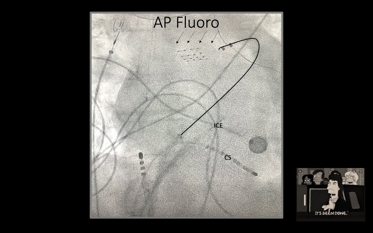
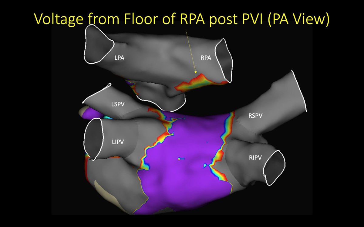
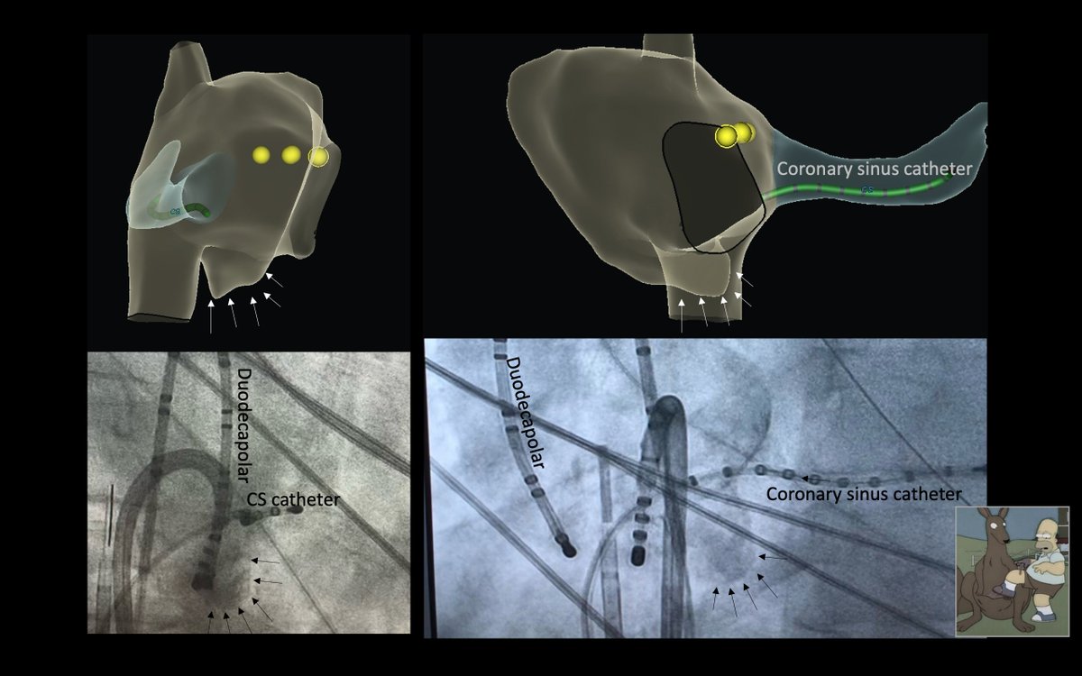
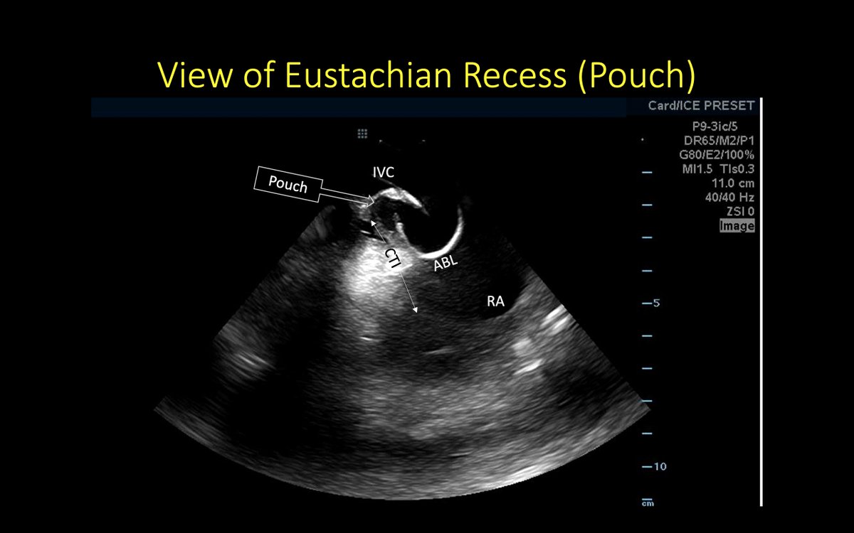
ICE was essential in recognizing the variant anatomy in this patient. Subsequently, we appreciated it in the other imaging modalities. Thanks @ryancolemaps@AbbottCardio