Starting the year with a recap on a recent challenging VT case at @mhshospital@Mapbastian@JC_EPMaps@mike_lean@joel_chic
A 3rd time redo in a non-ischemic VT patient.
Success in these cases requires the use of advanced techniques - however, advanced techniques do not mean unsafe procedures.
Some specific workflow highlights that led to a successful bipolar case without complications and 0 clinical VT on follow-up:
- Meticulous mapping was performed to fully understand each VT before delivering energy.
- Reentrant VTs are inherently 3D in nature, often revealing only a portion of the channel in the chamber mapped (in this case, only the exit for each VT).
- Entrainment at the presumed VT exit confirmed what the map suggested.
- Remote entrainment from the RV was helpful to confirm diastolic signals on mapping catheter were within circuit.
#AblateVT #EPpeeps #WIC #WomenInEP
I do like the method of starting with OT NF and switching to FD. Alternatively, I know @AMatthews0/@brysontindal9 tend to stick with OT NF and, if necessary, close their window when they see diastolics. Others will map with FD then TurboMap with NF rather than juggling two algorithms. Many ways to do it!
I know @DrRoderickTung has presented a slide about OT NF outperforming FD/LD in VT mapping, but I don’t have it handy.
Also worth mentioning at the HRS 2024 fellows mapping event, @RChung_EP and I found that OT NF accurately delineated the isthmus, while FD/LD alone seemed to under-represent the channel boundary and overestimate the width of the exit.
@pjsm83
Some post-case learning with @danealson (Dr. Neal Bhatia) and @_shannonmillard: following an ischemic VT case, together we compared how the clinical VT circuit presented when mapped with the First Deflection algorithm versus EnSite™ OT Near Field.
What we found brings up an interesting question. Is it better to….
- Force the system to visualize the full circuit with First Deflection, or
- Illustrate the potential midmyocardial bridges and touchdown points with OT NF?
While there may not be a "correct" answer, we think there is value in appreciating both pieces of information. And if nothing else, it makes for a fun discussion with talented colleagues like Dr. Bhatia and Shannon.
Here are LAT & voltage maps in VT with each detection algorithm.
I do agree about the need to appropriately delineate LOB/split signals. With that, we have actually found NF to work great in objectively annotating those signals. However to your point, it does come down to physician preference and many prefer to see the full circuit.
With that, in some cases we will switch to FD to force annotation of signals that are presumably deeper in the myocardium and not detected by OT NF. Either way, it is helpful to TurboMap and have both maps in a case.
Utilization of #AI based tools for delineating cardiac structures with ICE during #AblateVT cases as part of #FIH trials with new @AbbottCardio exceeded expectations - rapid definition of valves, paps in LV:
"Roads...where we are going, we don't need roads."
Happy to share our work @rimhalabymd@ChrisGBruce13 using VINTAGE for intramural VAs - first in human. We demonstrated that we can successfully navigate to "inaccessible" areas such as the LV summit, deep septum, and papillary muscles and deliver successful ablation lesions. More to come on this exciting new "space" in VT!
https://t.co/fYD0KvTeAl
@melchami99@FaisalMMerchant@BadhwarNitish
Challenging VT storm with multiple morphologies in a hemodynamically unstable patient for redo procedure. Substrate-guided strategy, focusing on areas of wavefront discontinuity with dual pacing wavefronts suggested a potential VT isthmus. Area corresponded nicely with @inheartmedical wall thinning. Non-inducible on PES after RF. @AbbottCardio@mike_lean@Mapbastian
Thankful for lessons learned @JRWinterfield@Davilandre@DrRoderickTung #AblateVT #NoExitBias #EPeeps #WADL
VT storm ablated with a first burn term over the weekend thanks to @JRWinterfield. #GridX with OT near field algorithm allowed for quick and accurate mapping in complex substrate.
Enjoyed assisting with some workflow and technology improvements in FIH trials of novel Viewflex X ICE catheter from @AbbottCardio -- we performed ablation of PVC from suspected intramural site with RF to septal RVOT and adjacent ASOV. Will be curious to see what @dhakalbish thinks with his @PennEPFellows based ICE approach in trials tomorrow. Contouring of key structures including pap muscles and ASOV shown below:
@AMatthews0@mikelean@Zachkoch13
It is amazing to reflect on how many specialties and technologies had to come together to make this case and collaboration happen. At the forefront of this is @FergieLosiniec1 - thank you for being a pioneer and a top-class operator!
Great case of ICE guided LAAO with @medinbox@AbbottCardio support. @medinbox allows for procedural support worldwide between physicians and key specialist that can assist with tips during complex cases. It was super easy to connect. Looking forward to sharing more cases with my peers all around the world. Thanks for the visit! @KahabkaMark@Austin_in_EP@mike_lean@Mapbastian@joel_chic
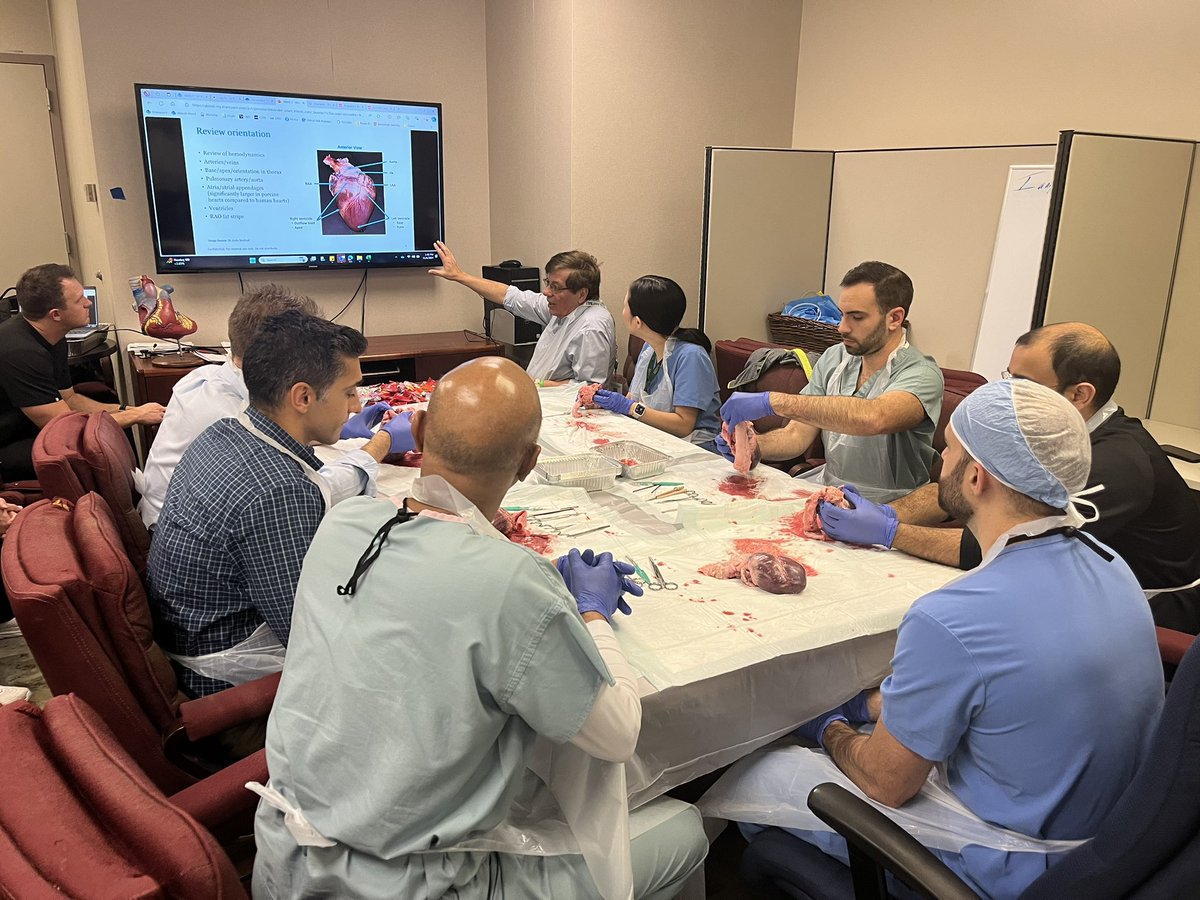
Great heart dissection with the EP fellows at @UABCardiology. Incredible insight from the very knowledgeable cardiac pathologist, Dr. Silvio Litovsky! @AbbottCardio @WilliamMaddoxMD @tommcelderry