Join us 10/19/20 for the second BCM pathology residency virtual open house! Main program is 5-6:30pm with option to join in on am lecture from 8-9am. We hope to see you there! https://t.co/XYadrEmkcS #pathmatch21#pathmatch2021#path2path
Spermatocytic tumor: 77 yo M, L testicular mass. Hx of R scrotal mass. Cellular tumor composed of three types of cells of varying sizes (small lymphocyte-like cells, intermediate cells and giant cells). Bilaterality is more common than in seminoma, affects older patients. CD117+
Ameloblastoma: 49 yo F, with jaw expansion. Variably sized epithelial islands composed of central loosely arranged stellate reticulum-like cellular areas, surrounded by columnar/cuboidal epithelial cells (ameloblast-like cells) with nuclear palisading and reverse polarization.
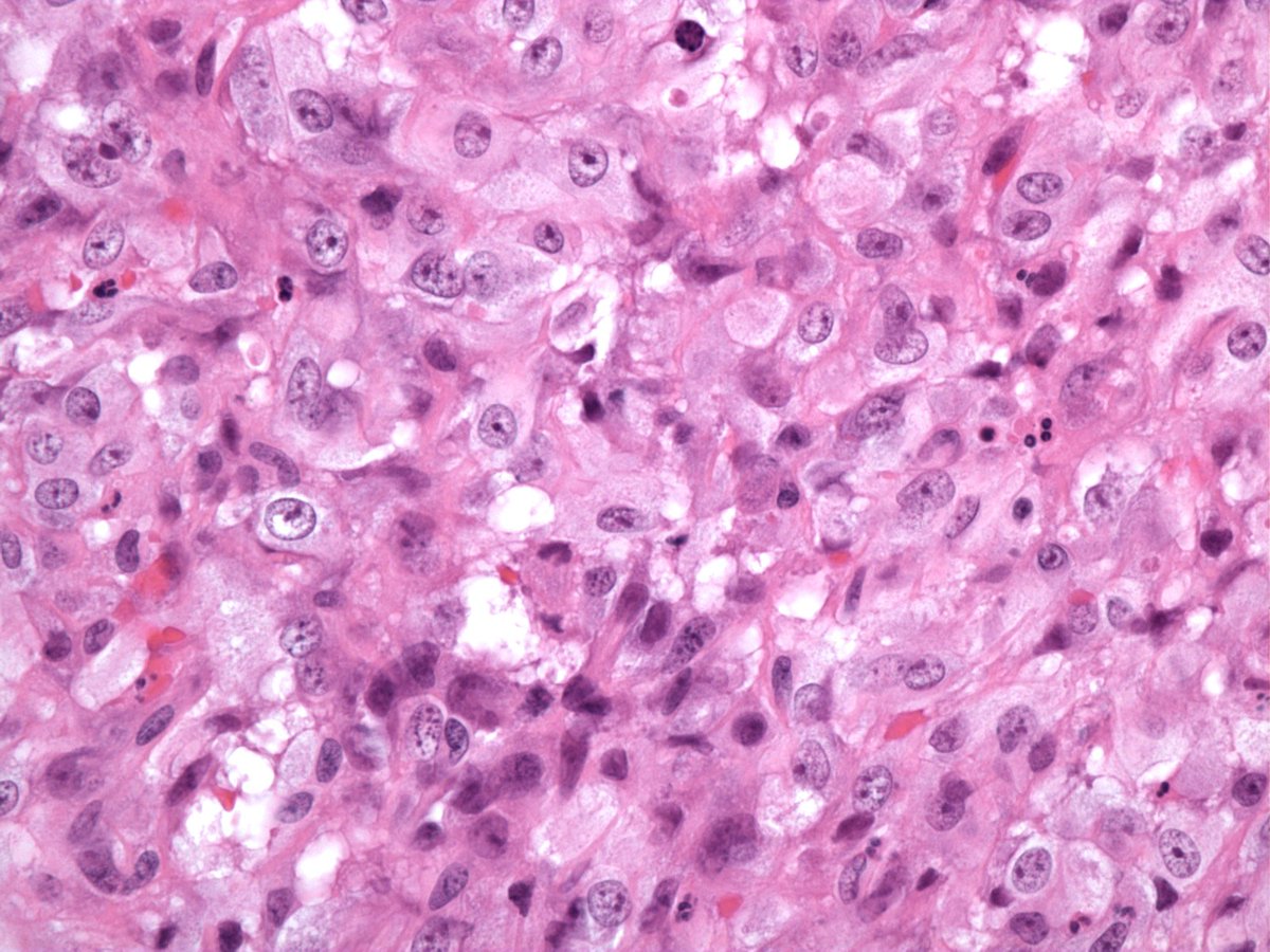
Malakoplakia: 56 yo M with nodule on bladder wall. Sheets of round histiocytes with abundant eosinophilic granular cytoplasm (von Hansemann cells) with rounded concentric basophilic intracytoplasmic inclusions (Michaelis-Guttman bodies). Admixed acute and chronic inflammation.
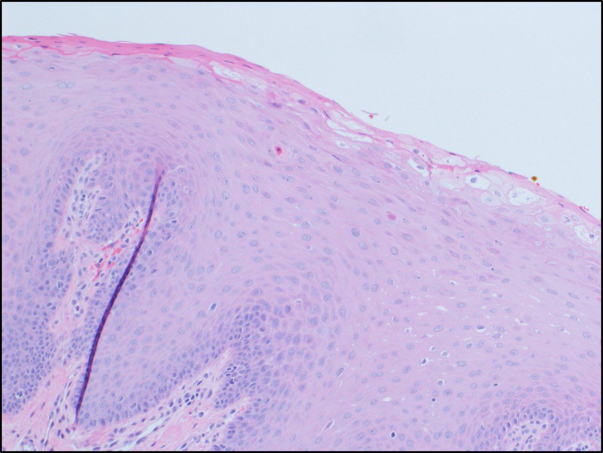
Heck's Disease (Focal Epithelial Hyperplasia) shows irregular hyperplasia with hyperkeratosis, parakeratosis, and acanthosis with endophytic growth and widened and fused rete ridges. Mitosoid cells (arrows) are characteristic feature. It's due to HPV 13 & 32 #pathology#dermpath
Angiomatoid Fibrous Histiocytoma: 28 yo man with 4.2 cm LUE mass. H&E showed nodules and sheets of bland spindled histiocyte-like cells with abundant blood-filled pseudovascular spaces. The mass was well-circumscribed and contained a peripheral cuff of lymphocytes. IHC: Desmin+
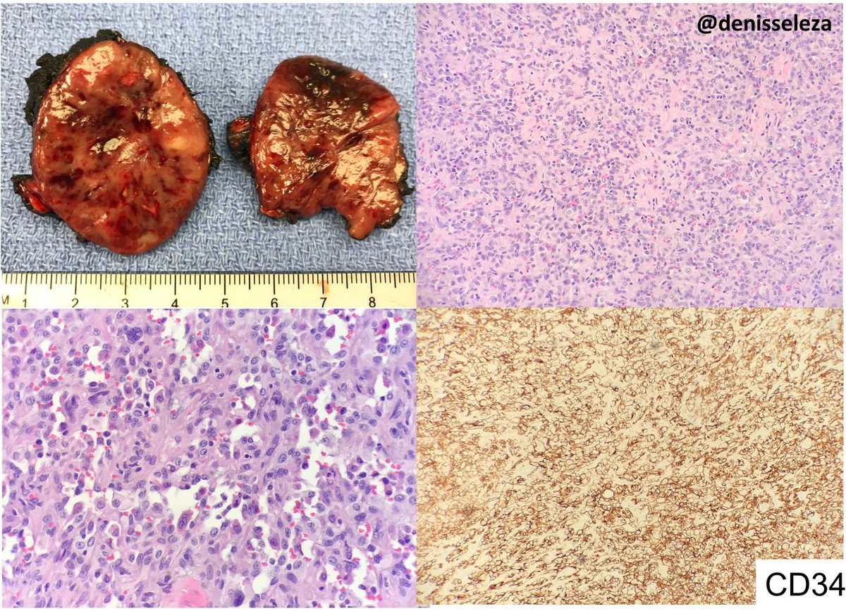
Epithelioid hemangioendothelioma: 5.5 cm mass, solid cut surface. H&E: large epithelioid cells with enlarged, vesicular nuclei and abundant eosinophilic cytoplasm. Anastomosing vascular spaces are also present. Tumor cells diffusely positive for CD34, CD31 and TFE3 @denisseleza
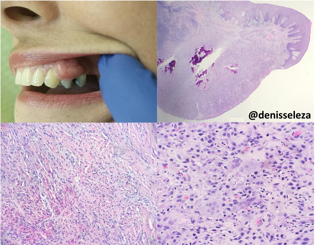
Peripheral ossifying fibroma: Gingival lesion. Cellular proliferation of spindled–fibroblast like cells with ovoid-nuclei and dispersed chromatin, osteoid or woven bone. May have multinucleated giant cells. Ddx pyogenic granuloma and peripheral giant cell granuloma.
@denisseleza
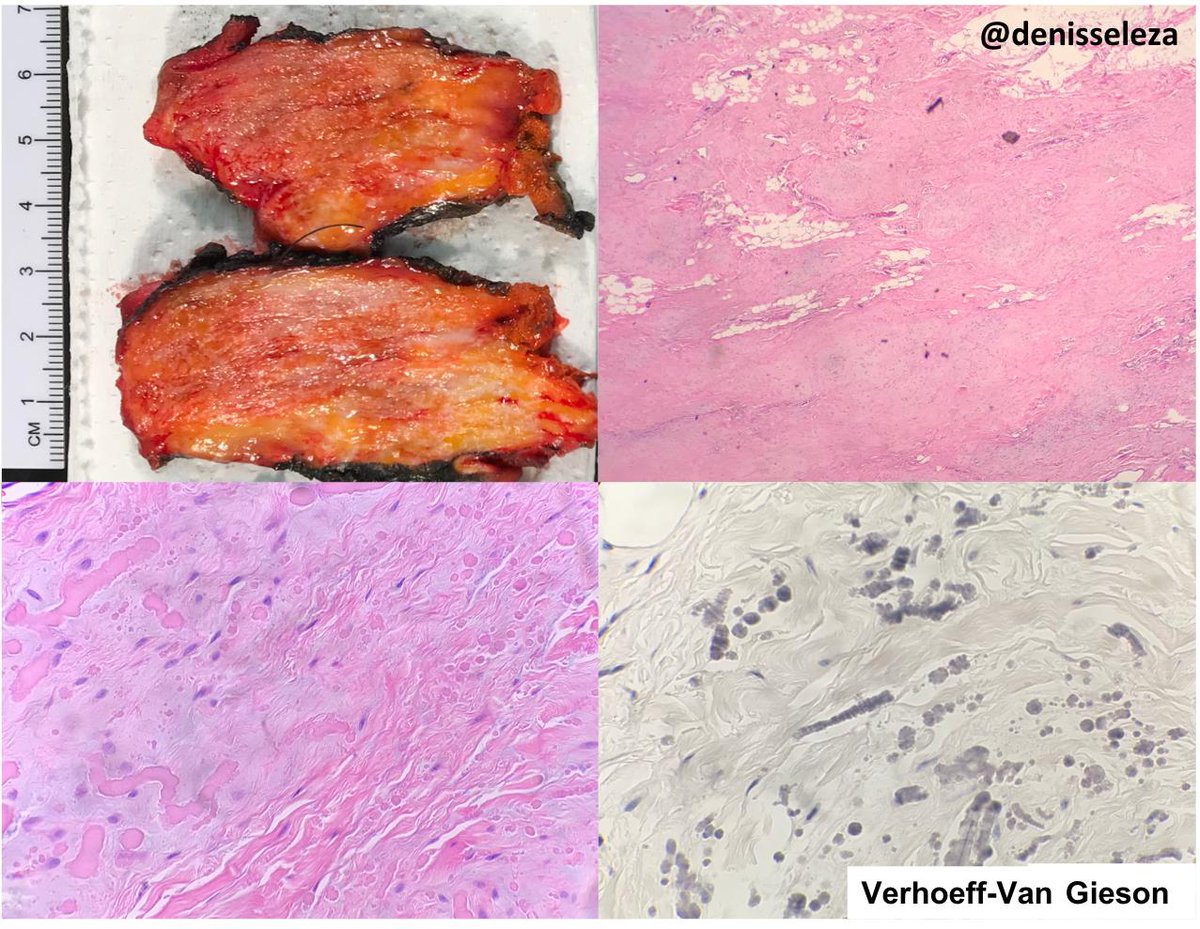
Elastofibroma: Gross: Fibrous areas interposed with adipose tissue. Low power: Elastic fibers within a collagenous stroma admixed with adipose tissue. High power and VVG stain: Elongated and globoid elastic fibers with a dense core (beads on string). @denisseleza
Cutaneous epithelioid angiomatous nodule. Benign vascular tumor composed of circumscribed sheet-like proliferation of epithelioid endothelial cells. First described by Brenn and Fletcher PubMed PMID: 14726818. #dermpath
I strongly condemn the use of force by TMC and the continuing killing of innocent people by the Janjaweed forces in Sudan.
I strongly urge the TMC to expedite the power transfer to Civilian Government without any delay or further bloodshed.
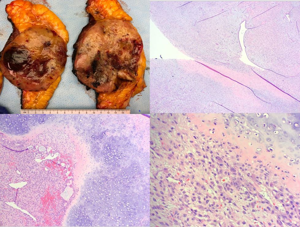
Breast mass with epithelial and stromal cells, with infiltrative borders, stromal overgrowth, nuclear atypia, abundant mitoses , necrosis, and heterologous elements in the form of chondrosarcoma and osteosarcoma were identified in this malignant phyllodes tumor. @HajaratTara
Interesting case of low grade endometrial stromal sarcoma with schwannoma-like pattern. The tumor is typically composed of fusiform cells resembling proliferative phase endometrial stromal cells whorling around arterioles. LVI is commonly seen in this tumor.
@HajaratTara
Malignant melanoma of the breast: sheets of neoplastic cells with intervening pigment-laden macrophages. The cells were positive for S100 & MART1, and negative for breast markers, cytokeratin, PAX-8, and HepPar-1. Patient has no previous history of melanoma. @HajaratTara
Lung adenocarcinoma metastasizing to a breast fibroadenoma. Glandular structures showing pleomorphic and hyperchromatic cells with prominent nucleoli. Neoplastic cells were positive for TTF-1 & Napsin A, and negative for GATA3 & ER. @HajaratTara
Sebaceous CIS: Multifocal nests of cells with abundant foamy vacuolated cytoplasm with sparing of the basal layer. Tumor cells are (+) for adipophilin (membranous vesicular pattern), p63 and p16; (-) for mucicarmine, CK7, CK20, SYN and CHR. No area of invasion is identified.
Reactive perforating collagenosis: A cup-shaped invagination with a plug of parakeratin with neutrophilic remnants and transepidermal elimination of collagen. Involves transepidermal elimination of type IV collagen, but mechanism remains unknown.
Pleomorphic fibroma: Polypoid hypocellular proliferation of spindle cells with stellate cells with enlarged, hyperchromatic nuclei. No mitotic figures. Stroma with hyalinized-appearing collagen fibers.
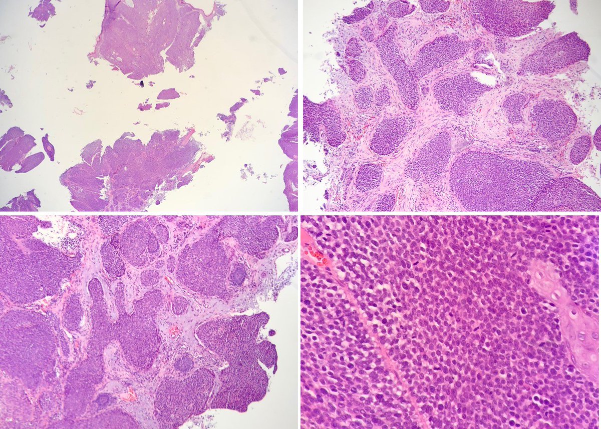
Porocarcinoma: Invasive nests and anastomosing cords of basaloid cells with focal squamous differentiation with numerous mitotic figures. Top differential diagnosis: Poroma, hidradenoma and hidradenocarcinomas, SCC, BCC.