📝 Narrative Review: #RightVentricle metrics, such as strain, fractional area change, and ejection fraction, offer mechanistic insight in cardiovascular trials but remain underused due to acquisition variability and lack of standardization.
🧠⚡ #Migraña en #Urgencias así quedan resumidas las guías actualizadas👇
✅ Que si debe ofrecerse
• Proclorperazina IV
• Bloqueo del nervio occipital mayor
✅ Que también debería ofrecerse
• Ketorolaco IV
• Metoclopramida IV
• Sumatriptán SC
• Bloqueo supraorbitario
⚠️ Que podría ofrecerse
• Clorpromazina / haloperidol / droperidol IV
• Dexametasona IV
• Valproato IV
🚫 Que No debería ofrecerse
• Paracetamol IV
• Difenhidramina IV
• Morfina IV
• Octreótido SC/IV
⛔ Y que definitivamente no
• Hidromorfona IV
❓ Sin recomendación
• Cafeína
• Granisetrón
• Ibuprofeno
• Ketamina
• Lidocaína
• Solución salina
• Propofol
• Bloqueo del ganglio esfenopalatino
• Eptinezumab (limitado a ensayos clínicos)
https://t.co/kRVQeqSnZj
Today's Paper of the Day is:
Optimising positive end-expiratory pressure in ARDS: a narrative review of approaches to titration
https://t.co/JKgcYjlUQ5
Join us to read 1 paper per day and stay up-to-date as we cover the spectrum of critical care across 2026
🩸🏥Manejo de la Sangre en el Paciente en Cuidados Intensivos
🔰📚Intensive Care Med 2026
https://t.co/0C4J3zRik1
Enlace a Artículo Completo👇🏻🆓✅
https://t.co/xumUMHaxVK
🫀Preeclampsia Is Not One Disease: Hemodynamics Matter
For decades, preeclampsia has been viewed primarily as a placental disorder characterized by hypertension, proteinuria, and endothelial dysfunction. However, accumulating evidence suggests that this framework may be incomplete.
In this comprehensive review, Masini and colleagues argue that preeclampsia is better understood as two distinct cardiovascular phenotypes, each with different hemodynamic profiles and potentially requiring different therapeutic approaches.
The traditional classification of early-onset versus late-onset disease may not be the most clinically relevant distinction.
Instead, the authors propose that the key differentiator is the presence or absence of fetal growth restriction (FGR).
Phenotype 1: Low Cardiac Output, High Vascular Resistance
This phenotype is typically associated with FGR and often presents earlier in pregnancy.
Hemodynamically, these women demonstrate:
• Reduced cardiac output
• Increased systemic vascular resistance
• Relative intravascular volume depletion
• Impaired uteroplacental perfusion
• Higher risk of maternal cardiovascular dysfunction after delivery
Importantly, abnormalities in cardiac output and vascular resistance may already be detectable before conception, suggesting that maternal cardiovascular dysfunction may precede clinical disease.
Phenotype 2: High Cardiac Output, Low Vascular Resistance
This phenotype is more frequently observed in preeclampsia without FGR and is often associated with maternal obesity.
Characteristics include:
• Elevated cardiac output
• Normal or reduced vascular resistance
• Relative intravascular volume overload
• Different pathophysiological mechanisms despite similar blood pressure values
These findings challenge the assumption that all women with preeclampsia should receive identical management.
Why This Matters Clinically
The review highlights a fundamental problem in obstetric medicine:
Current antihypertensive treatment is usually guided by blood pressure alone.
Yet two women with identical blood pressure values may have completely opposite hemodynamic states. One may be vasoconstricted and volume depleted, while the other may be volume overloaded with high cardiac output.
Treating both patients identically may therefore be physiologically inappropriate.
The authors suggest that noninvasive maternal hemodynamic assessment could help identify the dominant phenotype and guide therapy more rationally. Examples include cardiac output monitoring, assessment of vascular resistance, pulse wave velocity, and arterial stiffness measurements.
Reference 📚
Masini G, Foo LF, Tay J, et al. Preeclampsia has two phenotypes which require different treatment strategies. American Journal of Obstetrics & Gynecology. 2022;226(2S):S1006-S1018. DOI: 10.1016/j.ajog.2020.10.052.
🫁🩸Farmacoterapia en el Manejo Agudo del Embolismo Pulmonar
🔰📚A M E R I C A N C O L L E G E O F C A R D I O L O G Y F O U N D A T I O N
https://t.co/9979zI55jw
Enlace a Articulo Completo👇🏻🆓✅
https://t.co/xumUMHaxVK
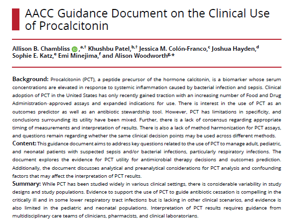
🧪📈 Hablemos de #Procalcitonina cuándo medirla?, cada cuánto? y qué cortes usar?
📌 Cuándo pedirla?
Al inicio, cuando tienes duda diagnóstica o quieres usarla como apoyo para decidir duración de antibióticos, sobre todo en sepsis o infecciones respiratorias.
📌 Cada cuánto repetirla?
Depende del contexto clínico de tu paciente, pero en general:
• si la sospecha de infección existe y el cuadro no mejora, puede repetirse en 6–24 horas
• si ya se inició tratamiento, suele revalorarse cada 2–3 días para buscar oportunidad de suspensión
• en UCI, muchos estudios la midieron diariamente
📌 Cómo interpretar la Proca al decidir si iniciar antibiótico?
• Menos de 0.1 ng/mL → el inicio de antibiótico está fuertemente desaconsejado
• De 0.1 a 0.25 ng/mL → el inicio de antibiótico está desaconsejado
• De 0.26 a 0.5 ng/mL → el inicio de antibiótico está favorecido
• Más de 0.5 ng/mL → el inicio de antibiótico está fuertemente favorecido
En si entre más baja la proca, menos apoya dar el antibiótico; entre más alta, más lo respalda.
PEro OJO.... la proca no decide sola.
Si el paciente está grave o la sospecha clínica es alta, manda la clínica.
Y aquí viene lo importante 👇
⚠️ No uses esos números como un numero mas en tu nota en "paraclinicos".
Si el paciente está muy grave, la clínica pesa más que cualquier corte.
📌 Cuándo si ayuda más de verdad?
Para suspender antibióticos.
La estrategia más útil es:
✅ suspender si la proca baja >80% desde su pico
o
✅ si cae a valores bajos (por ejemplo <0.25 ng/mL) junto con mejoría clínica
La proca sirve más como herramienta de seguimiento que como prueba única de decisión inicial.
Lo más valioso no siempre es el número absoluto… sino cómo cambia con el tiempo.
Y aunque también se ha estudiado para predecir mortalidad, los estudios usan cortes muy distintos y hay demasiada heterogeneidad como para quedarnos con un solo número universal.
Entonces ya sabes mi pana pídela con contexto, repítela con intención y nunca la interpretes sin clínica.
https://t.co/jyYQ47Ih3c
🫀⚠️ 𝗙𝗮𝗹𝗹𝗮 𝗵𝗲𝗺𝗼𝗱𝗶𝗻𝗮́𝗺𝗶𝗰𝗮 𝗲𝗻 𝘀𝗲𝗽𝘀𝗶𝘀: 𝗲𝗹 𝗽𝗿𝗼𝗯𝗹𝗲𝗺𝗮 𝗻𝗼 𝗲𝘀 𝘀𝗼𝗹𝗼 “𝘃𝗮𝘀𝗼𝗱𝗶𝗹𝗮𝘁𝗮𝗰𝗶𝗼́𝗻”, 𝘀𝗶𝗻𝗼 𝘂𝗻𝗮 𝗱𝗶𝘀𝗿𝘂𝗽𝗰𝗶𝗼́𝗻 𝗴𝗹𝗼𝗯𝗮𝗹 𝗱𝗲𝗹 𝗰𝗼𝗻𝘁𝗿𝗼𝗹 𝘃𝗮𝘀𝗰𝘂𝗹𝗮𝗿, 𝗰𝗮𝗿𝗱𝗶́𝗮𝗰𝗼 𝘆 𝗺𝗶𝗰𝗿𝗼𝗰𝗶𝗿𝗰𝘂𝗹𝗮𝘁𝗼𝗿𝗶𝗼🚨
@ElsevierConnect@sciencedirect
👇🏼👇🏼👇🏼👇🏼
📑🔗🔑🔓
https://t.co/89ht2mBRSA
⬇️⬇️⬇️⬇️
🧵👇
En sepsis la inestabilidad hemodinámica surge por la combinación de:
📉 Pérdida del tono vascular
🫀 Alteración de función cardíaca
🧬 Disfunción microcirculatoria y metabólica
👉 Todo esto es un determinante mayor de falla multiorgánica y mortalidad.
🧠 𝙇𝙖 𝙛𝙞𝙨𝙞𝙤𝙥𝙖𝙩𝙤𝙡𝙤𝙜𝙞́𝙖 𝙚𝙨 𝙢𝙪𝙘𝙝𝙤 𝙢𝙖́𝙨 𝙘𝙤𝙢𝙥𝙡𝙚𝙟𝙖 𝙦𝙪𝙚 “𝙛𝙖𝙡𝙩𝙖 𝙣𝙤𝙧𝙚𝙥𝙞𝙣𝙚𝙛𝙧𝙞𝙣𝙖”
La revisión resume varios mecanismos simultáneos:
✅ Exceso de 𝙤́𝙭𝙞𝙙𝙤 𝙣𝙞́𝙩𝙧𝙞𝙘𝙤 y prostaciclina
✅ Disfunción del 𝙨𝙞𝙨𝙩𝙚𝙢𝙖 𝙣𝙚𝙧𝙫𝙞𝙤𝙨𝙤 𝙖𝙪𝙩𝙤́𝙣𝙤𝙢𝙤
✅ Desensibilización catecolaminérgica
✅ Alteración del 𝙍𝘼𝘼𝙎
✅ 𝘿𝙚𝙛𝙞𝙘𝙞𝙚𝙣𝙘𝙞𝙖 𝙧𝙚𝙡𝙖𝙩𝙞𝙫𝙖 𝙙𝙚 𝙫𝙖𝙨𝙤𝙥𝙧𝙚𝙨𝙞𝙣𝙖
💧 𝙇𝙖 𝙛𝙡𝙪𝙞𝙙𝙤𝙩𝙚𝙧𝙖𝙥𝙞𝙖 𝙮𝙖 𝙣𝙤 𝙙𝙚𝙗𝙚 𝙚𝙣𝙩𝙚𝙣𝙙𝙚𝙧𝙨𝙚 𝙘𝙤𝙢𝙤 𝙪𝙣𝙖 𝙢𝙖𝙣𝙞𝙤𝙗𝙧𝙖 𝙖𝙪𝙩𝙤𝙢𝙖́𝙩𝙞𝙘𝙖 𝙣𝙞 𝙪𝙣𝙞𝙛𝙤𝙧𝙢𝙚
Los fluidos siguen siendo fundamentales al inicio, pero su beneficio disminuye rápido fuera de la fase precoz.
Mensajes clave:
📌 La 𝙧𝙚𝙨𝙥𝙪𝙚𝙨𝙩𝙖 𝙖 𝙛𝙡𝙪𝙞𝙙𝙤𝙨 debe valorarse activamente
📌 No basta con pensar en “respondedor a fluidos”; también importa la 𝙩𝙤𝙡𝙚𝙧𝙖𝙣𝙘𝙞𝙖 𝙖 𝙛𝙡𝙪𝙞𝙙𝙤𝙨
📌 El exceso de fluidos favorece edema intersticial, disfunción orgánica y peor pronóstico
👉 El enfoque actual es personalizar volumen, velocidad y momento de administración.
💉 𝙀𝙣 𝙫𝙖𝙨𝙤𝙥𝙧𝙚𝙨𝙤𝙧𝙚𝙨, 𝙡𝙖 𝙩𝙚𝙣𝙙𝙚𝙣𝙘𝙞𝙖 𝙖𝙘𝙩𝙪𝙖𝙡 𝙚𝙨 𝙢𝙚𝙣𝙤𝙨 𝙧𝙚𝙩𝙧𝙖𝙨𝙤 𝙮 𝙢𝙖́𝙨 𝙚𝙨𝙩𝙧𝙖𝙩𝙚𝙜𝙞𝙖 𝙢𝙪𝙡𝙩𝙞𝙢𝙤𝙙𝙖𝙡 𝙥𝙧𝙚𝙘𝙤𝙯
La revisión reafirma a la 𝙣𝙤𝙧𝙚𝙥𝙞𝙣𝙚𝙛𝙧𝙞𝙣𝙖 como vasopresor de primera línea, pero resalta que prolongar o escalar demasiado la monoterapia catecolaminérgica puede asociarse a peor evolución.
Por eso propone pensar antes en:
➕ 𝙑𝙖𝙨𝙤𝙥𝙧𝙚𝙨𝙞𝙣𝙖
➕ incluso 𝙖𝙣𝙜𝙞𝙤𝙩𝙚𝙣𝙨𝙞𝙣𝙖 𝙄𝙄 en escenarios seleccionados
Según una lógica de “soporte vasopresor multimodal”.
👉 El objetivo no es solo subir PAM, sino hacerlo con menor toxicidad catecolaminérgica y más racionalidad fisiológica.
📟 𝙀𝙡 𝙢𝙤𝙣𝙞𝙩𝙤𝙧𝙚𝙤 𝙝𝙚𝙢𝙤𝙙𝙞𝙣𝙖́𝙢𝙞𝙘𝙤 𝙪́𝙩𝙞𝙡 𝙣𝙤 𝙚𝙨 𝙚𝙡 𝙦𝙪𝙚 𝙙𝙖 𝙢𝙖́𝙨 𝙣𝙪́𝙢𝙚𝙧𝙤𝙨, 𝙨𝙞𝙣𝙤 𝙚𝙡 𝙦𝙪𝙚 𝙖𝙮𝙪𝙙𝙖 𝙖 𝙙𝙚𝙘𝙞𝙙𝙞𝙧 𝙢𝙚𝙟𝙤𝙧
El review enfatiza que la evaluación debe integrar:
🫀 𝙀𝙘𝙤𝙘𝙖𝙧𝙙𝙞𝙤𝙜𝙧𝙖𝙛𝙞́𝙖 𝙘𝙧𝙞́𝙩𝙞𝙘𝙖
📈 Parámetros dinámicos de respuesta a fluidos
🩸 Perfusión periférica, incluyendo 𝙩𝙞𝙚𝙢𝙥𝙤 𝙙𝙚 𝙡𝙡𝙚𝙣𝙖𝙙𝙤 𝙘𝙖𝙥𝙞𝙡𝙖𝙧
🧪 Lactato, pero interpretado con cautela
Además, subraya que no existe un biomarcador único perfecto de perfusión tisular.
👉 La resucitación efectiva sigue siendo multimodal y guiada por fisiología real, no por un solo marcador.
🎯 𝙏𝙖𝙠𝙚-𝙝𝙤𝙢𝙚: 𝙚𝙡 𝙛𝙪𝙩𝙪𝙧𝙤 𝙙𝙚𝙡 𝙨𝙝𝙤𝙘𝙠 𝙨𝙚́𝙥𝙩𝙞𝙘𝙤 𝙚𝙨𝙩𝙖́ 𝙚𝙣 𝙥𝙖𝙨𝙖𝙧 𝙙𝙚 𝙪𝙣𝙖 𝙧𝙚𝙖𝙣𝙞𝙢𝙖𝙘𝙞𝙤́𝙣 𝙧𝙚𝙖𝙘𝙩𝙞𝙫𝙖 𝙖 𝙪𝙣𝙖 𝙧𝙚𝙖𝙣𝙞𝙢𝙖𝙘𝙞𝙤́𝙣 𝙥𝙧𝙚𝙙𝙞𝙘𝙩𝙞𝙫𝙖 𝙮 𝙥𝙚𝙧𝙨𝙤𝙣𝙖𝙡𝙞𝙯𝙖𝙙𝙖
El artículo mira hacia adelante con varias líneas potentes:
🤖 Inteligencia artificial para anticipar deterioro hemodinámico
🧬 Biomarcadores más precisos, especialmente del eje 𝙧𝙚𝙣𝙞𝙣𝙖-𝙖𝙣𝙜𝙞𝙤𝙩𝙚𝙣𝙨𝙞𝙣𝙖
⚙️ Estrategias preventivas, no solo reactivas
📚📖 Más en 𝕏 @MarlonVFZR y en el blog 👉 [https://t.co/tojooaa9Gy]
‼️Si te sirve: ❤️ Me gusta | 🔁 Repost | ➕ Follow para más👇🏼👇🏼👇🏼👇🏼
📚📖#ClubCrit👨🏻⚕️👨🏻🏫🧠🫶
#Sepsis #SepticShock #Hemodynamics #Vasopressors
#FOAMed #FOAMcc #CriticalCare #CriticalCare #CuidadoCrítico #MedTwitter #CritCare #icu #intensivecare #diagnosis #management #MedicinaBasadaEnEvidencia #MedEd #MedX #IntensiveCare #MedIntensiva #MedXCommunity #MedED #ICUmanagement #MustRead #LecturaRecomendada
Acute Respiratory Failure 2026: What Should Change at the Bedside?
Despite advances in respiratory support, mortality from severe acute respiratory failure remains stubbornly high. This 2026 update argues that the future is not simply improving oxygenation, but improving patient phenotyping.
Several key messages emerge:
🔹 PaO₂/FiO₂ alone is an imperfect guide. Ventilator-induced lung injury is driven more by respiratory mechanics and patient effort than by oxygenation severity.
🔹 ARDS is not one disease. Inflammatory and non-inflammatory phenotypes may respond differently to corticosteroids, neuromuscular blockade, and other therapies.
🔹 Awake proning should not be viewed as a binary intervention. Early improvement in the ROX index may help identify responders and predict outcomes.
🔹 The goal is no longer only lung protection. Strategies that reduce sedation and preserve spontaneous breathing may also benefit brain function and overall recovery.
🔹 The next frontier may be biomarker-guided ventilation, using transcriptomic and microRNA signatures to identify patients at risk of ventilator-induced lung injury.
The future of respiratory critical care appears increasingly focused on precision medicine: matching the right therapy to the right physiological phenotype rather than treating all hypoxemic patients the same.
Reference 📚
Hernandez G, Muñiz Albaiceta G, Thille AW. Update on acute respiratory failure. Intensive Care Medicine. 2026. DOI: 10.1007/s00134-026-08308-6
🦀🏥Pronóstico del Paciente Oncológico en Estado Crítico
🔰📚Medicina Intensiva
https://t.co/VDHsRnLalG
Enlace a Artículo Completo👇🏻🆓✅
https://t.co/rPEDXf5xvO
We often take circulation for granted, yet it is rarely as simple as it seems. Grateful to these experts for providing fresh insights into hemodynamic management to help us better care for our patients. 🎩 tip to the authors, including @DrMCecconi
https://t.co/Sfr6vCn6le
Today's Paper of the Day is cardiac output estimation using pulse wave analysis—physiology, algorithms, and technologies
https://t.co/JKgcYjlUQ5
Join us to read 1 paper per day and stay up-to-date as we cover the spectrum of critical care across 2024
💜 Contemporary Management of Acute Heart Failure: From Emergency Presentation to Long-Term Remission
Acute heart failure (AHF) remains one of the leading causes of hospitalization worldwide, with persistently high mortality and readmission rates. This comprehensive 2026 JACC State-of-the-Art Review proposes a practical shift in our approach: hospitalization should no longer be viewed merely as a decongestion episode, but as a critical disease-modifying opportunity to optimize long-term outcomes.
Several key messages emerge.
First, congestion remains the primary therapeutic target. The authors emphasize rapid administration of intravenous loop diuretics based on clinical suspicion rather than waiting for confirmatory testing. Importantly, diuretic response should be evaluated within 2 hours using urine output (>300 mL) and, when available, spot urinary sodium (>70 mmol/L). Failure to achieve these targets should trigger immediate escalation of decongestive therapy.
Second, the review introduces the SPLASH framework for early bedside assessment:
• Symptoms
• Past medical history
• Life signs
• Assessment of congestion
• STEMI or equivalent exclusion
• Hypoperfusion
This structured approach aims to improve diagnostic accuracy and accelerate treatment initiation.
Third, the review strongly supports early implementation of guideline-directed medical therapy (GDMT). Hospitalization is described as the ideal moment to initiate or rapidly uptitrate the four pillars of heart failure treatment:
• ARNI/RAS inhibition
• Beta blockers
• Mineralocorticoid receptor antagonists
• SGLT2 inhibitors
The authors highlight evidence from STRONG-HF, EMPULSE, PIONEER-HF, and SOLOIST-WHF demonstrating that early initiation reduces mortality, rehospitalization, and improves quality of life.
Perhaps the most important practical message is that therapy optimization should begin before discharge rather than being deferred to outpatient follow-up. The review advocates frequent early post-discharge visits and rapid titration strategies to achieve full GDMT implementation within weeks rather than months.
Another notable concept is the emphasis on complete decongestion. Residual congestion at discharge remains common and is consistently associated with worse outcomes. Multimodal assessment combining clinical examination, natriuretic peptides, echocardiography, and lung ultrasound is recommended to avoid premature discharge.
The future of AHF management is not simply treating congestion. It is using hospitalization as a strategic opportunity to alter the disease trajectory.
Reference 📚
Bruno J, Arrigo M, Baudry G, et al. Contemporary Management of Acute Heart Failure: From Emergency Presentation to Long-Term Remission. Journal of the American College of Cardiology. 2026. doi:10.1016/j.jacc.2026.03.029
📱🫀Uso del Ultrasonido en el Abordaje del Paciente con Shock en Cuidados Críticos
🔰📚J. Clin. Med. 2024
https://t.co/zieQLQdUIA
Enlace a Artículo Completo👇🏻🆓✅
https://t.co/rPEDXf5xvO
☝🏻🤓Lactato en choque cardiogénico🫀: fisiopatología, valor pronóstico e interpretación clínica
➡️El lactato es uno de los biomarcadores más utilizados en medicina crítica, pero en el choque cardiogénico su interpretación es más compleja que en la sepsis. El incremento del lactato no refleja exclusivamente hipoxia tisular, sino una interacción entre disminución del aporte de oxígeno (DO₂), activación simpática, alteraciones microcirculatorias y disfunción mitocondrial.
🔰La fisiopatológica moderna, enfatizando que el lactato debe analizarse dentro del contexto hemodinámico y no como un marcador aislado.
🫀Fisiopatología del lactato en choque cardiogénico🔸️🔸️🔸️
🔹️1. Hipoperfusión tisular y metabolismo anaerobio
⬇️La reducción del gasto cardíaco disminuye el DO₂.
🔷️Cuando el aporte de oxígeno cae por debajo del umbral crítico:
➡️La fosforilación oxidativa es insuficiente.
▪️El piruvato se convierte en lactato.
▪️Se genera una acidosis metabólica asociada.
▪️Este mecanismo suele predominar en fases avanzadas del choque.
🔹️2. Producción aeróbica de lactato
La mayoría del lactato en el choque cardiogénico no proviene exclusivamente del metabolismo anaerobio.
✅️La activación β2-adrenérgica induce:
➡️Glucogenólisis.
➡️Aumento de la glucólisis.
➡️Producción acelerada de piruvato.
☝🏻🤓La velocidad de producción excede la capacidad mitocondrial para oxidarlo, produciendo hiperlactatemia incluso con oxigenación suficiente.
✴️El lactato es un combustible metabólico, no un producto de desecho.
🔹️3. Alteraciones microcirculatorias
➡️La vasoconstricción y la disfunción endotelial provocan:
➡️Heterogeneidad del flujo capilar.
➡️Hipoxia regional.
➡️Aumento de la extracción de oxígeno.
➡️Producción local de lactato.
🔹️4. Disfunción mitocondrial
En estados prolongados:
➡️Las mitocondrias pierden capacidad oxidativa.
➡️Disminuye la utilización del piruvato.
➡️Aumenta la producción de lactato aun con DO₂ aparentemente adecuado.
🔹️5. Disminución del aclaramiento
➡️El hígado elimina aproximadamente 60-70 % del lactato.
➡️La congestión venosa y la hipoperfusión hepática:
➡️Reducen su metabolismo.
➡️Favorecen la acumulación plasmática.
▪️El riñón aporta otro 20-30 % del aclaramiento, por lo que la lesión renal aguda también contribuye.
✅️Valor pronóstico
▪️La hiperlactatemia es uno de los predictores más robustos de mortalidad en choque cardiogénico.
☝🏻🤓Lactato inicial
▪️Valores >2 mmol/L se asocian con:
Mayor gravedad.🥴‼️
🔴Incremento de falla multiorgánica.
🟡Necesidad de soporte mecánico.
🔵Valores >4 mmol/L identifican pacientes con riesgo elevado.
🟣Valores >10 mmol/L se asocian con mortalidades superiores al 70-80 %.
🔴Importancia del aclaramiento del lactato
☝🏻🤓Más importante que el valor inicial es su evolución.
🔸️Una disminución del lactato durante las primeras 6-24 horas refleja:🔸️Mejoría del gasto cardíaco.
🔸️Recuperación de la perfusión tisular.
✅️Mejor pronóstico.
☝🏻🤓La persistencia de hiperlactatemia indica:
▪️Hipoperfusión residual.
▪️Fracaso del tratamiento.
▪️Peor supervivencia.💀
☝🏻🤓Debemos de tomar en cuenta que...
🔴El lactato no debe ser un objetivo terapéutico aislado
➡️No toda elevación del lactato significa:
▪️Hipovolemia.
▪️Necesidad de más líquidos.
▪️Administrar volumen indiscriminadamente puede:
▪︎Incrementar la presión venosa central.
▪︎Aumentar la congestión sistémica.
▪︎Empeorar la función renal y hepática.
☝🏻🤓Debe interpretarse junto con otros parámetros
▪️Macrocirculación
▪️Presión arterial media.
▪️Gasto cardíaco.
▪️Índice cardíaco.
🫀Ecocardiografía.
🔹️Perfusión periférica
🔹️Tiempo de llenado capilar.
🔹️Moteado cutáneo.
🔹️Temperatura de extremidades.
🔹️Variables metabólicas
🔹️ScvO₂/SvO₂.
🔹️Pv-aCO₂.
🔹️Pv-aCO₂/Ca-vO₂.
✅️Déficit de base.
💧Congestión
💧VExUS.
💧PVC.
🔰Ecografía venosa.
🔹️Función hepática y renal.
☝🏻🤓️Lactato y soporte circulatorio mecánico
▪️️Durante ECMO VA, Impella o balón intraaórtico:
▪️


![MarlonVFZR's tweet photo. 🫀⚠️ 𝗙𝗮𝗹𝗹𝗮 𝗵𝗲𝗺𝗼𝗱𝗶𝗻𝗮́𝗺𝗶𝗰𝗮 𝗲𝗻 𝘀𝗲𝗽𝘀𝗶𝘀: 𝗲𝗹 𝗽𝗿𝗼𝗯𝗹𝗲𝗺𝗮 𝗻𝗼 𝗲𝘀 𝘀𝗼𝗹𝗼 “𝘃𝗮𝘀𝗼𝗱𝗶𝗹𝗮𝘁𝗮𝗰𝗶𝗼́𝗻”, 𝘀𝗶𝗻𝗼 𝘂𝗻𝗮 𝗱𝗶𝘀𝗿𝘂𝗽𝗰𝗶𝗼́𝗻 𝗴𝗹𝗼𝗯𝗮𝗹 𝗱𝗲𝗹 𝗰𝗼𝗻𝘁𝗿𝗼𝗹 𝘃𝗮𝘀𝗰𝘂𝗹𝗮𝗿, 𝗰𝗮𝗿𝗱𝗶́𝗮𝗰𝗼 𝘆 𝗺𝗶𝗰𝗿𝗼𝗰𝗶𝗿𝗰𝘂𝗹𝗮𝘁𝗼𝗿𝗶𝗼🚨
@ElsevierConnect @sciencedirect
👇🏼👇🏼👇🏼👇🏼
📑🔗🔑🔓
https://t.co/89ht2mBRSA
⬇️⬇️⬇️⬇️
🧵👇
En sepsis la inestabilidad hemodinámica surge por la combinación de:
📉 Pérdida del tono vascular
🫀 Alteración de función cardíaca
🧬 Disfunción microcirculatoria y metabólica
👉 Todo esto es un determinante mayor de falla multiorgánica y mortalidad.
🧠 𝙇𝙖 𝙛𝙞𝙨𝙞𝙤𝙥𝙖𝙩𝙤𝙡𝙤𝙜𝙞́𝙖 𝙚𝙨 𝙢𝙪𝙘𝙝𝙤 𝙢𝙖́𝙨 𝙘𝙤𝙢𝙥𝙡𝙚𝙟𝙖 𝙦𝙪𝙚 “𝙛𝙖𝙡𝙩𝙖 𝙣𝙤𝙧𝙚𝙥𝙞𝙣𝙚𝙛𝙧𝙞𝙣𝙖”
La revisión resume varios mecanismos simultáneos:
✅ Exceso de 𝙤́𝙭𝙞𝙙𝙤 𝙣𝙞́𝙩𝙧𝙞𝙘𝙤 y prostaciclina
✅ Disfunción del 𝙨𝙞𝙨𝙩𝙚𝙢𝙖 𝙣𝙚𝙧𝙫𝙞𝙤𝙨𝙤 𝙖𝙪𝙩𝙤́𝙣𝙤𝙢𝙤
✅ Desensibilización catecolaminérgica
✅ Alteración del 𝙍𝘼𝘼𝙎
✅ 𝘿𝙚𝙛𝙞𝙘𝙞𝙚𝙣𝙘𝙞𝙖 𝙧𝙚𝙡𝙖𝙩𝙞𝙫𝙖 𝙙𝙚 𝙫𝙖𝙨𝙤𝙥𝙧𝙚𝙨𝙞𝙣𝙖
💧 𝙇𝙖 𝙛𝙡𝙪𝙞𝙙𝙤𝙩𝙚𝙧𝙖𝙥𝙞𝙖 𝙮𝙖 𝙣𝙤 𝙙𝙚𝙗𝙚 𝙚𝙣𝙩𝙚𝙣𝙙𝙚𝙧𝙨𝙚 𝙘𝙤𝙢𝙤 𝙪𝙣𝙖 𝙢𝙖𝙣𝙞𝙤𝙗𝙧𝙖 𝙖𝙪𝙩𝙤𝙢𝙖́𝙩𝙞𝙘𝙖 𝙣𝙞 𝙪𝙣𝙞𝙛𝙤𝙧𝙢𝙚
Los fluidos siguen siendo fundamentales al inicio, pero su beneficio disminuye rápido fuera de la fase precoz.
Mensajes clave:
📌 La 𝙧𝙚𝙨𝙥𝙪𝙚𝙨𝙩𝙖 𝙖 𝙛𝙡𝙪𝙞𝙙𝙤𝙨 debe valorarse activamente
📌 No basta con pensar en “respondedor a fluidos”; también importa la 𝙩𝙤𝙡𝙚𝙧𝙖𝙣𝙘𝙞𝙖 𝙖 𝙛𝙡𝙪𝙞𝙙𝙤𝙨
📌 El exceso de fluidos favorece edema intersticial, disfunción orgánica y peor pronóstico
👉 El enfoque actual es personalizar volumen, velocidad y momento de administración.
💉 𝙀𝙣 𝙫𝙖𝙨𝙤𝙥𝙧𝙚𝙨𝙤𝙧𝙚𝙨, 𝙡𝙖 𝙩𝙚𝙣𝙙𝙚𝙣𝙘𝙞𝙖 𝙖𝙘𝙩𝙪𝙖𝙡 𝙚𝙨 𝙢𝙚𝙣𝙤𝙨 𝙧𝙚𝙩𝙧𝙖𝙨𝙤 𝙮 𝙢𝙖́𝙨 𝙚𝙨𝙩𝙧𝙖𝙩𝙚𝙜𝙞𝙖 𝙢𝙪𝙡𝙩𝙞𝙢𝙤𝙙𝙖𝙡 𝙥𝙧𝙚𝙘𝙤𝙯
La revisión reafirma a la 𝙣𝙤𝙧𝙚𝙥𝙞𝙣𝙚𝙛𝙧𝙞𝙣𝙖 como vasopresor de primera línea, pero resalta que prolongar o escalar demasiado la monoterapia catecolaminérgica puede asociarse a peor evolución.
Por eso propone pensar antes en:
➕ 𝙑𝙖𝙨𝙤𝙥𝙧𝙚𝙨𝙞𝙣𝙖
➕ incluso 𝙖𝙣𝙜𝙞𝙤𝙩𝙚𝙣𝙨𝙞𝙣𝙖 𝙄𝙄 en escenarios seleccionados
Según una lógica de “soporte vasopresor multimodal”.
👉 El objetivo no es solo subir PAM, sino hacerlo con menor toxicidad catecolaminérgica y más racionalidad fisiológica.
📟 𝙀𝙡 𝙢𝙤𝙣𝙞𝙩𝙤𝙧𝙚𝙤 𝙝𝙚𝙢𝙤𝙙𝙞𝙣𝙖́𝙢𝙞𝙘𝙤 𝙪́𝙩𝙞𝙡 𝙣𝙤 𝙚𝙨 𝙚𝙡 𝙦𝙪𝙚 𝙙𝙖 𝙢𝙖́𝙨 𝙣𝙪́𝙢𝙚𝙧𝙤𝙨, 𝙨𝙞𝙣𝙤 𝙚𝙡 𝙦𝙪𝙚 𝙖𝙮𝙪𝙙𝙖 𝙖 𝙙𝙚𝙘𝙞𝙙𝙞𝙧 𝙢𝙚𝙟𝙤𝙧
El review enfatiza que la evaluación debe integrar:
🫀 𝙀𝙘𝙤𝙘𝙖𝙧𝙙𝙞𝙤𝙜𝙧𝙖𝙛𝙞́𝙖 𝙘𝙧𝙞́𝙩𝙞𝙘𝙖
📈 Parámetros dinámicos de respuesta a fluidos
🩸 Perfusión periférica, incluyendo 𝙩𝙞𝙚𝙢𝙥𝙤 𝙙𝙚 𝙡𝙡𝙚𝙣𝙖𝙙𝙤 𝙘𝙖𝙥𝙞𝙡𝙖𝙧
🧪 Lactato, pero interpretado con cautela
Además, subraya que no existe un biomarcador único perfecto de perfusión tisular.
👉 La resucitación efectiva sigue siendo multimodal y guiada por fisiología real, no por un solo marcador.
🎯 𝙏𝙖𝙠𝙚-𝙝𝙤𝙢𝙚: 𝙚𝙡 𝙛𝙪𝙩𝙪𝙧𝙤 𝙙𝙚𝙡 𝙨𝙝𝙤𝙘𝙠 𝙨𝙚́𝙥𝙩𝙞𝙘𝙤 𝙚𝙨𝙩𝙖́ 𝙚𝙣 𝙥𝙖𝙨𝙖𝙧 𝙙𝙚 𝙪𝙣𝙖 𝙧𝙚𝙖𝙣𝙞𝙢𝙖𝙘𝙞𝙤́𝙣 𝙧𝙚𝙖𝙘𝙩𝙞𝙫𝙖 𝙖 𝙪𝙣𝙖 𝙧𝙚𝙖𝙣𝙞𝙢𝙖𝙘𝙞𝙤́𝙣 𝙥𝙧𝙚𝙙𝙞𝙘𝙩𝙞𝙫𝙖 𝙮 𝙥𝙚𝙧𝙨𝙤𝙣𝙖𝙡𝙞𝙯𝙖𝙙𝙖
El artículo mira hacia adelante con varias líneas potentes:
🤖 Inteligencia artificial para anticipar deterioro hemodinámico
🧬 Biomarcadores más precisos, especialmente del eje 𝙧𝙚𝙣𝙞𝙣𝙖-𝙖𝙣𝙜𝙞𝙤𝙩𝙚𝙣𝙨𝙞𝙣𝙖
⚙️ Estrategias preventivas, no solo reactivas
📚📖 Más en 𝕏 @MarlonVFZR y en el blog 👉 [https://t.co/tojooaa9Gy]
‼️Si te sirve: ❤️ Me gusta | 🔁 Repost | ➕ Follow para más👇🏼👇🏼👇🏼👇🏼
📚📖#ClubCrit👨🏻⚕️👨🏻🏫🧠🫶
#Sepsis #SepticShock #Hemodynamics #Vasopressors
#FOAMed #FOAMcc #CriticalCare #CriticalCare #CuidadoCrítico #MedTwitter #CritCare #icu #intensivecare #diagnosis #management #MedicinaBasadaEnEvidencia #MedEd #MedX #IntensiveCare #MedIntensiva #MedXCommunity #MedED #ICUmanagement #MustRead #LecturaRecomendada](https://pbs.twimg.com/media/HLvd5KNXcAAco89.jpg)
![MarlonVFZR's tweet photo. 🫀⚠️ 𝗙𝗮𝗹𝗹𝗮 𝗵𝗲𝗺𝗼𝗱𝗶𝗻𝗮́𝗺𝗶𝗰𝗮 𝗲𝗻 𝘀𝗲𝗽𝘀𝗶𝘀: 𝗲𝗹 𝗽𝗿𝗼𝗯𝗹𝗲𝗺𝗮 𝗻𝗼 𝗲𝘀 𝘀𝗼𝗹𝗼 “𝘃𝗮𝘀𝗼𝗱𝗶𝗹𝗮𝘁𝗮𝗰𝗶𝗼́𝗻”, 𝘀𝗶𝗻𝗼 𝘂𝗻𝗮 𝗱𝗶𝘀𝗿𝘂𝗽𝗰𝗶𝗼́𝗻 𝗴𝗹𝗼𝗯𝗮𝗹 𝗱𝗲𝗹 𝗰𝗼𝗻𝘁𝗿𝗼𝗹 𝘃𝗮𝘀𝗰𝘂𝗹𝗮𝗿, 𝗰𝗮𝗿𝗱𝗶́𝗮𝗰𝗼 𝘆 𝗺𝗶𝗰𝗿𝗼𝗰𝗶𝗿𝗰𝘂𝗹𝗮𝘁𝗼𝗿𝗶𝗼🚨
@ElsevierConnect @sciencedirect
👇🏼👇🏼👇🏼👇🏼
📑🔗🔑🔓
https://t.co/89ht2mBRSA
⬇️⬇️⬇️⬇️
🧵👇
En sepsis la inestabilidad hemodinámica surge por la combinación de:
📉 Pérdida del tono vascular
🫀 Alteración de función cardíaca
🧬 Disfunción microcirculatoria y metabólica
👉 Todo esto es un determinante mayor de falla multiorgánica y mortalidad.
🧠 𝙇𝙖 𝙛𝙞𝙨𝙞𝙤𝙥𝙖𝙩𝙤𝙡𝙤𝙜𝙞́𝙖 𝙚𝙨 𝙢𝙪𝙘𝙝𝙤 𝙢𝙖́𝙨 𝙘𝙤𝙢𝙥𝙡𝙚𝙟𝙖 𝙦𝙪𝙚 “𝙛𝙖𝙡𝙩𝙖 𝙣𝙤𝙧𝙚𝙥𝙞𝙣𝙚𝙛𝙧𝙞𝙣𝙖”
La revisión resume varios mecanismos simultáneos:
✅ Exceso de 𝙤́𝙭𝙞𝙙𝙤 𝙣𝙞́𝙩𝙧𝙞𝙘𝙤 y prostaciclina
✅ Disfunción del 𝙨𝙞𝙨𝙩𝙚𝙢𝙖 𝙣𝙚𝙧𝙫𝙞𝙤𝙨𝙤 𝙖𝙪𝙩𝙤́𝙣𝙤𝙢𝙤
✅ Desensibilización catecolaminérgica
✅ Alteración del 𝙍𝘼𝘼𝙎
✅ 𝘿𝙚𝙛𝙞𝙘𝙞𝙚𝙣𝙘𝙞𝙖 𝙧𝙚𝙡𝙖𝙩𝙞𝙫𝙖 𝙙𝙚 𝙫𝙖𝙨𝙤𝙥𝙧𝙚𝙨𝙞𝙣𝙖
💧 𝙇𝙖 𝙛𝙡𝙪𝙞𝙙𝙤𝙩𝙚𝙧𝙖𝙥𝙞𝙖 𝙮𝙖 𝙣𝙤 𝙙𝙚𝙗𝙚 𝙚𝙣𝙩𝙚𝙣𝙙𝙚𝙧𝙨𝙚 𝙘𝙤𝙢𝙤 𝙪𝙣𝙖 𝙢𝙖𝙣𝙞𝙤𝙗𝙧𝙖 𝙖𝙪𝙩𝙤𝙢𝙖́𝙩𝙞𝙘𝙖 𝙣𝙞 𝙪𝙣𝙞𝙛𝙤𝙧𝙢𝙚
Los fluidos siguen siendo fundamentales al inicio, pero su beneficio disminuye rápido fuera de la fase precoz.
Mensajes clave:
📌 La 𝙧𝙚𝙨𝙥𝙪𝙚𝙨𝙩𝙖 𝙖 𝙛𝙡𝙪𝙞𝙙𝙤𝙨 debe valorarse activamente
📌 No basta con pensar en “respondedor a fluidos”; también importa la 𝙩𝙤𝙡𝙚𝙧𝙖𝙣𝙘𝙞𝙖 𝙖 𝙛𝙡𝙪𝙞𝙙𝙤𝙨
📌 El exceso de fluidos favorece edema intersticial, disfunción orgánica y peor pronóstico
👉 El enfoque actual es personalizar volumen, velocidad y momento de administración.
💉 𝙀𝙣 𝙫𝙖𝙨𝙤𝙥𝙧𝙚𝙨𝙤𝙧𝙚𝙨, 𝙡𝙖 𝙩𝙚𝙣𝙙𝙚𝙣𝙘𝙞𝙖 𝙖𝙘𝙩𝙪𝙖𝙡 𝙚𝙨 𝙢𝙚𝙣𝙤𝙨 𝙧𝙚𝙩𝙧𝙖𝙨𝙤 𝙮 𝙢𝙖́𝙨 𝙚𝙨𝙩𝙧𝙖𝙩𝙚𝙜𝙞𝙖 𝙢𝙪𝙡𝙩𝙞𝙢𝙤𝙙𝙖𝙡 𝙥𝙧𝙚𝙘𝙤𝙯
La revisión reafirma a la 𝙣𝙤𝙧𝙚𝙥𝙞𝙣𝙚𝙛𝙧𝙞𝙣𝙖 como vasopresor de primera línea, pero resalta que prolongar o escalar demasiado la monoterapia catecolaminérgica puede asociarse a peor evolución.
Por eso propone pensar antes en:
➕ 𝙑𝙖𝙨𝙤𝙥𝙧𝙚𝙨𝙞𝙣𝙖
➕ incluso 𝙖𝙣𝙜𝙞𝙤𝙩𝙚𝙣𝙨𝙞𝙣𝙖 𝙄𝙄 en escenarios seleccionados
Según una lógica de “soporte vasopresor multimodal”.
👉 El objetivo no es solo subir PAM, sino hacerlo con menor toxicidad catecolaminérgica y más racionalidad fisiológica.
📟 𝙀𝙡 𝙢𝙤𝙣𝙞𝙩𝙤𝙧𝙚𝙤 𝙝𝙚𝙢𝙤𝙙𝙞𝙣𝙖́𝙢𝙞𝙘𝙤 𝙪́𝙩𝙞𝙡 𝙣𝙤 𝙚𝙨 𝙚𝙡 𝙦𝙪𝙚 𝙙𝙖 𝙢𝙖́𝙨 𝙣𝙪́𝙢𝙚𝙧𝙤𝙨, 𝙨𝙞𝙣𝙤 𝙚𝙡 𝙦𝙪𝙚 𝙖𝙮𝙪𝙙𝙖 𝙖 𝙙𝙚𝙘𝙞𝙙𝙞𝙧 𝙢𝙚𝙟𝙤𝙧
El review enfatiza que la evaluación debe integrar:
🫀 𝙀𝙘𝙤𝙘𝙖𝙧𝙙𝙞𝙤𝙜𝙧𝙖𝙛𝙞́𝙖 𝙘𝙧𝙞́𝙩𝙞𝙘𝙖
📈 Parámetros dinámicos de respuesta a fluidos
🩸 Perfusión periférica, incluyendo 𝙩𝙞𝙚𝙢𝙥𝙤 𝙙𝙚 𝙡𝙡𝙚𝙣𝙖𝙙𝙤 𝙘𝙖𝙥𝙞𝙡𝙖𝙧
🧪 Lactato, pero interpretado con cautela
Además, subraya que no existe un biomarcador único perfecto de perfusión tisular.
👉 La resucitación efectiva sigue siendo multimodal y guiada por fisiología real, no por un solo marcador.
🎯 𝙏𝙖𝙠𝙚-𝙝𝙤𝙢𝙚: 𝙚𝙡 𝙛𝙪𝙩𝙪𝙧𝙤 𝙙𝙚𝙡 𝙨𝙝𝙤𝙘𝙠 𝙨𝙚́𝙥𝙩𝙞𝙘𝙤 𝙚𝙨𝙩𝙖́ 𝙚𝙣 𝙥𝙖𝙨𝙖𝙧 𝙙𝙚 𝙪𝙣𝙖 𝙧𝙚𝙖𝙣𝙞𝙢𝙖𝙘𝙞𝙤́𝙣 𝙧𝙚𝙖𝙘𝙩𝙞𝙫𝙖 𝙖 𝙪𝙣𝙖 𝙧𝙚𝙖𝙣𝙞𝙢𝙖𝙘𝙞𝙤́𝙣 𝙥𝙧𝙚𝙙𝙞𝙘𝙩𝙞𝙫𝙖 𝙮 𝙥𝙚𝙧𝙨𝙤𝙣𝙖𝙡𝙞𝙯𝙖𝙙𝙖
El artículo mira hacia adelante con varias líneas potentes:
🤖 Inteligencia artificial para anticipar deterioro hemodinámico
🧬 Biomarcadores más precisos, especialmente del eje 𝙧𝙚𝙣𝙞𝙣𝙖-𝙖𝙣𝙜𝙞𝙤𝙩𝙚𝙣𝙨𝙞𝙣𝙖
⚙️ Estrategias preventivas, no solo reactivas
📚📖 Más en 𝕏 @MarlonVFZR y en el blog 👉 [https://t.co/tojooaa9Gy]
‼️Si te sirve: ❤️ Me gusta | 🔁 Repost | ➕ Follow para más👇🏼👇🏼👇🏼👇🏼
📚📖#ClubCrit👨🏻⚕️👨🏻🏫🧠🫶
#Sepsis #SepticShock #Hemodynamics #Vasopressors
#FOAMed #FOAMcc #CriticalCare #CriticalCare #CuidadoCrítico #MedTwitter #CritCare #icu #intensivecare #diagnosis #management #MedicinaBasadaEnEvidencia #MedEd #MedX #IntensiveCare #MedIntensiva #MedXCommunity #MedED #ICUmanagement #MustRead #LecturaRecomendada](https://pbs.twimg.com/media/HLvd5ItWUAAP0T_.jpg)


![MarlonVFZR's tweet photo. 🫀⚠️ 𝗙𝗮𝗹𝗹𝗮 𝗵𝗲𝗺𝗼𝗱𝗶𝗻𝗮́𝗺𝗶𝗰𝗮 𝗲𝗻 𝘀𝗲𝗽𝘀𝗶𝘀: 𝗲𝗹 𝗽𝗿𝗼𝗯𝗹𝗲𝗺𝗮 𝗻𝗼 𝗲𝘀 𝘀𝗼𝗹𝗼 “𝘃𝗮𝘀𝗼𝗱𝗶𝗹𝗮𝘁𝗮𝗰𝗶𝗼́𝗻”, 𝘀𝗶𝗻𝗼 𝘂𝗻𝗮 𝗱𝗶𝘀𝗿𝘂𝗽𝗰𝗶𝗼́𝗻 𝗴𝗹𝗼𝗯𝗮𝗹 𝗱𝗲𝗹 𝗰𝗼𝗻𝘁𝗿𝗼𝗹 𝘃𝗮𝘀𝗰𝘂𝗹𝗮𝗿, 𝗰𝗮𝗿𝗱𝗶́𝗮𝗰𝗼 𝘆 𝗺𝗶𝗰𝗿𝗼𝗰𝗶𝗿𝗰𝘂𝗹𝗮𝘁𝗼𝗿𝗶𝗼🚨
@ElsevierConnect @sciencedirect
👇🏼👇🏼👇🏼👇🏼
📑🔗🔑🔓
https://t.co/89ht2mBRSA
⬇️⬇️⬇️⬇️
🧵👇
En sepsis la inestabilidad hemodinámica surge por la combinación de:
📉 Pérdida del tono vascular
🫀 Alteración de función cardíaca
🧬 Disfunción microcirculatoria y metabólica
👉 Todo esto es un determinante mayor de falla multiorgánica y mortalidad.
🧠 𝙇𝙖 𝙛𝙞𝙨𝙞𝙤𝙥𝙖𝙩𝙤𝙡𝙤𝙜𝙞́𝙖 𝙚𝙨 𝙢𝙪𝙘𝙝𝙤 𝙢𝙖́𝙨 𝙘𝙤𝙢𝙥𝙡𝙚𝙟𝙖 𝙦𝙪𝙚 “𝙛𝙖𝙡𝙩𝙖 𝙣𝙤𝙧𝙚𝙥𝙞𝙣𝙚𝙛𝙧𝙞𝙣𝙖”
La revisión resume varios mecanismos simultáneos:
✅ Exceso de 𝙤́𝙭𝙞𝙙𝙤 𝙣𝙞́𝙩𝙧𝙞𝙘𝙤 y prostaciclina
✅ Disfunción del 𝙨𝙞𝙨𝙩𝙚𝙢𝙖 𝙣𝙚𝙧𝙫𝙞𝙤𝙨𝙤 𝙖𝙪𝙩𝙤́𝙣𝙤𝙢𝙤
✅ Desensibilización catecolaminérgica
✅ Alteración del 𝙍𝘼𝘼𝙎
✅ 𝘿𝙚𝙛𝙞𝙘𝙞𝙚𝙣𝙘𝙞𝙖 𝙧𝙚𝙡𝙖𝙩𝙞𝙫𝙖 𝙙𝙚 𝙫𝙖𝙨𝙤𝙥𝙧𝙚𝙨𝙞𝙣𝙖
💧 𝙇𝙖 𝙛𝙡𝙪𝙞𝙙𝙤𝙩𝙚𝙧𝙖𝙥𝙞𝙖 𝙮𝙖 𝙣𝙤 𝙙𝙚𝙗𝙚 𝙚𝙣𝙩𝙚𝙣𝙙𝙚𝙧𝙨𝙚 𝙘𝙤𝙢𝙤 𝙪𝙣𝙖 𝙢𝙖𝙣𝙞𝙤𝙗𝙧𝙖 𝙖𝙪𝙩𝙤𝙢𝙖́𝙩𝙞𝙘𝙖 𝙣𝙞 𝙪𝙣𝙞𝙛𝙤𝙧𝙢𝙚
Los fluidos siguen siendo fundamentales al inicio, pero su beneficio disminuye rápido fuera de la fase precoz.
Mensajes clave:
📌 La 𝙧𝙚𝙨𝙥𝙪𝙚𝙨𝙩𝙖 𝙖 𝙛𝙡𝙪𝙞𝙙𝙤𝙨 debe valorarse activamente
📌 No basta con pensar en “respondedor a fluidos”; también importa la 𝙩𝙤𝙡𝙚𝙧𝙖𝙣𝙘𝙞𝙖 𝙖 𝙛𝙡𝙪𝙞𝙙𝙤𝙨
📌 El exceso de fluidos favorece edema intersticial, disfunción orgánica y peor pronóstico
👉 El enfoque actual es personalizar volumen, velocidad y momento de administración.
💉 𝙀𝙣 𝙫𝙖𝙨𝙤𝙥𝙧𝙚𝙨𝙤𝙧𝙚𝙨, 𝙡𝙖 𝙩𝙚𝙣𝙙𝙚𝙣𝙘𝙞𝙖 𝙖𝙘𝙩𝙪𝙖𝙡 𝙚𝙨 𝙢𝙚𝙣𝙤𝙨 𝙧𝙚𝙩𝙧𝙖𝙨𝙤 𝙮 𝙢𝙖́𝙨 𝙚𝙨𝙩𝙧𝙖𝙩𝙚𝙜𝙞𝙖 𝙢𝙪𝙡𝙩𝙞𝙢𝙤𝙙𝙖𝙡 𝙥𝙧𝙚𝙘𝙤𝙯
La revisión reafirma a la 𝙣𝙤𝙧𝙚𝙥𝙞𝙣𝙚𝙛𝙧𝙞𝙣𝙖 como vasopresor de primera línea, pero resalta que prolongar o escalar demasiado la monoterapia catecolaminérgica puede asociarse a peor evolución.
Por eso propone pensar antes en:
➕ 𝙑𝙖𝙨𝙤𝙥𝙧𝙚𝙨𝙞𝙣𝙖
➕ incluso 𝙖𝙣𝙜𝙞𝙤𝙩𝙚𝙣𝙨𝙞𝙣𝙖 𝙄𝙄 en escenarios seleccionados
Según una lógica de “soporte vasopresor multimodal”.
👉 El objetivo no es solo subir PAM, sino hacerlo con menor toxicidad catecolaminérgica y más racionalidad fisiológica.
📟 𝙀𝙡 𝙢𝙤𝙣𝙞𝙩𝙤𝙧𝙚𝙤 𝙝𝙚𝙢𝙤𝙙𝙞𝙣𝙖́𝙢𝙞𝙘𝙤 𝙪́𝙩𝙞𝙡 𝙣𝙤 𝙚𝙨 𝙚𝙡 𝙦𝙪𝙚 𝙙𝙖 𝙢𝙖́𝙨 𝙣𝙪́𝙢𝙚𝙧𝙤𝙨, 𝙨𝙞𝙣𝙤 𝙚𝙡 𝙦𝙪𝙚 𝙖𝙮𝙪𝙙𝙖 𝙖 𝙙𝙚𝙘𝙞𝙙𝙞𝙧 𝙢𝙚𝙟𝙤𝙧
El review enfatiza que la evaluación debe integrar:
🫀 𝙀𝙘𝙤𝙘𝙖𝙧𝙙𝙞𝙤𝙜𝙧𝙖𝙛𝙞́𝙖 𝙘𝙧𝙞́𝙩𝙞𝙘𝙖
📈 Parámetros dinámicos de respuesta a fluidos
🩸 Perfusión periférica, incluyendo 𝙩𝙞𝙚𝙢𝙥𝙤 𝙙𝙚 𝙡𝙡𝙚𝙣𝙖𝙙𝙤 𝙘𝙖𝙥𝙞𝙡𝙖𝙧
🧪 Lactato, pero interpretado con cautela
Además, subraya que no existe un biomarcador único perfecto de perfusión tisular.
👉 La resucitación efectiva sigue siendo multimodal y guiada por fisiología real, no por un solo marcador.
🎯 𝙏𝙖𝙠𝙚-𝙝𝙤𝙢𝙚: 𝙚𝙡 𝙛𝙪𝙩𝙪𝙧𝙤 𝙙𝙚𝙡 𝙨𝙝𝙤𝙘𝙠 𝙨𝙚́𝙥𝙩𝙞𝙘𝙤 𝙚𝙨𝙩𝙖́ 𝙚𝙣 𝙥𝙖𝙨𝙖𝙧 𝙙𝙚 𝙪𝙣𝙖 𝙧𝙚𝙖𝙣𝙞𝙢𝙖𝙘𝙞𝙤́𝙣 𝙧𝙚𝙖𝙘𝙩𝙞𝙫𝙖 𝙖 𝙪𝙣𝙖 𝙧𝙚𝙖𝙣𝙞𝙢𝙖𝙘𝙞𝙤́𝙣 𝙥𝙧𝙚𝙙𝙞𝙘𝙩𝙞𝙫𝙖 𝙮 𝙥𝙚𝙧𝙨𝙤𝙣𝙖𝙡𝙞𝙯𝙖𝙙𝙖
El artículo mira hacia adelante con varias líneas potentes:
🤖 Inteligencia artificial para anticipar deterioro hemodinámico
🧬 Biomarcadores más precisos, especialmente del eje 𝙧𝙚𝙣𝙞𝙣𝙖-𝙖𝙣𝙜𝙞𝙤𝙩𝙚𝙣𝙨𝙞𝙣𝙖
⚙️ Estrategias preventivas, no solo reactivas
📚📖 Más en 𝕏 @MarlonVFZR y en el blog 👉 [https://t.co/tojooaa9Gy]
‼️Si te sirve: ❤️ Me gusta | 🔁 Repost | ➕ Follow para más👇🏼👇🏼👇🏼👇🏼
📚📖#ClubCrit👨🏻⚕️👨🏻🏫🧠🫶
#Sepsis #SepticShock #Hemodynamics #Vasopressors
#FOAMed #FOAMcc #CriticalCare #CriticalCare #CuidadoCrítico #MedTwitter #CritCare #icu #intensivecare #diagnosis #management #MedicinaBasadaEnEvidencia #MedEd #MedX #IntensiveCare #MedIntensiva #MedXCommunity #MedED #ICUmanagement #MustRead #LecturaRecomendada](https://pbs.twimg.com/media/HLvd5KiXUAAVfe6.jpg)