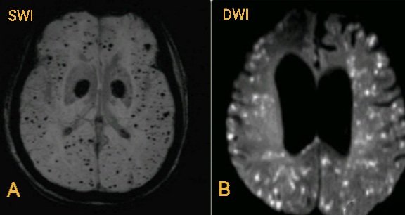
Image A(SWI MRI) belongs to an old lady. Image B(DWI MRI) belongs to a middle-aged man. Both with long standing hypertension present with altered sensorium. Patient B also has fever,tachycardia and recent pancreatitis? What's your opinion about each? Which one is emergency? Why?
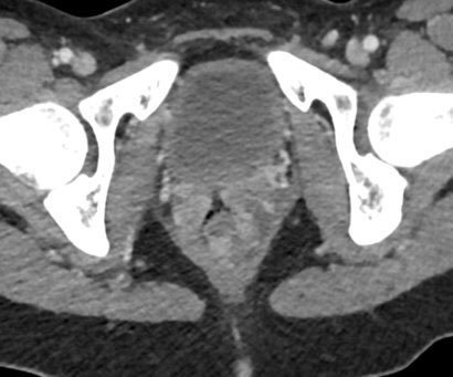
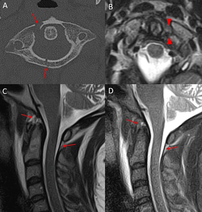
High velocity CCJ #Trauma on #MRI varies by age:
✅ Adults = tectorial membrane tears; hematoma in supradental space (blue).
✅ Children = tectorial membrane is stripped off bone; hematoma in retroclival space (blue).
🔐: lig. laxity ⬇️ w/ age
#NeuroRad#RadRes#Spine
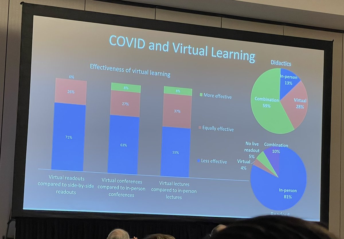
#AUR22 reveal: for those who maintain that remote readouts area just as good/accepted by #radres the data says otherwise. More than 70% say negative impact on learning!
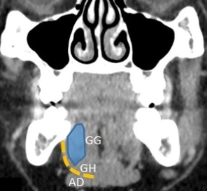
Best way to learn H&N #anatomy#RadRes ➡️ Study your coronal & sagittal imaging!
✅ Mylohyoid (yellow) forms a "sling" separating SL & SM spaces (difficult to appreciate on axials)
GG = genioglossus, GH = geniohyoid, AD = ant. digastric
#Radiology#FOAMRad#MedEd
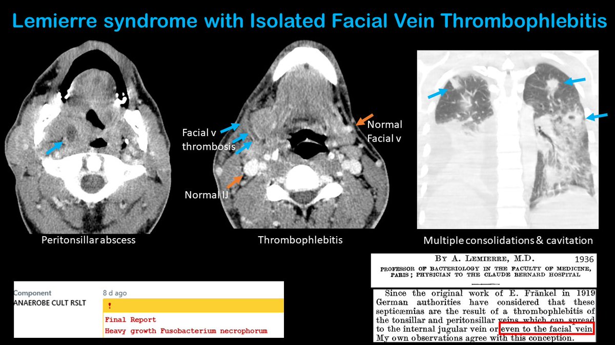
Be on the look out for thrombophlebitis of other veins besides the IJ in the setting of a peritonsillar abscess. This finding is also a feature of Lemierre syndrome and was described by Dr Lemierre himself in 1936! @ASHNRSociety