What happens when allergy meets cardiology? Experts explore Type I #KounisSyndrome in a 52M following a bee swarm attack. A potentially underrecognized condition, it can create a diagnostic blind spot in emergency care
Watch the full discussion: https://t.co/Ky4dDxIe6p #JACCAsia
Neo-LVOT obstruction ends TMVR cases before they start. Automated LVOT analysis + virtual device placement show you the risk before the lab does.
Start a free trial → https://t.co/RRsVnbvyiT
#TMVR#StructuralHeart
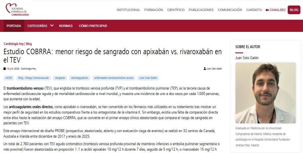
Juan Soto: "El estudio COBRRA cierra una brecha de evidencia de más de una década y refuerza el posicionamiento del apixabán como la opción preferente en el TEV agudo cuando la seguridad hemorrágica constituye la prioridad clínica". #BlogSEC@jovenesSEC https://t.co/TUCSOujofg
#echofirst
✅ Time to close- 🙏 to all comments
✅ Saline contrast study shows prompt contrast filling of RV inflow followed by stalled forward movement beyond mid RV. Color Doppler of apex (see ⬇️) confirms presence of apical ventricular septal rupture with large Left-Right shunt (many of you called it!). Flow through VSR from RV apex to base impedes forward movement of saline contrast & explains why contrast failed to fill entire RV
✅ RVSP was ⬆️ but does not explain by itself the stalled saline contrast movement
@argulian@Hragy@HeartOTXHeartMD@DrRajeshG1@MKaldas@bwoody58
How is a MitraClip implanted?
Transcatheter edge-to-edge repair (MitraClip) is performed under echocardiographic guidance to reduce mitral regurgitation without open-heart surgery.
The procedure follows these key steps:
Cross the interatrial septum to access the left atrium with the delivery system
⬇️
Advance the MitraClip into the left atrium and position it above the mitral valve
⬇️
Open the clip arms and align them perpendicular to the mitral valve coaptation line
⬇️
Advance the clip into the left ventricle through the area of the largest regurgitant jet
⬇️
Retract the clip to grasp both the anterior and posterior mitral leaflets
⬇️
Close the clip, creating a double-orifice mitral valve and reducing mitral regurgitation
Throughout the procedure, transesophageal echocardiography (TEE) is essential for guiding clip positioning, confirming adequate leaflet grasp, and assessing the final reduction in mitral regurgitation.
Reference: Catherine M. Otto, Textbook of Clinical Echocardiography.
Fascicular Ventricular Tachycardias: Potential Role of the Septal Fascicle
*
Read about the supposed anatomic nature of slow conduction area in the left lower corner (click the pic).
https://t.co/z8efJxQjp6
Dosis de radiación & eventos cardíacos. ☢️⚡️❤️🔥
🔷️La dosis recibida por subestructuras cardíacas predice mejor qué complicación CV desarrollará el paciente. 🤔🩻
🔹️Arterias coronarias: mayor dosis → mayor riesgo de infarto y MACE. Umbral sugerido: ADA V15 Gy <10% del volumen o <1 cc, ACX V15 Gy <14% o <1cc.
🔹️Ventrículo izquierdo: la irradiación del miocardio se asocia con insuficiencia cardíaca y deterioro de la función ventricular. Umbral sugerido: V15 Gy <1%.
🔹️Venas pulmonares: su exposición predice mejor la aparición de FA que la dosis recibida por la aurícula.
🔹️Válvulas y pericardio: dosis elevadas incrementan el riesgo de valvulopatía y pericarditis tardías.
🔷️Se propone un enfoque de 3 pilares: 👨⚕️🔎⤵️
✅ Evaluar el riesgo cardiovascular basal antes de iniciar radioterapia.
✅ Optimizar la planificación para reducir la dosis a coronarias izquierdas y ventrículo izquierdo.
✅ Incorporar la dosis cardíaca al seguimiento cardio-oncológico para personalizar la vigilancia.
*V15 Gy significa el porcentaje (o volumen absoluto) de una estructura cardíaca que recibe al menos 15 Gy.
📄🆓️⤵️ State-Of-The-Art Review 2026 @JACCJournals 💯
https://t.co/q2o823ZNdr
https://t.co/XJyYKaKwWR
#JADEL
We've heard about atrial septal pouches.
We may have even seen one.
But, did you ever think you'd see a thrombus-in-situ within an atrial pouch?
CASE reports help expand our understanding of uncommon events.
New in CASE @CASEfromASE@ASE360
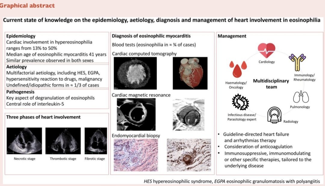
Afección Cardíaca en la Eosinofilia. 🫀💥
🔴La afección cardíaca en la eosinofilia oscila entre el 13%-50% (poco frecuente pero potencialmente mortal). Edad media de aparición de la miocarditis eosinofílica: 41 años. Prevalencia similar en ambos sexos. 🧐🔎
🔴Etiología multifactorial: Síndrome hipereosinofílico, granulomatosis eosinofilica con poliangeitis, reacción de hipersensibilidad a fármacos, maligno/idiopático (1/3 casos). 📊🔥
🔴La enfermedad progresa en 3 fases: dase necrótica (miocarditis eosinofílica), fase trombótica (formación de trombos intracavitarios) y fase fibrótica (fibrosis endomiocárdica con miocardiopatía restrictiva). ❤️🩹❤️🔥
🔴Eco♡: trombos apicales (frecuentemente biventriculares), engrosamiento endomiocárdico, fibrosis apical, ins mitral/tricuspídea secundaria, disf diastólica restrictiva en fases avanzadas. 💢💔
🔴RMC: el hallazgo más característico es el realce tardío subendocárdico difuso, además de edema, fibrosis y trombos intracavitarios.💥
🔴Tto: para la IC (GDMT) + anticoagulación si trombos + Corticoides ± inmunosupresor (ciclofosfamida, rituximab, imatinib). 💊💉
📄🆓️⤵️Position Statement 2026 @ESC_Journals 💯
https://t.co/4Ax1YSqnrl
https://t.co/8JIcu6XRIc
ATHEROSCLEROTIC CORONARY PLAQUE IS THE TARGET OF DIAGNOSIS AND THERAPY
We are experiencing (finally) a major paradigm shift in the world of cardiovascular medicine. Our ability to to detect, characterize and follow-up over time Coronary Artery Disease with non invasive tools has reached clinical feasibility levels.
We are there. And we should really embrace this shift to the effect that it may one of the biggest strategic shift in the management of Cardiovascular Disease of the past decades. But this is not happening just because of the technology associated with imaging (in particular Photon Counting CT).
It is happening because of the major leap forward in the area of pharmacologic treatment of Cardiovascular Diseases and associated Complications and because previous models, especially the one based on ischemia showed critical limitations.
We are at the beginning of this phase and it's very exciting. We have to be cautious and careful in what we promise or forecast. However, the almost 3 decades of R&D and scientific data that led to the ultimate deployment of Photon Counting CT gave us the framework and the roots into which anchor the evidence we are going to use and produce in the near future.
Identification and management of non-obstructive high-risk coronary artery plaque
Please read the article and enjoy an in-depth interview between @jhfrudd and the first author, University of Edinburgh's Dr Craig Balmforth
Publication - https://t.co/j3ZQk2CNPa
Podcast - https://t.co/qPPLSvTdhj
In selected LVAD candidates (e.g. redo cardiac surgery following CABG), the outflow graft may be anastomosed to the descending aorta - making it visible in the descending aortic LAX TEE view #echofirst
🚨 Check out the recently published ULYSSES Trial in Europace! @dvidschaack
Read here 👉 https://t.co/toV3Ur6HCE
🔹 Multicenter randomized trial (6 centers, 968 patients)
🔹 Patients undergoing AF/AT ablation randomized to ultrasound-guided vs conventional (palpation-guided) femoral venous access
🔹 Trial stopped early for efficacy after enrollment of half of the planned study population
🔹 Primary endpoint: composite of venous access-site complications within 30 days (AV fistula, pseudoaneurysm, access-site bleeding requiring intervention or prolonged hospitalization)
🔹 Primary endpoint: 0.6% in the ultrasound-guided group vs 3.3% in the conventional group (HR 0.18, 95% CI 0.05–0.63; P=0.02)
Ultrasound-guided vascular access should become the standard of care in EP labs.
@escardio #EHRA_ESC @ehj_ed
Poner número al riesgo cambia la forma de tratarlo: así lo demuestra la nueva declaración AHA/ACC sobre el síndrome cardiovascular-renal-metabólico.
🔗 Descubre el marco C-P-R y todos los umbrales clave: https://t.co/IO3sjSc7Jb
🔢 PREVENT-CVD ≥7,5% a 10 años activa el tratamiento cardiorrenoprotector en diabetes tipo 2.
🫀 PREVENT-IC ≥5% indica medir biomarcadores cardíacos para detectar pre-insuficiencia cardíaca.
⚖️ A mayor riesgo basal, menor número de pacientes a tratar y mayor beneficio absoluto del tratamiento.
MRI late gadolinium enhancement (LGE) and STIR sequences show concordant pericardial thickening in a patient with myopericarditis refractory to three lines of treatment for Still’s disease.
📌PCSK9 inhibitor treatment & outcomes in 👥 with ASCVD but without prior ischaemic events
👉In ASCVD without prior events in clinical practice, PCSK9i mAb was associated w/ lower ischaemic event & mortality rates
#PCSK9i#LLT
Mechanical circulatory support for patients with infarct-related cardiogenic shock: a state-of-the-art review
In this episode of the Heart podcast, Digital Media Editor @jhfrudd is joined by Prof. Jacob Eifer Moller from Odense, Denmark. They discuss his review paper on mechanical circulatory support, along with supporting guidelines and papers in this area.
Podcast: https://t.co/aQQGioFhRI
Paper: https://t.co/DjFxj3J5ei