Research group interested in neuroinflammation, de- and regeneration in the injured or aged CNS. Headed by Prof. Dr. Moons @BiologyKULeuven @KU_Leuven, Belgium
The registration deadline for #EMBORegeneration2026 is approaching: May 20 !!! Don't miss this amazing meeting on the banks of the Danube in Austria: https://t.co/ApxR4HaVtv
🚨Paper Alert!
Check out our latest work, now published in @FrontNeurosci. We show how this remarkable fish surprisingly displays mammalian-like injury responses after optic nerve transection.
🔗 https://t.co/KJtP2gkrYe
@JulieDeSchutter@StevenBergmans@LucaMasin@AnyiZhang2
To reduce the extensive scarring, we performed a partial optic nerve transection in young adults, leaving a portion of the nerve uninjured.
This approach indeed limited scar formation, and it allowed regenerating axons to reach the brain and reinnervate their targets. ✅
A new preprint from our lab is out! 🥳 Huge congrats to @AnyiZhang2 and @LucaMasin for this great paper titled: Repressed mTORC1 signaling and transient dendritic pruning support axonal regeneration! Grateful for the fruitful collaboration with the @filodelbene and @Poulain lab!
🚨New preprint out!🚨
I’m excited to share the latest work by @AnyiZhang2 and me, which shows that mTORC1-dependent transient dendritic pruning is essential for efficient axonal regeneration in spontaneously regenerating zebrafish neurons.
https://t.co/InNouAxI8o
With transcriptomic and functional data, @lucamasin.bsky.social et al. @labmoons.bsky.social show that local glycolysis supports injury-induced axonal regeneration. https://t.co/egNcPKTOSE
📕 In Cellular Neurobiology collection https://t.co/PmLbkZc1SW
#SfN25
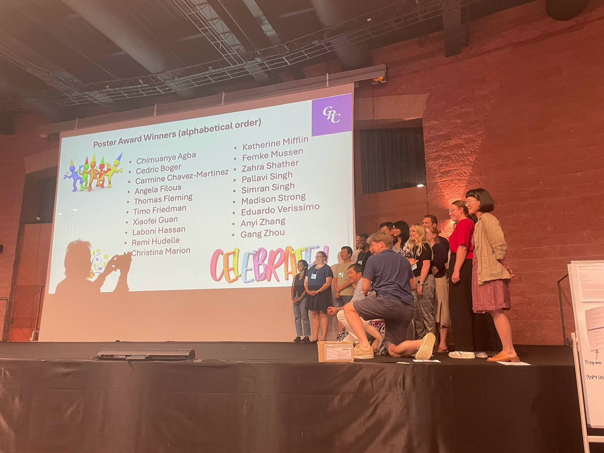
Proud to be one of the best poster presenters of GRC CNS injury and repair meeting 🥳 Big thanks to the organisers giving me this opportunity! Also big thanks to our lab members @LabMoons, especially our excellent post doc @LucaMasin. I could not achieve this without him 😊
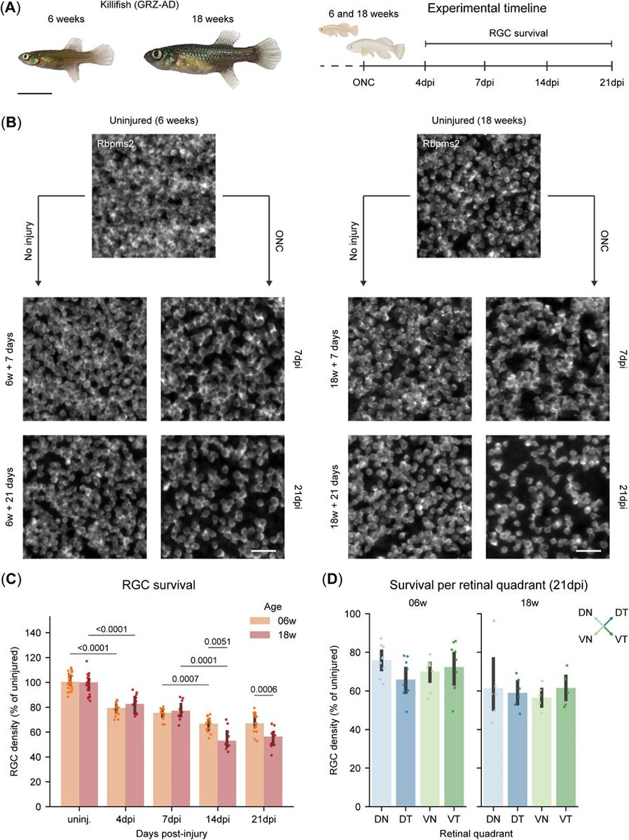
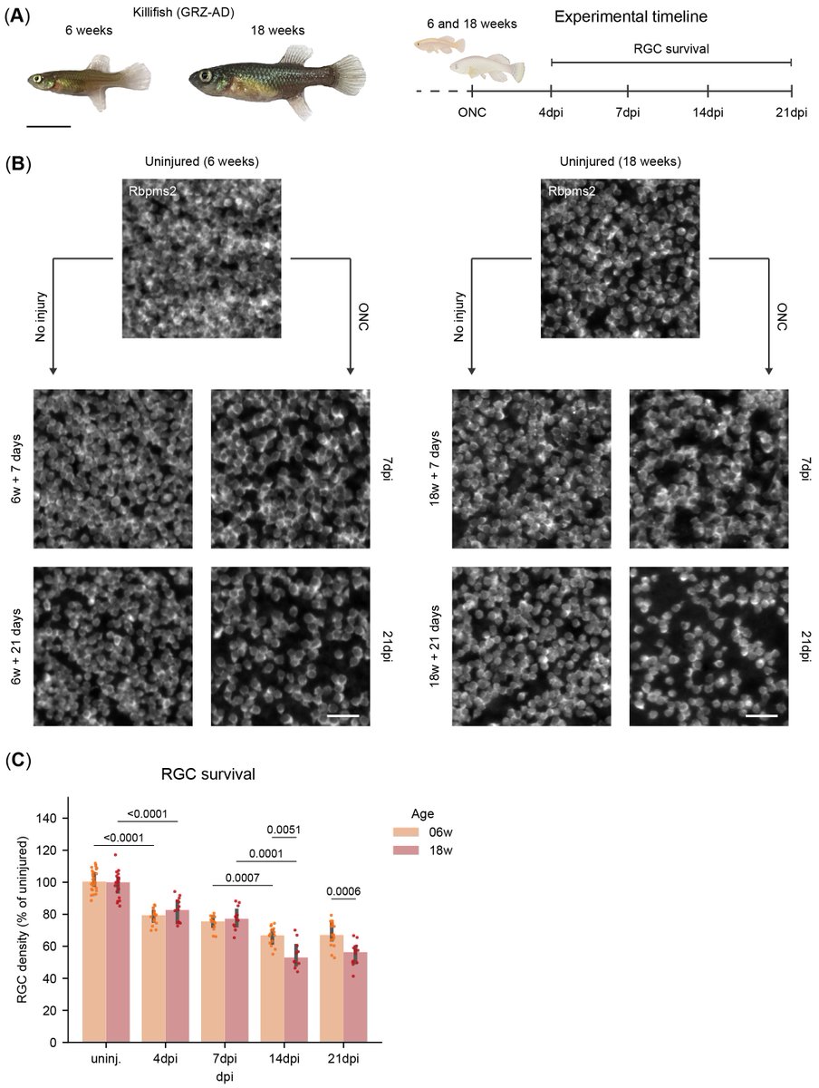
All these data were generated by an updated version of RGCode, RGCode2. An automated cell counting platform able to reliable count murine, zebrafish and killifish retinal ganglion cells using RBPMS and Rbpms2 as marker! RGCode2 is publicly available https://t.co/HTwin6wwxF
Killifish, an aging model shown to lose its regenerative capacity with age, displays a unique biphasic RGC loss profile. Both young adult and old killifish present with a similar degree of RGC loss during the first wave. Old fish, however, lose more RGCs during the second wave.
We have a 3 year postdoc position using the killifish to study retinal ageing available in my lab. Based in London, UK and visa eligible. We’re interested in glial mechanisms underpinning age-related neurodegeneration. If you, or someone you know, is interested please reach out.
Excited to share our new preprint: Successful axonal regeneration is driven by evolutionarily conserved metabolic reprogramming. A joint effort by me, @LucaMasin; @StevenBergmans; @vandyckannelies; AnBeckers; @LabMoons
Killifish, an aging model shown to lose its regenerative capacity with age, displays a unique biphasic RGC loss profile. Both young adult and old killifish present with a similar degree of RGC loss during the first wave. Old fish, however, lose more RGCs during the second wave.
❓ Interested in how different species respond to a central nervous system injury?
💡 Check our latest pre-print on bioRxiv: https://t.co/JZ604St9tu
🧑🔬Wonderful teamwork of the entire lab
@JulieDeSchutter@AnyiZhang2 @PieterJanSerneels @LucaMasin@StevenBergmans
After optic nerve crush (ONC), mice lose ~80% of their RGC by 14 days post injury (dpi). In contract, zebrafish retain nearly all RGC during the first two weeks, before presenting with late RGC degeneration (~13%) by 21dpi.