INTERNISTA 👨⚕️⚕🏥
.W.Osler In Memory. amor por la asistencia, docencia y la investigación. Honrando a mis profesores de semiologia.MIR25
Hoy estudiante de CTO
🚨 The surgeon opened the abdomen—and froze.
The liver was strikingly black, yet smooth and structurally normal. The patient had lifelong mild conjugated hyperbilirubinemia with otherwise normal liver function.
What is the diagnosis?
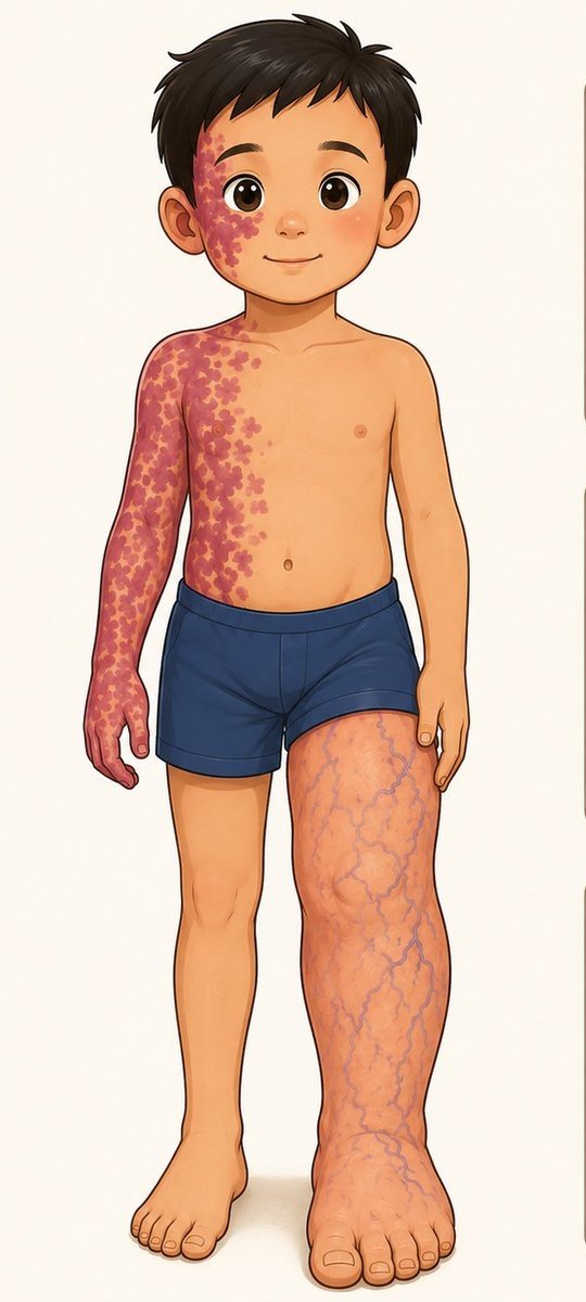
A 7-year-old boy was brought to clinic because his parents noticed that his left leg had been “growing bigger and longer every year.”
What is the diagnosis?
A 1-year-old baby presents with widespread monomorphic, painful vesicles and fever for 4 days. What is the most likely on-the-spot diagnosis?
A) Varicella
B) Eczema herpeticum (Kaposi–Juliusberg syndrome)
C) Bullous impetigo
D) Hand, foot and mouth disease
A 39 yo man presents with 1 week of fever, cough, and progressive shortness of breath despite taking oral antibiotics. He has a history of intravenous drug use and is hypotensive, tachycardic, and tachypneic on admission.
Cardiac examination reveals a new systolic murmur. This is his chest X-ray.
What is the diagnosis, and what is the underlying cause?
A man in his 20s with no past medical and family history presented with dyspnea during strenuous activities.
Denied any drug or substance abuse.
Thoughts?
Spot Diagnosis ❓
A 27-year-old patient with diabetes mellitus presented with diabetic ketoacidosis (DKA).
Despite receiving appropriate DKA management, the patient showed minimal clinical improvement.
Additional findings:
Persistent high-grade fever
During oral cavity examination, the lesion shown in the image below was noteed
❓ What is the most likely diagnosis
A woman in her 70s presented with a “non-growing” palpable mass of 10 × 10 cm2 on the right side of her chest.
This tumor was reported to have been stable for over more than 20 years.
Thoughts?
A man in his 60s w/ CGD (dx in childhood), on TMP-SMX, azithromycin, & itraconazole prophylaxis, presents w/ scattered erythematous scaly pustular plaques on the abdomen & a solitary 1–2 cm well-circumscribed hyperkeratotic, verrucous red–violaceous plaque w/ peripheral erythema & scale on the L lateral lower leg, slowly enlarging over 3 years & asymptomatic.
CT chest shows reticulonodular and alveolar infiltrates with RUL bronchitic/infectious-inflammatory changes.
What’s your DDx❔
A man with diabetes on insulin kept waking up around 2 to 3 am with sweating and trembling.
By morning, his glucose levels were even higher.
Reason?
Should his insulin dose be increased or reduced?
A classic example of the Somogyi phenomenon.
The patient experiences nocturnal hypoglycemia (typically around 2–3 AM) due to excessive evening insulin, manifested by sweating, tremors, and palpitations. In response, the body releases counter-regulatory hormones glucagon, epinephrine, cortisol, and growth hormone which trigger hepatic glucose production, resulting in rebound morning hyperglycemia.
Key Points:-
🔹 Nighttime hypoglycemia → Morning hyperglycemia
🔹 Symptoms at 2–3 AM: sweating, trembling, nightmares
🔹 Caused by excess evening/night insulin
🔹 Management: Reduce the evening insulin dose, not increase it
🚨 MEDICAL CASE CHALLENGE 🚨
Calling all medical students, residents, and doctors! 🩺🧠 Can you identify this definitive dermatological condition?
Case Details:
A young adult male presents with a chronic, severe, and painful inflammatory eruption localized primarily to his hands, distal phalanges, and periungual regions. The examination reveals confluent, pustular lakes, scaly plaques, and significant subungual involvement leading to nail dystrophy. The condition is characteristically resistant to standard topical therapies and slowly progressive, localized to the extremities.
What is the most definitive clinical diagnosis? 👇
A) Acrodermatitis Continua of Hallopeau
B) Impetigo Herpetiformis
C) Von Zumbusch Pustular Psoriasis
D) Palmo-plantar Pustular Psoriasis
Drop your answer and your clinical reasoning in the comments below!
#MedicalCase #Dermatology #MedEd
A woman in her 70s w/ no prior derm hx presented w/ several months of asthenia, weight loss, depression, & a progressive eruption.
Lesions began on the LLE & spread to the gluteal/perineal regions, groin, lower back, & perioral skin.
O/E: annular/polycyclic erythematous plaques w/ advancing erythematous borders, central clearing/healing, scale, crusting, erosions, & residual PIH. Lesions were nonpruritic & nonpainful. She also had glossitis and B/L proximal femoral DVTs.
What’s your DDx❓
A Young patient with huge soft head and neck mass.
Marked eosinophilia.
Biopsy showed aggregate of eosinophilia and lymphatics.
Considered idiopathic disease inflammation of Asian population.
What is the diagnosis?